Introduction
The preimplantation period encompasses a critical time window during which important developmental and molecular events occur in the embryo (Duranthon et al., Reference Duranthon, Watson and Lonergan2008; Palmer & Kaldis, Reference Palmer and Kaldis2016). During this period, the maternal microenvironment plays an active role providing the right physiological conditions to promote developmental potential and pregnancy success (Rizos et al., Reference Rizos, Maillo, Sanchez-Calabuig and Lonergan2017). Within this context, the oviduct provides the first maternal microenvironment that makes contact with the embryo during the early days of embryo development. There is growing evidence that several regulatory proteins secreted by the oviductal cells can perform paracrine actions and activate specific signalling pathways that, in turn, can regulate the gene expression and the developmental potential of the embryo both under in vivo and in vitro conditions (Lee & Yeung, Reference Lee and Yeung2006; Aviles et al., Reference Aviles, Gutierrez-Adan and Coy2010). Among these proteins are a wide range of growth factors that are known mediators of communication between the maternal tract and the embryo (Buhi et al., Reference Buhi, Alvarez and Kouba2000; Robertson et al., Reference Robertson, Chin, Schjenken and Thompson2015).
In the present study, the attention was particularly focused on transforming growth factor-β1 (TGF-β1), a secreted protein that belongs to the TGF-β superfamily. The TGF-β factors exert their biological action as disulfide-linked dimers that bind to cell surface receptor complexes including two type I and two type II serine–threonine kinase receptors (Weiss & Attisano, Reference Weiss and Attisano2013). Activation of the receptors leads to activation of the canonical SMAD signalling pathways through phosphorylation of SMAD2 and SMAD3 proteins (Weiss & Attisano, Reference Weiss and Attisano2013). Subsequently, these SMAD proteins form a heterotrimeric complex with SMAD4, after which they translocate to the nucleus to regulate transcriptional responses (Weiss & Attisano, Reference Weiss and Attisano2013). It has also been shown that TGF-β factors can activate non-SMAD signalling pathways, demonstrating a versatile and complex mechanism of action (Zhang, Reference Zhang2017).
Members of the TGF-β superfamily, including TGF-β1, play an important role in mammalian fertility (Ingman & Jones, Reference Ingman and Jones2008; Ingman & Robertson, Reference Ingman and Robertson2009). These factors act as regulators in ovarian function and oocyte competence (Knight & Glister, Reference Knight and Glister2006). They are also involved in remodelling male and female reproductive tissues, promoting pre- and post-implantation embryo development and mediating trophoblast invasion into the endometrium during implantation (Jones et al., Reference Jones, Stoikos, Findlay and Salamonsen2006; Li, Reference Li2014; Monsivais et al., Reference Monsivais, Matzuk and Pangas2017). Recent studies have demonstrated that TGF-β1 is expressed in the bovine oviduct (Cometti et al., Reference Cometti, Dubey, Imthurn and Rosselli2018) and shows higher transcriptional levels during the postovulatory stage (Tribulo et al., Reference Tribulo, Siqueira, Oliveira, Scheffler and Hansen2018). In addition, the presence of this growth factor in the bovine oviductal fluid and in secretions of oviductal epithelial cells cultured in vitro supports its physiological action as an autocrine/paracrine modulator in the oviductal context (Pillai et al., Reference Pillai, Weber, Phinney and Selvaraj2017). Although it has been evidenced that TGF-β1 alone or in combination with other growth factors and cytokines supports bovine embryo development (Larson et al., Reference Larson, Ignotz and Currie1992; Neira et al., Reference Neira, Tainturier, Pena and Martal2010; Moreno et al., Reference Moreno, Neira, Dubreil, Liegeois, Destrumelle, Michaud, Thorin, Briand-Amirat, Bencharif and Tainturier2015), its action in the early stages during which the embryo transits through the oviduct has not been studied in detail.
Information derived from transcriptome analysis has demonstrated changes in the dynamics of the TGF-β signalling pathway during early embryo development in different species including mouse, human, cattle, sheep and goat (Sudheer & Adjaye, Reference Sudheer and Adjaye2007; Kues et al., Reference Kues, Sudheer, Herrmann, Carnwath, Havlicek, Besenfelder, Lehrach, Adjaye and Niemann2008; Hajian et al., Reference Hajian, Hosseini, Ostadhosseini and Nasr-Esfahani2016; HosseinNia et al., Reference HosseinNia, Tahmoorespur, Hosseini, Hajian, Ostadhosseini, Nasiri and Nasr-Esfahani2016; Zuo et al., Reference Zuo, Su, Wang, Yang, Liao, Wei, Bai and Li2016). These findings suggest that TGF-β signalling plays an important role during earlier, rather than more advanced stages of, preimplantation development. In fact, inhibition of TGF-β signalling in zygotes markedly reduced the subsequent development into the blastocyst stage in bovine species (Zhang et al., Reference Zhang, Rajput, Lee, Wang, Huang, Folger, Knott, Zhang and Smith2015). Moreover, TGF-β ligands together with transcription factors OCT4 and NANOG are responsible for maintaining the pluripotent state in murine and human embryonic stem cells (Beyer et al., Reference Beyer, Narimatsu, Weiss, David and Wrana2013). An interesting fact to emphasize is that TGF-β signalling is also able to modulate gene expression changes through epigenetic regulation mediated by DNA methyltransferase (DNMT) enzymes both in normal and tumour cells (Zhang et al., Reference Zhang, Chen, Helfand, Jang, Sharma, Kozlowski, Kuzel, Zhu, Yang, Javonovic, Guo, Lonning, Harper, Teicher, Brendler, Yu, Catalona and Lee2011; Pan et al., Reference Pan, Chen, Huang, Yao and Ma2013; Cardenas et al., Reference Cardenas, Vieth, Lee, Segar, Liu, Nephew and Matei2014). Hence, the crosstalk between TGF-β signalling and methylation marks could serve as a versatile fine-tuning mechanism for transcriptional regulation during embryonic development.
Despite the evidence described above, there is little information on the effect of exogenous TGF-β factors during the early stages of preimplantation development. Given that TGF-β1 forms part of the oviductal microenvironment, in which the first embryonic stages take place, and that this growth factor is a potential modulator of the pluripotency and epigenetic marks in embryonic stem cells, the objective of the current study was to evaluate whether the addition of TGF-β1 to the embryo culture medium during the first period of development has any effect on embryo development and expression of genes associated with pluripotency and DNA methylation in bovine embryos produced in vitro.
Materials and Methods
Chemicals
All reagents were purchased from the Sigma Chemical Company (St. Louis, MO, USA) unless otherwise stated. Human recombinant TGF-β1 (H8541) was reconstituted according to the manufacturer's recommendations in sterile 4 mM HCl containing 0.1% endotoxin-free recombinant human serum albumin to give a stock concentration of 20 μg/ml. This stock solution was aliquoted and stored at −80°C. The stock solution was diluted before the experiments and a volume of 5 or 10 μl was added to 50 μl microdrops of culture medium (CR1aa) to achieve a final concentration of 50 or 100 ng TGF-β1/ml medium and 0.04 mM HCl (vehicle).
Experimental design
Experiment 1: Gene expression of type I and type II TGF-β receptors in bovine embryos produced in vitro
Considering that TGF-β1 was going to be added to the embryo culture medium during the first stages of development, it was determined whether early bovine embryos could respond to this growth factor. For this purpose, mRNA expression levels of type I and type II TGF-β receptors were measured by RT-qPCR in three pools of 10 embryos harvested at the 2-cell (28 h post insemination, hpi), 4-cell and 8-cell stages (48 hpi), and in three pools of four embryos at the expanded blastocyst stage (8 days pi), obtained by in vitro fertilization (IVF) and cultured in CR1aa medium. Biological replicates were obtained from three independent IVF sessions.
Experiment 2: Development of bovine embryos cultured with or without exogenous TGF-β1 during the first stages of preimplantation development
To evaluate the effect of early addition of TGF-β1 to the culture medium on embryo development, presumptive zygotes were randomly cultured in serum-free CR1aa culture medium for 2 days in the absence or presence of recombinant TGF-β1 and using different experimental groups: I) control (CR1aa supplemented with vehicle; 0.04 mM HCl; n = 236); II) TGF-β1 50 (CR1aa supplemented with 50 ng/ml of recombinant TGF-β1; n = 245) and III) TGF-β1 100 (CR1aa supplemented with 100 ng/ml of recombinant TGF-β1; n = 255). The cleavage rate was recorded 48 hpi (day 2) and then embryos were transferred to CR1aa supplemented with 10% fetal bovine serum (FBS) and cultured up to day 8 pi, maintaining the different experimental groups separately. The cumulative blastocyst rate was determined on days 7 to 8 pi. Development rates were obtained from five replicates performed under the same assay conditions. The experiments were conducted using TGF-β1 concentrations (50 ng/ml and 100 ng/ml) that had been proven to support bovine embryo development in serum-free culture medium in previous studies (Neira et al., Reference Neira, Tainturier, Pena and Martal2010).
Experiment 3: Effect of early TGF-β1 supplementation on mRNA abundance of genes related to pluripotency and DNA methylation in bovine embryos
To evaluate whether the addition of TGF-β1 to the embryo culture medium during the first stages of development had any effect at molecular level, three pools of 10 embryos at the 8-cell stage (48 hpi) and three pools of four embryos at the expanded blastocyst stage (day 8 pi), derived from control and TGF-β1-treated groups, were collected to assess mRNA expression levels of genes related to pluripotency (OCT4, NANOG) and DNA methylation (DNMT1, DNMT3A, DNMT3B) using RT-qPCR. The embryonic stages analyzed (8-cell stage and blastocyst stage) were selected in order to evaluate if early addition of TGF-β1 is able to induce changes in gene expression before and/or after the main phase of embryonic genome activation (EGA) (Graf et al., Reference Graf, Krebs, Zakhartchenko, Schwalb, Blum and Wolf2014).
Oocyte recovery and in vitro maturation
In vitro maturation of bovine cumulus–oocyte complexes (COCs) was carried out as described previously by García et al. (Reference García, Miceli, Rizo, Valdecantos and Barrera2015). Briefly, immature COCs were recovered from heifer ovaries collected at a slaughterhouse by aspiration of follicles (2–8 mm) with an 18-G needle and a 5 ml syringe containing ~1 ml of HEPES-buffered Tyrode's albumin lactate pyruvate medium (H-TALP) plus 10% (v/v) FBS. Groups of 50 COCs containing homogenous oocyte cytoplasm and more than three layers of compact cumulus cells were transferred to 4-well plates (NUNC, Roskilde, Denmark) containing 500 μl of TCM-199 maturation medium (Gibco product 11150–059, Grand Island, NY, USA) supplemented with 10% (v/v) FBS (Internegocios, Buenos Aires, Argentina), 0.05 IU rFSH/ml (Puregon, Organon, Dublin, Ireland), 0.1 mM sodium pyruvate (P4562), 20 μM cysteamine (M9768), and 2% antibiotic–antimycotic solution (Gibco product 15240). Plates were cultured for 22 h at 38.5°C under an atmosphere of 5% CO2 in air with maximum humidity.
In vitro fertilization
For IVF, frozen semen from one bull, previously proven suitable for IVF assays, was thawed in a 35°C water bath for 40 s. Spermatozoa were washed twice in 5 ml of Brackett and Oliphant (BO) medium supplemented with 5 mM caffeine (C4144) and 20 μg/ml heparin (H3149) (BO wash medium), and centrifuged at 1000 rpm for 5 min. After the final wash, the spermatozoa concentration was determined and the pellet was resuspended in 50% (v/v) BO wash medium and 50% (v/v) BO medium with 20 mg/ml fatty acid-free bovine serum albumin (FAF BSA, A6003) as previously described by García et al. (Reference García, Miceli, Rizo, Valdecantos and Barrera2015). Sperm concentration was adjusted to 15 × 106 cells/ml and 100 μl droplets were placed under mineral oil (Fisher Scientific, Fair Lawn, NJ, USA) in 60-mm culture dishes. Matured COCs were washed three times with H-TALP and co-incubated in groups of 10 COCs with sperm for 5 h at 38.5°C under 5% CO2 in humidified air.
In vitro embryo culture
Following the IVF period, presumptive zygotes were removed from the insemination droplets and washed with H-TALP. Cumulus cells were removed by repeated pipetting and presumptive zygotes were washed three times with H-TALP and subsequently cultured in groups of 20 in 50 μl of serum-free CR1aa medium [CR1 stock supplemented with BME essential amino acids (20 μl/ml), MEM non-essential amino acids (10 μl/ml), 1 mM l-glutamine, 1% antibiotic–antimycotic solution (Gibco, 15240) and fatty acid free (FAF) BSA (3 mg/ml)], supplemented or not with the recombinant growth factor up to 48 hpi under mineral oil at 38.5°C and a humidified 5% CO2 atmosphere. Next, embryos were transferred and cultured in 50 μl of fresh CR1aa supplemented with 10% (v/v) FBS up to day 8 pi.
RNA extraction, cDNA synthesis and quantitative real-time polymerase chain reaction (qPCR)
To determine mRNA expression levels of the selected genes in in vitro produced bovine embryos, total RNA from pooled embryos was extracted using a NucleoSpin® RNA XS kit (Macherey-Nagel, Düren, Germany) according to the manufacturer's protocol. Briefly, embryos were collected from the culture medium, washed in H-TALP and immediately placed in sterile RNase and DNase-free tubes (0.6 ml) containing 100 μl lysis solution, supplemented with 20 ng of carrier RNA [poly(A) RNA]. Contaminating genomic DNA in each sample was removed from the total RNA preparations by digestion with recombinant RNase-free DNase, which was directly added to the extraction column during the purification process. RNA was then eluted from the purification column with 10 μl of RNase-free H2O and immediately used for cDNA synthesis.
RNA was reverse-transcribed into cDNA using Moloney Murine Leukemia Virus (M-MLV) enzyme (Promega, Madison, WI, USA), oligo(dT)17 primer and random primers. The reaction mixture (25 μl) consisted of 8 μl of total embryonic RNA (heat denatured), 50 mM Tris–HCl (pH 8.3), 75 mM KCl, 3 mM MgCl2, 0.5 mM of each dNTP, 25 pmol of oligo(dT)17, 25 pmol of random primers and 200 units of reverse transcriptase and RNase-free water. Reactions were performed by incubating the mixture in a thermal cycler at 42°C for 90 min followed by a reverse transcriptase inactivation at 94°C for 5 min. The remaining 2 μl of each embryonic RNA sample was incubated without reverse transcriptase (RT) enzyme under the same assay conditions as described above (but scaling the reaction to a lower volume) to create RT-negative controls in order to confirm the absence of genomic contamination. cDNA (or RNA in the case of the RT-negative controls) was stored at −20°C until RT-qPCR amplification.
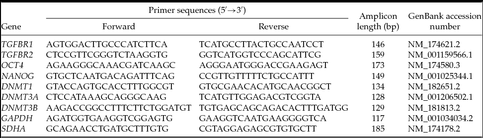
Gene expression levels were determined using specific primers (Table 1). Primers were previously validated for adequate primer efficiency and specificity of their PCR products was confirmed by electrophoresis on a 2.0% agarose gel. All target genes showed acceptable efficiency (97–100%) and correlation coefficients (close to 1.0). PCR reactions were performed in a CFX96™ real-time PCR detection System (Bio-Rad, Hercules, CA, USA) and had a final volume of 20 μl, containing 5 μl of embryonic cDNA template (diluted 1:5), 0.25 mM of forward and reverse primers and 10 μl of Fast EvaGreen qPCR Master Mix (Biotium, Hayward, CA, USA). The PCR program consisted of an initial step of 2 min at 95°C, followed by 48 cycles of 15 s at 95°C and 30 s at 58°C for annealing and extension. At the end of each PCR run, melt curve analysis was performed for all genes to ensure single product amplification and exclude any possible interference of dimers. The melting protocol was a step cycle starting at 60°C and increasing to 95°C with a transition rate of +0.5°C/s increments. Negative controls (sterile RNase and DNase-free water) and RT-negative controls were included in each run. For each embryonic stage, three biological repetitions were performed and the mean Ct value for each repetition was obtained from a technical duplicate. Relative expression levels were quantified by the ΔΔCt method using CFX Manager Software version 3.0 (Bio-Rad Laboratories, Hercules, CA, USA). Values were normalized to the geometric mean of two housekeeping genes, GAPDH and SDHA, which were assayed in previous studies (García et al., Reference García, Miceli, Rizo, Valdecantos and Barrera2015). In our determinations, GAPDH and SDHA produced uniform expression levels varying less than 0.5 Ct between control and treated cDNA samples. The target stability function of the CFX96 software determined that the combined M-value for GAPDH and SDHA was M = 0.37 (CV = 0.13). The entire study was carried out according to the Minimum Information for Publication of Quantitative Real-Time PCR Experiments (MIQE) guidelines (Bustin et al., Reference Bustin, Benes, Garson, Hellemans, Huggett, Kubista, Mueller, Nolan, Pfaffl, Shipley, Vandesompele and Wittwer2009).
Primers used for RT-qPCR analysis

bp: base pairs.
Statistical analysis
Data were analyzed using SigmaStat 3.5 and SigmaPlot 10.0 statistical software (Systat Software, Richmond, CA, USA). One-way analysis of variance (ANOVA) and subsequent multiple pair-wise comparisons using Tukey's test when applicable, were used to analyzed cleavage and blastocyst rates and to detect differences in mRNA abundance between the different experimental groups. P-values < 0.05 were considered statistically significant.
Results
Relative mRNA abundance of TGF-β receptors in bovine embryos during preimplantation development
As a first approach to confirm expression of TGF-β receptors in early stage bovine embryos, the relative abundance of TGFBR1 and TGFBR2 mRNA was evaluated by RT-qPCR in IVF embryos at the 2-cell, 4-cell and 8-cell stages. For comparative purposes, expanded blastocysts were also evaluated.
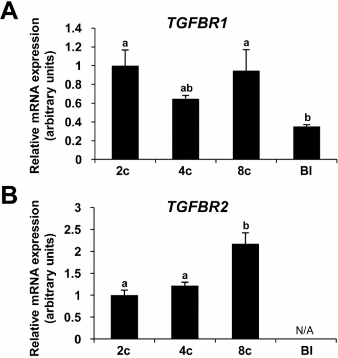
As shown in Fig. 1A, TGFBR1 mRNA was detected during all embryonic stages examined, showing a higher relative abundance during the early stages of preimplantation development than during the blastocyst stage (P < 0.05). Conversely, TGFBR2 expression was detected from the 2-cell to 8-cell stage with a greater relative abundance during the 8-cell stage, but transcripts were not detected during the blastocyst stage (Fig. 1B).
Relative mRNA abundance of type I and type II TGF-β receptors in bovine embryos during preimplantation development using quantitative real-time PCR. (A) Bars represent TGFBR1 mRNA levels during early development stages normalized to the geometric mean of GAPDH and SDHA housekeeping genes. (B) Bars represent TGFBR2 mRNA levels during early development stages normalized to the geometric mean of GAPDH and SDHA housekeeping genes. Results are expressed as means ± standard error of the mean (SEM). Significant differences (P < 0.05) are indicated with different letters. TGFBR1 and TGFBR2: type I and type II transforming growth factor-beta receptors; mRNA, messenger RNA; 2c, 2-cell stage; 4c, 4-cell stage; 8c, 8-cell stage; Bl, expanded blastocyst stage.

Development rate of bovine embryos cultured in the presence or absence of TGF-β1 during the first embryonic stages
After confirming the expression of TGF-β receptors in early stage embryos, the next objective was to evaluate the effect of TGF-β1 supplement during the initial days of embryo culture. Addition of 50 ng/ml or 100 ng/ml of TGF-β1 to the embryo culture medium during the first 48 hpi did not affect the cleavage rate when compared with the control group (Table 2, P > 0.05). Similarly, the proportion of embryos that reached the first, second and third cell cycles evaluated at 48 hpi did not show any significant difference between TGF-β1 treated and control groups (Table 2, P > 0.05). Moreover, the addition of TGF-β1 to the embryo culture medium during the first stages of development did not show any significant effect on blastocyst yield when compared with the control group (Table 2, P > 0.05).
Development rate of in vitro fertilized bovine embryos cultured in the absence or presence of 50 or 100 ng/mL TGF-β1 during the first 48 hpi

The table shows the total number of oocytes used and embryos developed in a total of five different IVF experiments. Zygotes were cultured in CR1aa medium assaying three different treatments: (I) control (CR1aa + vehicle); (II) TGF-β1 50 (CR1aa + 50 ng/mL TGF-β1) and (III) TGF-β1 treatment (CR1aa + 100 ng/mL TGF-β1). At 48 hpi (day 2), developing embryos were transferred to CR1aa supplemented with 10% FBS and cultured until day 8 pi.
a Percentage of presumptive zygotes that undergo cleavage evaluated at day 2 pi.
b Percentage of cleaved embryos that developed to the 2-, 4- and 8-cell stages at day 2 pi.
c Cumulative blastocyst rate evaluated on days 7 and 8 pi.
Values are expressed as mean ± standard error of the mean (SEM).
Effect of early addition of TGF-β1 on expression levels of genes associated with pluripotency and DNA methylation in bovine embryos
Relative transcriptional levels of OCT4 and NANOG (pluripotency related genes) and DNMT1, DNMT3A, and DNMT3B (DNA methylation related genes) were determined in 8-cell stage embryos and expanded blastocysts derived from control and TGF-β1 treated groups according to the experimental design.
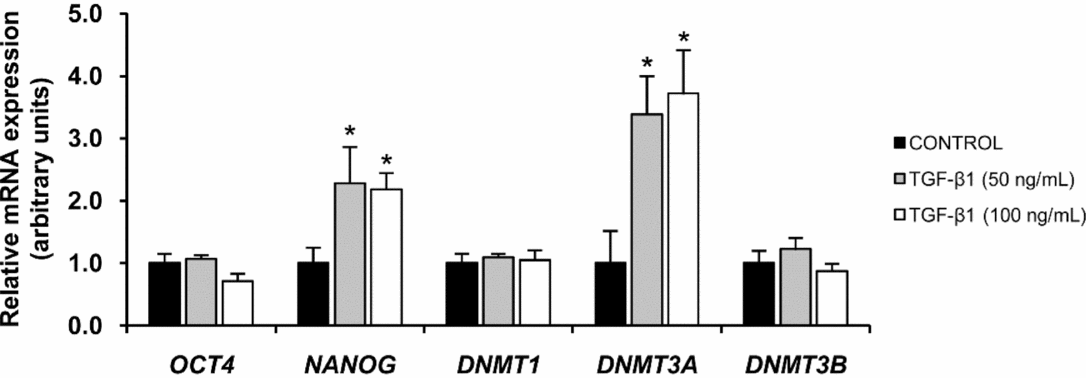
During the 8-cell stage, transcript abundance of OCT4 mRNA did not significantly differ among embryos derived from the different experimental groups (Fig. 2). However, the transcription level for NANOG significantly increased in embryos exposed at an earlier stage to both TGF-β1 concentrations compared with control embryos (Fig. 2, P < 0.05). Relative mRNA expression for DNMT3A also increased in 8-cell embryos cultured in the presence of 50 and 100 ng/ml of TGF-β1 compared with transcript levels in controls (Fig. 2, P < 0.05). Nevertheless, relative mRNA abundance of DNMT1 and DNMT3B gene transcripts of 8-cell stage embryos from TGF-β1 treated groups and the control group was similar (Fig. 2).
Quantitative real-time PCR analysis of relative abundance of genes related to pluripotency and DNA methylation in 8-cell embryos cultured in the presence or absence of TGF-β1. Bars represent relative abundance of OCT4, NANOG, DNMT1, DNMT3A and DNMT3B transcripts. Relative gene expression levels were normalized to the geometric mean of the endogenous controls GAPDH and SDHA. The control group is represented by black columns and TGF-β1 treated embryos are represented by grey and white columns. Results are expressed as means ± standard error of the mean (SEM). Asterisks indicate significant differences (P < 0.05) between TGF-β1 treated and control groups. Data were obtained from three replicates of independent groups of 10 embryos.

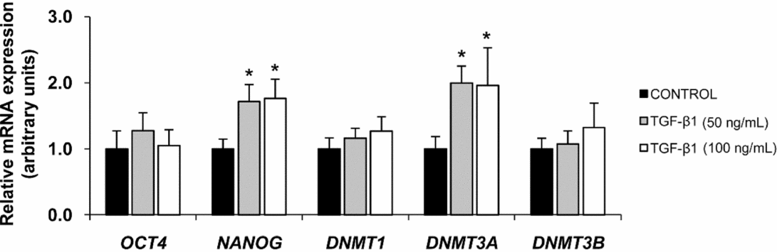
Gene expression analysis during the blastocyst stage showed that OCT4 transcript levels from TGF-β1 treated groups and the control were similar (Fig. 3). In contrast, the relative abundance of NANOG mRNA was higher in expanded blastocysts developed from embryos cultured in medium with TGF-β1 than blastocysts from the control group (Fig. 3; P < 0.05). Similarly, the early addition of TGF-β1 to the culture medium significantly increased DNMT3A mRNA levels in expanded blastocysts from the different treatment groups compared with blastocysts from the control group (Fig. 3; P < 0.05). However, mRNA expression levels for DNMT1 and DNMT3B were not altered after the addition of TGF-β1 (Fig. 3).
Quantitative real-time PCR analysis of relative abundance of genes related to pluripotency and DNA methylation in expanded blastocysts derived from embryos cultured in the presence or absence of TGF-β1. Bars represent the relative abundance of OCT4, NANOG, DNMT1, DNMT3A and DNMT3B transcripts. Relative gene expression levels were normalized to the geometric mean of two endogenous control genes: GAPDH and SDHA. The control group is represented by black columns and TGF-β1 treated embryos are represented by grey and white columns. Results are expressed as means ± standard error of the mean (SEM). Asterisks indicate significant differences (P < 0.05) between TGF-β1 treated and control groups. Data were obtained from three replicates of independent groups of four expanded blastocysts.

Discussion
The present study has demonstrated that bovine embryos can respond to TGF-β1 action during the first stages of preimplantation development. A high abundance of TGFBR1 and TGFBR2 mRNA was observed between the 2-cell and 8-cell stages before maternal–embryonic transition. In contrast, a significant decrease in TGFBR1 mRNA levels was determined during the blastocyst stage, and expression of TGFBR2 was not detected, which may indicate that transcript products for this gene reflect a maternal origin. These results together manifest a stage-specific regulation of transcript abundance for both receptors during bovine early embryogenesis. In addition, the expression of TGF-β receptor mRNA in all preimplantation embryo stages examined supports the hypothesis that TGF-β1 could interact with the embryo while traversing the oviduct and during its further development in the uterus. Consistent with these results, genome-wide transcriptome analysis in mouse, human, cattle, sheep and goat embryos has demonstrated a similar expression profile for these TGF-β transcripts and others (Sudheer & Adjaye, Reference Sudheer and Adjaye2007; Kues et al., Reference Kues, Sudheer, Herrmann, Carnwath, Havlicek, Besenfelder, Lehrach, Adjaye and Niemann2008; Hajian et al., Reference Hajian, Hosseini, Ostadhosseini and Nasr-Esfahani2016; HosseinNia et al., Reference HosseinNia, Tahmoorespur, Hosseini, Hajian, Ostadhosseini, Nasiri and Nasr-Esfahani2016; Zuo et al., Reference Zuo, Su, Wang, Yang, Liao, Wei, Bai and Li2016), strongly suggesting that TGF-β signalling is a key regulator of early embryonic development.
Given the temporal gene expression pattern of TGF-β receptors in bovine embryos produced in vitro, the addition of TGF-β1 to the culture medium during the first 48 hpi was assessed to determine its early action on embryonic development. Our findings show that the addition of 50 or 100 ng/ml of TGF-β1 had no effect on the cleavage rate and developmental kinetics 48 hpi. These observations are in agreement with other studies that showed that the addition of TGF-β1 to the embryo culture medium at concentrations between 50 and 100 ng/ml did not alter the first cleavage rates (Neira et al., Reference Neira, Tainturier, Pena and Martal2010). Similarly, TGF-β1 treatment did not have any apparent effect on blastocyst rates. Previous studies have provided evidence that the addition of TGF-β1 to serum-free embryo culture medium produced a beneficial effect on bovine embryo development (Larson et al., Reference Larson, Ignotz and Currie1992; Neira et al., Reference Neira, Tainturier, Pena and Martal2010). However, other studies have determined that TGF-β1 did not affect bovine blastocysts development when 2-cell stage embryos were cultured in the presence of this growth factor (Keefer et al., Reference Keefer, Stice, Paprocki and Golueke1994). Probably, the timing of TGF-β1 addition to the embryo culture medium is a critical variable to be considered before evaluating and comparing these results.
In most studies, growth factors such as TGF-β1 are usually added during the entire period of embryo culture (7–8 days). Taking into account that TGF-β superfamily members are also present in the uterine fluid (Jones et al., Reference Jones, Stoikos, Findlay and Salamonsen2006) and that bovine blastocysts express TGF-β receptors, as was shown in the present study, it is possible to suggest that the embryo can interact and respond to TGF-β1 in the uterine environment. This fact could explain why an extended period of culture with TGF-β1 promotes blastocyst development. It is important to note that TGF-β1 addition in the present study was carried out during the first 2 days of embryo culture. Considering that the bovine embryo spends 3.5–4 days in the oviduct (Maillo et al., Reference Maillo, Lopera-Vasquez, Hamdi, Gutierrez-Adan, Lonergan and Rizos2016), the time period analyzed in the current study partially mimics this time window, focusing attention on developmental stages before the main phase of EGA. Although no effect was observed in the embryo development rates, it cannot be ruled out that certain indicators of embryo quality could be influenced by the exogenous action of TGF-β1. Previous studies have demonstrated that TGF-β1 increases the total cell number in mouse blastocysts derived from embryos cultured from the 8-cell stage onwards in the presence of this growth factor (Lim et al., Reference Lim, Bongso and Ratnam1993). Moreover, the addition of growth factors and cytokines, such as TGF-β1, IGF-I, IGF-II, bFGF, LIF and GM-CSF to embryo culture medium has been shown to increase the cell number of the inner cell mass and trophoblast in bovine blastocysts (Neira et al., Reference Neira, Tainturier, Pena and Martal2010). However, further studies are required to elucidate the precise effect of TGF-β1 on embryo quality parameters of bovine blastocysts produced in vitro.
Substantial evidence provided in several studies supports the pivotal role of TGF-β signalling in different cellular processes including proliferation, migration, differentiation, pluripotency, and even epigenetic modifications (Beyer et al., Reference Beyer, Narimatsu, Weiss, David and Wrana2013; Bai & Xi, Reference Bai and Xi2018). Clearly, all these process are critical to early embryo development. Considering this factor, it was decided to examine if the early addition of TGF-β1 to the embryo culture medium would affect mRNA abundance of developmentally important genes during embryonic stages before and after EGA. The first genes assayed were OCT4 and NANOG, two important transcription factors that play a key role in embryonic development and stem cell pluripotency (Loh et al., Reference Loh, Wu, Chew, Vega, Zhang, Chen, Bourque, George, Leong, Liu, Wong, Sung, Lee, Zhao, Chiu, Lipovich, Kuznetsov, Robson, Stanton, Wei, Ruan, Lim and Ng2006). It has been shown that TGF-β family signals play a critical role in the maintenance of the pluripotent state of ES cells by inducing Nanog and Oct4 expression (Brons et al., Reference Brons, Smithers, Trotter, Rugg-Gunn, Sun, Chuva de Sousa Lopes, Howlett, Clarkson, Ahrlund-Richter, Pedersen and Vallier2007; Itoh et al., Reference Itoh, Watabe and Miyazono2014). Our findings have revealed that the addition of 50 or 100 ng/ml of TGF-β1 to the culture medium up to 48 hpi produced a significant increase in the relative abundance of NANOG mRNA, both in 8-cell stage embryos and expanded blastocysts. These results indicated that exogenous addition of TGF-β1 has a stimulatory effect on the gene expression of specific pluripotency markers in the early stages of bovine embryo development, and also in the more advanced stages after EGA (6 days after TGF-β1 treatment). Interestingly, SMAD2/3, signal transducers involved in TGF-β signalling, can bind directly to the NANOG promoter, thus enhancing its activity and translates to an increase in NANOG expression in human embryonic stem cells (Xu et al., Reference Xu, Sampsell-Barron, Gu, Root, Peck, Pan, Yu, Antosiewicz-Bourget, Tian, Stewart and Thomson2008). It is important to note that NANOG is synthesized by the embryo from the 8-cell stage onwards and has been proposed as a candidate factor for early inner cell mass specification and maintenance of pluripotency during bovine preimplantation development (Khan et al., Reference Khan, Dube, Gall, Peynot, Ruffini, Laffont, Le Bourhis, Degrelle, Jouneau and Duranthon2012). Therefore, our findings suggest that exogenous activation of TGF-β signalling during the first stages of embryo development could subsequently contribute to pluripotency mechanisms that need to be activated to initiate the first step of differentiation.
Expression of three key epigenetic regulators of gene expression (DNMT1, DNMT3A and DNMT3B) was also evaluated. DNA methyltransferases are known to be involved in the establishment of genomic methylation markers, which are essential for normal embryo development (Edwards et al., Reference Edwards, Yarychkivska, Boulard and Bestor2017). Recent studies have demonstrated the existence of an active crosstalk between TGF-β signalling and the methylation machinery that could serve as a versatile fine-tuning mechanism for transcriptional regulation during embryonic development (Bai & Xi, Reference Bai and Xi2018). Within this context, our results have shown that the early addition of 50 and 100 ng/ml TGF-β1 to the culture medium produced a significant increase in the relative abundance of DNMT3A mRNA in 8-cell and blastocyst stage embryos. In agreement with these results, studies in ovarian cancer cells have shown that 48 h after TGF-β stimulation, mRNA and protein levels of DNMT3A were increased significantly (Cardenas et al., Reference Cardenas, Vieth, Lee, Segar, Liu, Nephew and Matei2014). Considering our results, early exposure of bovine embryos to TGF-β1 could modify the transcriptional levels of DNMT3A before and after EGA. DNA methylation levels are known to dynamically change in bovine embryos during preimplantation development, showing remethylation of the genome from the 8-cell to the 16-cell stage (Dobbs et al., Reference Dobbs, Rodriguez, Sudano, Ortega and Hansen2013). Therefore, TGFβ-1 could be able to modulate mRNA abundance of DNMT3A to contribute to the establishment of new methylation patterns in the early embryo. However, further studies are required to confirm this hypothesis.
In conclusion, the results of the current study have demonstrated temporal changes in mRNA abundance for TGF-β receptors (TGFBR1 and TGFBR2) with a higher relative abundance during early stages (2-cell, 4-cell and 8-cell stages) compared with the blastocyst stage. This finding suggests that the bovine embryo could be the target of TGF-β during early embryogenesis. Although early addition of TGF-β1 to the culture medium did not have any effect on embryo development, the addition of this growth factor can affect the molecular characteristics of developing embryos by inducing changes in the transcript abundance of NANOG and DNMT3A during early and more advanced stages of preimplantation development. Consequently, TGF-β1 could act selectively during specific embryonic stages with a positive effect on developmentally related genes. Therefore, our results highlight the importance of providing a more comprehensive understanding of the role of this signalling pathway during early embryogenesis, particularly in farm animals.
Acknowledgements
The authors would like to thank Calchaquí and Industrial del Norte S.A. slaughterhouses in Tucumán, Argentina, for providing biological material. The authors would also like to thank Cabaña ‘La Lilia’ (Colonia Aldao, Santa Fe, Argentina) for providing bull semen samples.
Financial support
This study was supported by research grants from Agencia Nacional de Promoción Científica y Tecnológica of Argentina (A.D.B., grant BID PICT 2012 No. 0401), Consejo Nacional de Investigaciones Científicas y Técnicas (A.D.B., grant PIP No. 724) and Consejo de Investigaciones de la Universidad Nacional de Tucumán, Argentina (D.C.M., grant 26/D525).
Competing interests
The authors declare that they have no competing interests.
Ethical standards
Not applicable.