

1. Introduction: The Structure of Biological Theories
Until fairly recently, many of the analyses of theories in the biological and biomedical sciences had subscribed to what I term the “Euclidean ideal.” This notion assumes that the ideal structure of a scientific theory resembles Euclid's approach to geometry: a small number of fundamental definitions and axioms constitute the essence of a theory. The axioms are formulated in mathematical symbols and are then elaborated deductively in the form of theorems and applications that cover a broad (scientific) domain. This view of theory structure obtains fairly strong support in the physical sciences—in, say, mechanics, thermodynamics, electromagnetics, and quantum mechanics (see, e.g., Stratton Reference Stratton1941; von Neumann Reference von Neumann and Beyer1955). But a similar orientation toward general theory in biology also can be found in the work of von Bertalanffy (Reference von Bertalanffy1968) on general system theory and his sets of multiple partial differential equations and in the kinds of theories found in the work of Waddington (Reference Waddington and Waddington1968).
These biologists, as well as some philosophers of biology, such as the early Michael Ruse (Reference Ruse1973), had maintained that the laws and theories of biology have the exact same logical structure as do those of the physical sciences (though some changes in this approach began to surface in the 1980s; see Schaffner Reference Schaffner1980; Kitcher Reference Kitcher1984; Rosenberg Reference Rosenberg1985; Culp and Kitcher Reference Culp and Kitcher1989; and my elaboration on these developments in Schaffner Reference Schaffner1993, esp. Chapter 3). This simple unity view is actually supportable only with the use of almost atypical examples, such as certain formulations of Mendelian genetics and of population genetics, especially Jacquard's axiomatization of Sewell Wright's theory, as summarized in Schaffner Reference Schaffner1993, 341–343. But a deeper analysis of even these theories will disclose difficulties with a strong methodological parallelism with the physical sciences (see Schaffner Reference Schaffner1980, Reference Schaffner1986; Kitcher Reference Kitcher1984). A close examination of a wide variety of other biological theories in genetics, immunology, physiology, embryology, and the neurosciences suggests that the typical theory in the biomedical sciences is a structure of overlapping interlevel, causal, and temporal prototypical models.
The models of such a structure usually constitute a series of idealized prototypical mechanisms and variations (some of which may be mutants) that bear family or similarity resemblances to each other, and characteristically each has a (relatively) narrow scope of straightforward application to (few) pure types. The models (or mechanisms) are typically interlevel, in the sense of levels of aggregation containing component parts that are often specified in intermingled body part (e.g., head or tail), cellular (e.g., neuron or axon), and biochemical (e.g., receptor or ions) terms. I have suggested that this different type of theory might be termed a “theory of the middle range” (with apologies to Robert K. Merton [Reference Merton and Merton1968], who first used that term in a somewhat different context) and that it both is found and should be expected to be found in the biomedical sciences (Schaffner Reference Schaffner1980).
Although the Waddington and von Bertalanffy programs have been confirmed in neither biology in general nor molecular genetics in particular, there are interesting advances that fall between those searches for broad theories couched in mathematically precise differential equation form and the narrow classes of mechanisms, usually described in qualitative multilevel causal language, that constitute the vast majority of current biomedical explainers.
There are several other equation-based theories that can be identified in contemporary biomedicine, and in the remainder of this article I discuss one of these in detail. They can disclose some important ways that very general and quantitative principles can be applied fruitfully in biology and medicine. They also disclose the limitations of this kind of physics-oriented approach to biology, and a comparison of those areas where mathematical modeling works and at what points it begins to fail may even indicate ways that what is now referred to by the buzzword “systems biology” can approach the issues of theories, models, and equations in this nascent area.Footnote 1
I now turn to a brief account of the development of the Hodgkin-Huxley (HH) giant squid model for action potentials, a stunning accomplishment for which Hodgkin and Huxley shared the Nobel Prize in Physiology or Medicine in 1963. One of the current standard textbooks of neuroscience (Kandel, Schwartz, and Jessell Reference Kandel, Schwartz and Jessell2000, 156) states that, 50 years after its publication, “the Hodgkin-Huxley model stands as the most successful quantitative computational model in neural sciences if not all of biology.”Footnote 2
2. The Development of the HH Giant Squid Model for Action Potentials
Action potentials are waves of potential difference (or voltage) that move down nerve axons, communicating the effect of a stimulus from the receptors located near the beginning of the neuron to the termination of the nerve cell. To a first approximation, action potentials are the result of rapid (millisecond) changes in the membrane's permeabilities to sodium and potassium ions, changes that underlie the wave of potential difference. Hodgkin and Huxley's work culminated in their extraordinary 1952 article in the Journal of Physiology, in which they systematically lay out the steps and their reasoning that culminates in the classic action-potential model of nerve transmission (Hodgkin and Huxley Reference Hodgkin and Huxley1952).
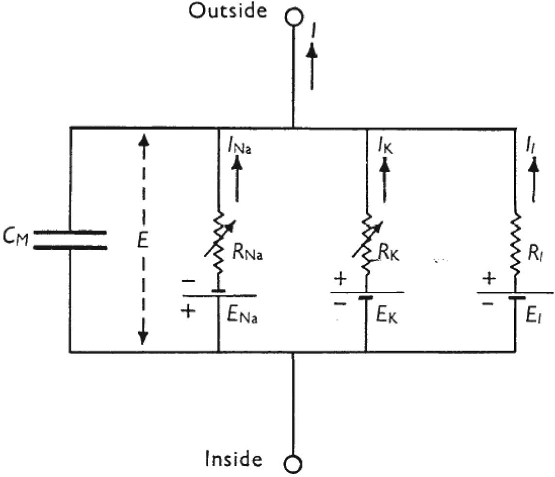
The 1952 paper closely parallels their more historical account of their steps toward their quantitative model that appears in the two Nobel Prize lectures (Hodgkin Reference Hodgkin1964; Huxley Reference Huxley1964). They begin by first discussing their careful experimental results, which had employed the voltage clamp apparatus developed in 1949 by Kenneth Cole. This experimental device permits the establishment of a set of different potential differences across the squid nerve cell membrane and a recording of the effects that the different membrane potentials have on the state of the cell. (A detailed description of the apparatus and technique can be found in the text box in Kandel et al. Reference Kandel, Schwartz and Jessell2000, 152.) Their earlier papers had indicated that the movement of currents based on ions across the nerve cell membrane could be well represented by an “equivalent circuit” involving a capacitor and three resistors, all in parallel, and with each resistor in series with a source of an electrical potential difference. This circuit captures the sodium (Na) and potassium (K) currents, as well as a small leakage current (l). This equivalent circuit, adapted from their 1952 paper, is shown in Figure 1 (cf. Huxley Reference Huxley1964). The “laws of working” (a term originally used by John Mackie, but see my discussion of the phrase in Schaffner Reference Schaffner1993, 287, 306–307) that govern this circuit are the standard physical laws, including Ohm's law as noted in the legend of Figure 1. Additionally, the potential difference across the membrane established by differences in the Na and K ions is as required by the Nernst equation

where V is the potential difference (voltage), R and F are the universal Boltzmann and Faraday constants, T is the temperature, z is the valence of the ion, and X o and X i are the concentrations of the ion outside and inside the cell. (Such laws are constraints and foundations but are not the complete derivational source for the later HH equations I introduce further below [also see Bogen Reference Bogen2005 and Craver Reference Craver2007 on this point, as well as the other articles in this symposium issue].)
Equivalent electrical circuit of a small area of membrane of the giant axon. R K and R Na obey Ohm's law for rapid changes in the potential difference across the membrane but change their values in times of the order of a millisecond if the membrane potential is held at a new value.  $R_{l}$ is constant.
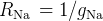 $R_{\mathrm{Na}\,}=1/ g_{\mathrm{Na}\,}$;
 $R_{\mathrm{K}\,}=1/ g_{\mathrm{K}\,}$;
 $R_{l}=1/ g_{l}$.
 $R_{\mathrm{Na}\,}$ and
 $R_{\mathrm{K}\,}$ vary with time and membrane potential; the other components are constant. From Hodgkin and Huxley Reference Hodgkin and Huxley1952, 501.

Part II of the HH paper is a “mathematical description of membrane current during a voltage clamp.” Equations for the sodium and potassium currents as conductances are developed. The equations do not come from “first principles” but rather are empirical equations fitted from the voltage clamp data. They are typically chosen on the basis of simplicity, with a first-order equation being preferred over a second-order one, and so on. A first-order equation is satisfactory to represent a portion of the time course of nerve depolarization (a rapid change of voltage across the membrane), but a fourth-order equation is needed to represent the beginning of the potassium depolarization process. The equation for potassium conductance, in the form that it could be compared with the empirical results, was chosen as

It is a theoretical equation, to use Hodgkin and Huxley's language, based on the equivalent circuit and the general empirically found form of the rise and fall of ion conduction during depolarization and repolarization. Hodgkin and Huxley doubt it gives a “correct picture” of the membrane, though they do provide a possible physical basis for the equation (see Reference Hodgkin and Huxley1952, 506–507). The equation contains a constant,  $\tau _{n}$, that can then be specified to be the best fit to experimentally determined depolarizations of different potential membrane differences. Hodgkin and Huxley found that there was reasonable agreement between theoretical and experimental curves. They then go on to develop the somewhat more complex reasoning leading to the equation for sodium conductance, which I shall not discuss but which can be found in Hodgkin and Huxley Reference Hodgkin and Huxley1952, 512–515. They also develop equations for rate constants α and β and the dimensionless proportions n, m, and h of ions inside and outside the membrane, in Part II as well.
At the beginning of Part III, titled “Reconstruction of Nerve Behavior,” Hodgkin and Huxley summarize the equations they developed in Part II. That summary is reproduced here, with the equations numbered according to their original equation numbers in Hodgkin and Huxley Reference Hodgkin and Huxley1952:


The first four of these equations are the differential equations that govern the system's behavior. The many computer simulations of the HH model involve programs that repeatedly step through those first four equations (see Fodor Reference Fodor2007 for one example).
Equation (26) is then applied to the action potential. We are most interested in the “propagated action potential,” as distinguished from a uniform membrane action potential. In the propagated action potential, the local circuit currents have to be provided by the net membrane current. At this point in their paper, Hodgkin and Huxley (Reference Hodgkin and Huxley1952) appeal to a well-known partial differential equation from cable theory (which is a variant of Laplace's well-known heat-diffusion partial differential equation) relating the current to the second partial derivative of the potential difference (V) with respect to distance (x). This equation is given by the expression

There are some simplifications then invoked; for example, since  $r_{1}\ll r_{2}$,
 $r_{1}$ can be dropped. The expression for the current density for the fiber with a radius of a then allows the equation to be rewritten as

This relation is then substituted into equation (26), which yields a partial differential equation that is “not practical to solve as it stands” (Reference Hodgkin and Huxley1952, 522). But a similarity is noted for the condition of steady propagation, one that permits the equation to be converted into an ordinary differential equation that can be solved numerically, if laboriously, given the computational tools available in Hodgkin and Huxley Reference Hodgkin and Huxley1952. This is the propagated action potential equation and was written as

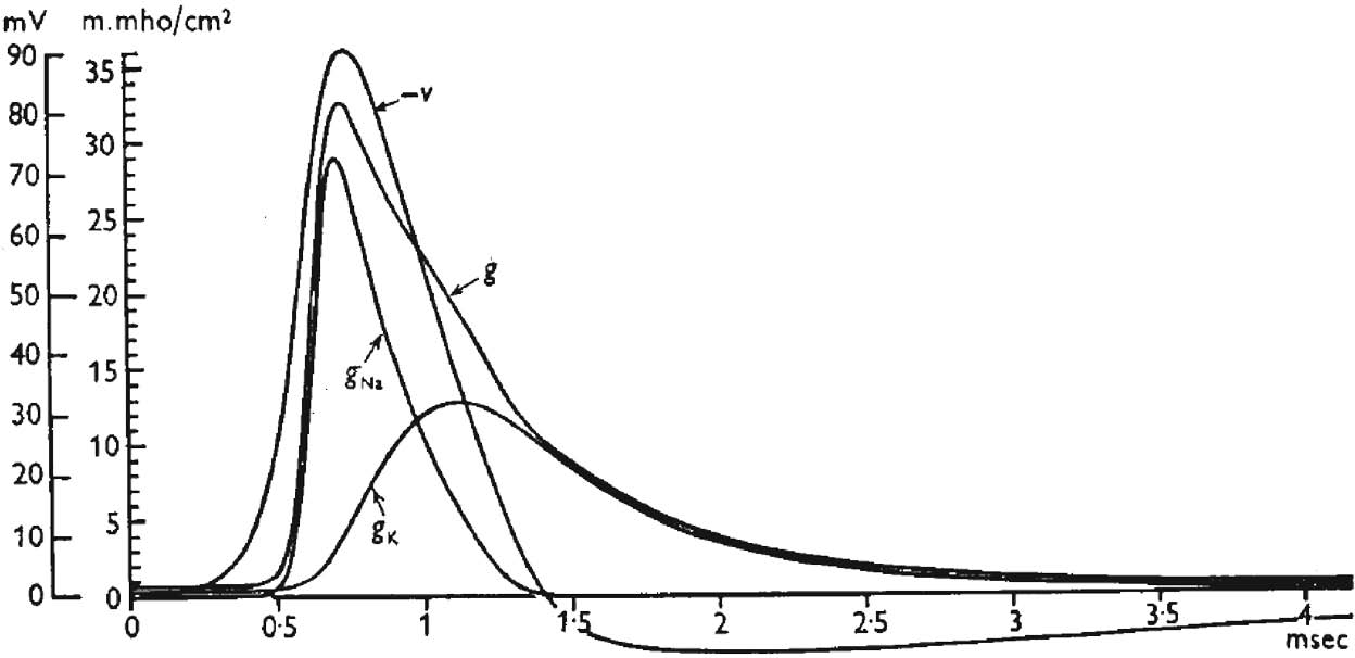
where θ is a parameter that has to be estimated numerically, on the basis of the behavior of the equation at extreme boundary conditions (see Reference Hodgkin and Huxley1952, 522, for details). A section on numerical methods of solution of such equations is interpolated in the 1952 article, and, after a minor (abbreviational) substitution, equation (30) is rewritten as equation (31) (not shown here, but see Reference Hodgkin and Huxley1952, 524). This equation (either [30] or the equivalent [31]) is solved numerically, and graphs of the membrane conductances during a propagated action potential are depicted. Hodgkin and Huxley's graphical results were shown in their Figure 17 and were based on numerical solutions of equation (31) showing components of membrane conductances (g) during propagated action potential (V). This figure is widely reproduced in standard neuroscience texts (see Figure 2 in this article; also see Hodgkin and Huxley Reference Hodgkin and Huxley1952, 530). Readers will also recognize such graphs of the conductances as the classic action-potential result, which is represented, on the basis of largely qualitative considerations, in typical neuroscience textbooks.
Numerical solution of Hodgkin and Huxley's equation (31) showing conductances (g) of sodium g Na, potassium g K, and total g, as well as the propagated action potential (−V) from Hodgkin and Huxley's original Figure 17 (Reference Hodgkin and Huxley1952).

3. Implications of the HH Model and Their Methodology
3.1 One Basic Mechanism with Many Types of Molecular Realizations?
An examination of the form of the key equations, especially the batch summarized beginning with equation (26) and then equations (30) and (31), might suggest that Hodgkin and Huxley's accomplishment is not that different than, say, Maxwell's articulation of the electromagnetic theory of light and Maxwell's derivation of the wave equation for an electromagnetic disturbance. But the HH equations are not universal equations as were Maxwell's—the HH equations were empirically generated from curve fittings to the squid action-potential changes read using the voltage clamp technique. Hodgkin and Huxley remarked on the limitations of their model a number of times during the course of their 1952 article, limitations that are well summarized by Bogen (Reference Bogen2005) and Craver (Reference Craver2007).
Toward the very end of their 1952 paper, Hodgkin and Huxley wrote:
Applicability to other tissues. The similarity of the effects of changing the concentrations of sodium and potassium on the resting and action potentials of many excitable tissues (Hodgkin, 1951) suggest that the basic mechanism of conduction may be the same as implied by our equations, but the great differences in the shape of action potentials show that even if equations of the same form as ours are applicable in other cases, some at least of the parameters must have very different values. (Reference Hodgkin and Huxley1952, 542; emphasis mine)
In addition, toward the end of his Nobel lecture, Hodgkin returned to this issue and the related theme of a specific or “definite” model of the membrane when he wrote:
To begin with we hoped that the analysis might lead to a definite molecular model of the membrane. However, it gradually became clear that different mechanisms could lead to similar equations and that no real progress at the molecular level could be made until much more was known about the chemistry and fine structure of the membrane. On the other hand, the equations that we developed proved surprisingly powerful and it was possible to predict much of the electrical behaviour of the giant axon with fair accuracy. Examples of some of the properties of the axon which are fitted by the equations are: the form, duration and amplitude of the action potential; the conduction velocity; impedance changes; ionic movements; and subthreshold phenomena including oscillatory behaviour. (Reference Hodgkin1964, 42)
A review of contemporary molecular models of various ion channels capable of supporting action potentials suggests that Hodgkin and Huxley happened on a most remarkable level of abstraction/aggregation that would support very broad generalization in terms of the specificity of membrane currents, though not any specific molecular mechanisms. For example, the chapter on “Propagated Signaling: The Action Potential” in Kandel et al. states somewhat “teleologically” that
The squid axon can generate an action potential with just two types of voltage-gated channels. Why then are there so many different types of voltage-gated channels found in the nervous system? The answer is that neurons with the expanded set of voltage-gated channels have much more complex information-processing abilities than those with only two types of channels. (Reference Kandel, Schwartz and Jessell2000, 159; emphasis mine)
The number and types of ion channels are explained, and to an extent unified, by the underlying genetics (and epigenetics) of ion channel diversity, a topic to which I turn next.
3.2. Genetic and Epigenetic Diversity Accounts for Ion Channel Diversity
Hille recounts the history of ion channel research over the course of the past half century following Hodgkin and Huxley's classic paper. The progress, he writes, has been “phenomenal,” and “the field has become highly interdisciplinary” (Hille Reference Hille2001, 61). Several recent Nobel prizes have been awarded for ion channel research, including to Neher and Sakmann in 1991, who developed the “patch clamp method” that provided direct evidence of ion channels, and to MacKinnon in 2003, for structural and mechanistic studies of ion channels, including his pore model.
Genetic studies that began in the 1980s have indicated that there are three general genetic “superfamilies” of ion channels, comprising ligand-gated, gap-junction, and the HH type of action potential–generating voltage-gated channels. This last class, which is activated by depolarization, also contains three subclasses of channels selective for Na+, K+, and Ca++ (Siegelbaum Reference Siegelbaum and Koester2000). Siegelbaum describes the similar architecture of this class of channels:
They contain four repeats of a basic motif composed of six transmembrane segments [known as] (S1–S6). The S5 and S6 segments are connected by a loop, through the extracellular face of the membrane, the P-region, that forms the selectivity filter of the channel. A single subunit of voltage gated Na+ and Ca++ channels contains four of these repeats. Potassium channels are composed of four subunits, each containing one repeat. (Reference Siegelbaum and Koester2000, 119)
Additional ion family channels are in the process of being discovered and characterized, including a class of Cl− channels. But already the number of different channel types is, according to Siegelbaum (Reference Siegelbaum and Koester2000), “enormous.” The diversity is accounted for in part because “most channels are made up of multiple subunits that can be combined in different permutations to produce channels with different functional properties” (119). Additionally, the variability is “produced by differential expression of two or more closely related genes, by alternative splicing of mRNA transcribed from the same gene, or by editing of mRNA” (120).
Some simplification of this extensive diversity occurs in the axonal region of the neuron where just the two major channel types, Na+ and K+ are involved. However, even here, Hille (Reference Hille2001) also describes an extensive “diversity of K channels” in different tissues and even within single cells. He sums up this “microheterogeneity of K channels,” noting that “such results are typical of experimental discoveries today. The finer the method of analysis, the more apparent subtypes of channels are discovered” (74). In spite of this extensive diversity and variation, genetics can provide a rationale for generalization, at least involving similarity modeling. On this point, Hille writes, “the Na, Ca, and K families of voltage gated channels form a homologous gene superfamily, as may be expected from their broad apparent functional similarity. This means that many findings for one type of channel can be generalized to the others” (85).
3.3. The HH “Basic Mechanism” as an Emergent Simplification
The account of the extensive diversity of specific mechanisms of ion channel types just summarized raises the question of how unity can be effectively achieved amid such natural variation. In a significant sense, Hodgkin and Huxley achieved that unification and simplification in advance of the more recent molecular knowledge by working at a higher level of abstraction. Their accomplishment suggests that in certain areas of biology, investigators can capture what might be termed “emergent simplifications” that transcend the specific workings of the molecular details. In a way, a more abstract mechanism can be a “basic mechanism,” even if it is clearly realized that there are as yet unknown molecular details of the mechanism. Possibly such a basic mechanism is more like a “prototypical” mechanism, which identifies and characterizes salient core features of a biological entity and its actions.
In some circumstances, the core features of those simplifications can be generated by quantitative investigations and represented by mathematical equations that are formally analogous to what we find in the Euclidean types of theories discussed in Section 1. But they lack that very broad universality and instead serve their functions by being prototypes for analogical modeling to similar prototypes—albeit, in this case, analogical modeling to other quantitative prototypes. In addition, they are not usually unilevel but instead mix levels of aggregation. In Hodgkin and Huxley's work, the discussion is focused on current flows and potential difference changes due to ions and inferred ion channels, but as situated in an axon of a particular species. Further reflection of Hodgkin and Huxley's systems-level methodology may provide important generalizable heuristics that can inform biology pursued at the level of general systems.
4. Emergent Simplification and a Reverse Application of Wimsatt's Reductionistic Heuristics
In a series of papers beginning in 1980 and represented in his 2007 book, Bill Wimsatt analyzed a series of “heuristics,” thought of as guides or “rules of thumb,” which are employed when scientists proceed in a reductionist manner (Reference Wimsatt1980, Reference Wimsatt2007). In a recent paper, Wimsatt (Reference Wimsatt2006) summarized these reductionistic heuristics under some 20 headings. These range from bias toward a unitary and monadic level to inappropriate simplifications—or even total disregard—of the environment. In general, Wimsatt views the simplifying reductionistic heuristics as accounting for the power of reductionistic approaches but as paying for this power by shortchanging the roles of the environment and actual complex multilevel interactions.
The emergent simplification of Hodgkin and Huxley described above also uses simplifying heuristics that shield us from more complex interactions. Ironically, the complexity that Hodgkin and Huxley decided not to pursue was at more reductionistic levels, such as the actual mechanisms of ion transport.
Some of what Wimsatt describes as reductionistic heuristics were adopted by Hodgkin and Huxley in service of their research at an emergent simplified level. For example, Wimsatt's reductionistic heuristic of “contextual simplification” states the reductionist should “simplify environment before simplifying system.” Hodgkin and Huxley followed this strategy by controlling for, and systematically varying, the external ionic concentrations. Also, the theory was initially tested using their preferred “model organism,” the squid, though Hodgkin and Huxley did speculate on generalization of their results as noted above. And the absolutely key voltage clamp tool also represents the application of a third of Wimsatt's heuristics of “tool binding.”
But reductionistic heuristics are also not followed by Hodgkin and Huxley, as in their recognition that they did not describe their system exhaustively (Wimsatt's “extra-perspectival blindness or perceptual focus”). Similarly, Hodgkin and Huxley did not attempt to refer all descriptions and processes “to entities at a given level”—their analysis is multilevel, as described in the analysis earlier in this article and in the companion articles in this symposium issue. Additional Wimsattian heuristics could be reviewed and commented on in terms of whether Hodgkin and Huxley followed them, but space does not allow additional discussion in the present article.
In summary, Hodgkin and Huxley use heuristics in the Wimsatt sense, and the heuristics fall both into what Wimsatt calls reductionistic heuristics and also nonreductionistic heuristics. But for Hodgkin and Huxley, both types of heuristics are used to develop an emergentist model. This suggests that the classification of heuristics into reductionistic versus nonreductionist might be rethought in relation to strategies for searching for emergentist simplifications.
Suffice it to say that we desperately need simplifications to make sense of neural processes, and in the above sections we see the actions of such simplifications in one of the most fundamental, central, and, many would say, simplest of the types of processes encountered in the neurosciences—the generation of action potentials.Footnote 3 But the simplifications are in the service of producing results at an emergent level, not a reductionistic level. As one seeks to analyze neural connections related to behavior, even in such simple neural networks as one finds in C. elegans and Drosophila, the need for simplifications becomes evident (Schaffner Reference Schaffner2006). And as one attempts to analyze human neuroscience, strategies for simplifications become all the more urgent (Schaffner Reference Schaffner, Kendler and Parnas2008).