Introduction
The abomasal parasite Haemonchus contortus is one of the main limitations to efficient sheep and goat production, particularly under low-input systems. The most common clinical signs are failure to thrive, weight loss, anaemia, hypoproteinemia, submandibular oedema (bottle jaw), ascites, diarrhoea, lethargy and death (Puspitasari et al., Reference Puspitasari, Farajallah and Sulistiawati2016). Modern control methods for worm parasites of livestock need to shift away from the reliance on anthelmintics to a more integrated approach for pest management (Santos et al., Reference Santos2017).
With the current move towards ‘sustainable’ agriculture, there is an ever increasing interest worldwide in natural products and molecules that are in tune with the move towards sustainable, ecologically and environmentally acceptable systems of livestock management and disease control (Santos et al., Reference Santos2017).
Interest in fermented milk beverages containing probiotics has increased greatly in recent years because of the increasing demand for safe and high-quality products (Rosa et al., Reference Rosa2017). Kefir is a sour, slightly carbonated fermented milk product with a low alcohol content, obtained by the fermentation of milk with activated kefir grains (El Mogheth & El-Gendy, Reference El Mogheth and El-Gendy2017). Kefir differs from other fermented products because it is produced from kefir grains comprising a specific and complex mixture of lactic acid- and acetic acid-producing bacteria as well as lactose-fermenting and non-fermenting yeast, which have a symbiotic association (Arslan, Reference Arslan2015; Rosa et al., Reference Rosa2017).
The micro-organisms present in kefir have probiotic potential (Arslan, Reference Arslan2015). Numerous bacterial species isolated from kefir comprise lactobacilli, such as Lactobacillus acidophilus, Lactobacillus brevis, Lactobacillus kefir and Lactobacillus casei; lactococci, including Lactococcus lactis ssp. lactis, Lactococcus lactis ssp. cremoris, Streptococcus salivarius ssp. thermophilus, Leuconostoc mesenteroides and Leuconostoc cremoris; and a variety of yeast species, such as Candida, Kluyveromyces and Saccharomyces spp. (Rosa et al., Reference Rosa2017). Additionally, the microbiota present in kefir can produce antagonistic substances, such as organic acids and bacteriocins; the latter interfere with the adherence of pathogenic bacteria to the intestinal mucosa, potentially contributing to the improvement of gut health (Prado et al., Reference Prado2015).
According to the Codex Alimentarius (CODEX STAN 243-2003), a typical kefir (fermented milk obtained from kefir grains) should contain at least 2.7% protein, 0.6% lactic acid and <10% fat. Kefir is traditionally produced from cow milk but can also be made with milk from other species, such as goat, sheep and buffalo, or other sources, such as soy milk (Arslan, Reference Arslan2015). In addition to beneficial bacteria, kefir contains vitamins, minerals and essential amino acids that are beneficial in healing, promoting longevity and homeostasis (El Mogheth & El-Gendy, Reference El Mogheth and El-Gendy2017).
Recently, kefir has raised interest in the scientific community because of its numerous beneficial effects on health, including antibacterial (Ismaiel et al., Reference Ismaiel, Ghaly and El-Naggar2011), antifungal (Gamba et al., Reference Gamba2015), antimutagenic and antioxidant properties (Yilmaz-Ersan et al., Reference Yilmaz-Ersan2016).
Plant extracts, bee-derived products and probiotics are safe and inexpensive options with great potential to treat intestinal parasite infections (Yilmaz-Ersan et al., Reference Yilmaz-Ersan2016). However, there are few scientific reports about the effects of fermented kefir on parasites. Franco et al. (Reference Franco2013) suggested the ability of kefir-fermented milk to protect mice from Giardia intestinalis infection. Furthermore, it was reported that kefir could help in reducing the effect of Entamoeba histolytica or in treating immune-suppressed patients for the parasite (Mohamed, Reference Mohamed2016). The role of kefir milk for control of Leishmania donovani was reported by Ali et al. (Reference Ali, Essa and Mohamed2016).
Research on kefir use in ruminants is rather limited. However, Daş et al. (Reference Daş2007) demonstrated that supplementation of goat kids with kefir shortly after weaning reduced the incidence of coccidial oocysts. Recently, the effect of kefir as a probiotic on the performance and health status of calves has been investigated by Satik & Günal (Reference Satık and Günal2017). There are no published data, however, on the anthelmintic effect of kefir against gastrointestinal nematodes in sheep.
Therefore, the current study aimed to assess the in vitro nematicidal potential of kefirs made from camel, goat, ewe and cow milk against H. contortus, a gastrointestinal nematode in sheep.
Materials and methods
Kefir grains
Kefir grains were obtained from the National School of Veterinary Medicine of Sidi Thabet, Tunisia. The grains were propagated in pasteurized raw milk at room temperature (27–28°C) for 24 h and renewed daily for a period of seven days. The grains were then separated from the fermented milk by filtration through a plastic sieve, washed with distilled water and then maintained at 4°C for short storage until the next production (Yilmaz-Ersan et al., Reference Yilmaz-Ersan2016).
Milk samples
Milk samples of camel, goat, ewe and cow were obtained from a private farm in central Tunisia. Milk was recovered by hand milking. Samples were collected in sterile screw bottles, kept in cooling boxes (4°C) and transported to our laboratory within 6 h.
Kefir preparation
Milk samples were pasteurized at 90°C for 10 minutes and cooled to 25°C in a water bath before inoculation. A litre of each sample was inoculated with 30 g of active kefir grains. Fermentation was carried out at 25°C for 24 h. Subsequently, all the fermentates were transferred to cold storage at 4°C until laboratory analysis (Yilmaz-Ersan et al., Reference Yilmaz-Ersan2016).
Physicochemical analysis
The pH was determined using a digital pH-meter (model no. 3505; Jenway, UK), and acidity by titration with N/10 NaOH in the presence of phenolphthalein. Acidity was expressed as % lactic acid (AFNOR, 1993). Total nitrogen was quantified by CHN analysis. Freeze-dried samples were combusted in a CHN elemental analyser (model 2400, series II-CHNS; PerkinElmer, Waltham, USA) (Jimenez & Ladho, Reference Jimenez and Ladho1993).
Total amino acid content was determined after hydrolysis of milk samples by 6N hydrochloric acid (HCl, 6N) at 110°C for 24 h in vacuum-sealed glass tubes. Reaction was stopped by adding NaOH (6 M). Mixtures were filtered through a membrane with a pore size of 0.45 μM before injection in a reversed-phase high-performance liquid chromatography (RP-HPLC) system of 100 μL (Davies & Thomas, Reference Davies and Thomas1973). Separation of amino acids was performed using an Agilent 1200 Chromatograph (Agilent Technologies, Waldbronn, Germany) with a C18 reverse-phase column (250 × 4.6 mm) and a fluorescence detector (FLD). Two solvents were used: solvent A comprised ACN/MeOH/H2O (45:45:10) and solvent B comprised Na2HPO4 (2.75 g/l; pH = 6.5). The solvent gradient was formed by increasing the proportion of solvent A from 10% to 90% during the first 30 minutes, and increasing solvent B to 100%. An aliquot (100 μl) of the filtrate was injected into the HPLC system. Excitation was at 340 nm and emission at 450 nm. The quantification of amino acids in samples was performed by measuring peak areas of samples, and plotting against the calibration curves of each amino acid.
Concentrations of the minerals calcium (Ca), magnesium (Mg), sodium (Na), iron (Fe) and potassium (K) were determined by atomic absorption spectrometry (SAA) (NOVA400, Analytik Jena AG, Jena, Germany), and phosphorus (P), copper (Cu), manganese (Mn), molybdenum (Mo), zinc (Zn) and iron (Fe) were analysed by inductively coupled plasma atomic emission spectrometry (ICP-AES) (ACTIVA; Horiba Scientific, Longjumeau, France) according to AOAC (2000) standards.
In vitro assays
Two different tests were performed: the egg hatching assay and the adult worm motility assay. For each test, faecal matter of Barbarine lambs experimentally infected with H. contortus was collected to obtain the eggs and L3 larvae of H. contortus, according to the World Association for the Advancement of Veterinary Parasitology (WAAVP) guidelines (Coles et al., Reference Coles1992).
Egg hatch assay
This assay was performed according to the method described by Coles et al. (Reference Coles1992). The fresh faecal matter was recovered, crushed and filtered four times on 500, 250, 125, 63, 50 and 30 μm sieves. Eggs collected from the 30 μm sieve were centrifuged at 1344 g for 10 minutes and the supernatant was removed and replaced by an NaCl solution (density: 1.2), mixed and centrifuged at 3024 g for 15 minutes. The recovered supernatant was filtered with a 30 μm sieve, washed and centrifuged three times at 2000 rpm for 10 minutes. The quantity was adjusted to reach a concentration of 200 eggs per ml.
Kefir, produced by using four different milks (cow, ewe, goat and camel), was used as the active treatment. Albendazole, reference drug (99.8% pure standard reference, Médivét, S.A., Tunisia) was dissolved in dimethyl sulfoxide (DMSO) and diluted at three concentrations (0.06, 0.25, 0.5 μg/ml). Albendazole served as a positive control, and untreated eggs in phosphate buffered saline (PBS) were used as a negative control.
In the assay, approximately 200 eggs in 1 ml of PBS were placed in each 24-well plate. Serial increasing concentration of each kefir in a total volume of 1 ml of PBS was added to make concentrations of 0.125, 0.25 and 0.5 mg/ml for both cow and ewe milk kefir, 0.03, 0.06, 0.125 and 0.25 mg/ml for goat milk kefir and 0.015, 0.03, 0.06 and 0.125 mg/ml for camel milk kefir. Interspecies differences in the used concentrations were set after preliminary observations to define the range of concentrations inducing in vitro inhibition of H. contortus.
Four replicates per concentration were performed. The plates were covered and incubated at 27°C for 48 h. After 48 h of incubation, hatching was stopped by adding Lugol's iodine solution, and the number of L1 larvae and eggs per well was counted under a dissecting microscope at 40× magnification. Thereafter, the percentage of hatched eggs was determined using the following ratio: number of L1/(number of eggs + number of L1) × 100.
Adult worm motility assay
Six weeks after infection, the lambs were euthanized. Immediately, the abomasum was removed, opened and placed in 9‰ sodium chloride solution at 37°C. The parasites were collected, washed and kept in PBS. The experiment was conducted according to Hounzangbe-Adote et al. (Reference Hounzangbe-Adote2005).
Eight actively moving worms were placed in Petri dishes containing 2, 1 and 0.5 mg/ml of cow milk kefir and ewe milk kefir, respectively; 0.5, 0.25 and 0.125 mg/ml of goat milk kefir; and 0.25, 0.125 and 0.06 mg/ml of camel milk kefir in a total volume of 3 ml.
PBS was used as a negative control. Albendazole dissolved in DMSO then in PBS at a final concentration of 1 mg/ml was used as a positive control.
The measurements were performed in triplicate. Inhibition of worm motility was the criterion for anthelmintic drug activity. The required time for paralysis or complete inactivity and immobility was recorded at 0, 2, 4, 6 and 8 h. To test the worm motility, after 8 h, possible parasite motility was checked under a dissecting microscope by suspending parasites in PBS for 30 minutes after washing away kefirs and the positive control. Live (motile) and dead (immotile) worms were counted and recorded in each concentration. Death or paralysis of worms was inferred from lack of motility for 5–6 s and from touching the parasites. The immobility index was calculated as the number of immotile worms divided by the total number of worms per Petri dish.
Statistical analysis
Statistical analysis was performed using Statview v. 5.0.1 (SAS Institute, Cary, North Carolina, USA). Mean values were used, and all analyses were carried out in triplicate. IC50 for egg hatch inhibition was calculated by probit analysis. Regression was used for evaluation of the dose–response relationship using Minitab® Release 14 (Minitab Inc., State College, Pennsylvania, USA). The worm motility inhibition was expressed as mean ± SE. Significant differences in means were compared using analysis of variance (ANOVA) followed by Fisher's least significant difference tests. The difference between the means was considered to be significant at P < 0.05.
Results
Physico-chemical analysis
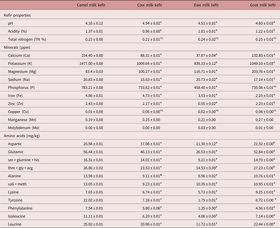
The physico-chemical compositions of kefirs produced from camel, cow, ewe and goat milk are reported in table 1. No significant changes in total nitrogen protein (0.21–0.25%) and pH (4.10–4.60) of kefir samples were observed. However, the total titratable acidity of kefir from camel milk has higher lactic acid than kefir from cow and ewe milk (P < 0.001) and from goat milk (P < 0.05).
Mean chemical composition of samples of kefir made from camel, cow, ewe and goat milk.

Significance: a, P < 0.0001 compared to camel milk kefir; b, P < 0.05 compared to camel milk kefir; ns, P > 0.05 compared to camel milk kefir
Overall, the kefirs had different mineral profiles. Kefir from camel milk was found to be a richer source of minerals than other kefir samples (P < 0.001). Large variations were also found in the amino acid content of the different kefirs. Kefir produced from camel milk had a significantly higher content of amino acids than other kefir samples (P < 0.001).
In vitro assays
Egg hatch assay
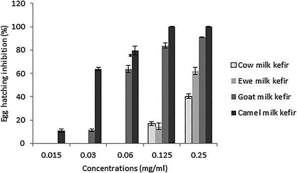
Kefir made from camel, goat, ewe and cow milk induced egg hatching inhibition in a dose-dependent manner (fig. 1). Compared to all other types of kefirs, kefir from camel milk showed higher inhibitory effects on egg hatching (P < 0.001) at all tested concentrations (IC50 = 0.024 mg/ml, Y = 34.31x − 41.50; R2 = 0.937).
Profile of dose-dependent egg hatching inhibition with increasing concentrations of kefir made from cow, ewe, goat and camel milk. * P < 0.0001 compared to camel milk kefir.

Albendazole inhibited 91.48% of egg hatching at 0.5 μg/ml (IC50 = 0.108 μg/ml, Y = 21.07x – 22.13, R2 = 0.951). The inhibitory effect of cow milk kefir was the lowest, as it induced 40.30% egg-hatching inhibition at 0.25 mg/ml (fig. 1). Kefirs made from cow and ewe milk did not induce any inhibitory activity at the lowest tested concentrations. In contrast, kefir from camel milk showed an ovicidal activity even at low concentrations (63.64% at 0.03 mg/ml) (fig. 1). The maximum concentration required to induce total (100%) egg-hatch inhibition for kefir produced from cow and ewe milk was 0.5 mg/ml.
Adult worm motility
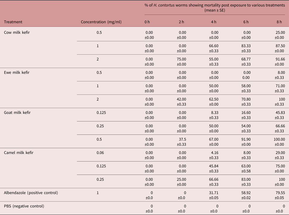
All kefirs showed concentration-dependent effects on adult parasites. The kefir produced from camel milk, however, killed more worms than the other kefirs, even at the lowest tested concentrations. After 8 h post exposure, it induced 100% mortality at 0.25 mg/ml, while at the same concentration, goat milk kefir induced only 66.66% mortality (table 2). Kefirs made from cow and ewe milk killed the parasites in a concentration-dependent manner, and all worms were dead at a concentration of 2 mg/ml within 8 h (table 2).
In vitro anthelmintic efficacy of kefir samples on Haemonchus contortus.

There was 79.55% mortality of worms in albendazole within 8 h post exposure. However, the worms in the DMSO negative control solution showed neither paralysis nor mortality. No worms recovered motility in the PBS revival test.
Discussion
Kefir, the traditional beverage, is now recognized as a potential source of probiotics and molecules with interesting health properties (Rosa et al., Reference Rosa2017). As such, the composition of kefir has been widely studied throughout the world (Ismaiel et al., Reference Ismaiel, Ghaly and El-Naggar2011; Pogačić et al., Reference Pogačić2013; Ünsal & Arslanoğlu, Reference Ünsal and Arslanoğlu2013; Arslan, Reference Arslan2015). The findings of the present study agree with most of the above reported studies. Variability in the composition of kefir from milks may be due to differences in milk quality associated with particular animal species, feeding management, lactation period, and processing methods, including the amount of kefir grains used, the time and temperature of incubation, and the micro-organisms occurring in the kefir grains (Rosa et al., Reference Rosa2017).
Based on the results of the present study, kefir produced from camel, goat, ewe and cow milk showed significant concentration-dependent in vitro anthelmintic activity against H. contortus. Our findings also provide evidence that compared with cow, ewe and goat milk kefir, camel milk kefir has an ovicidal (100% at 0.125 mg/ml) and adulticidal impact (100% at 0.25 mg/ml) against the tested parasite even at low concentrations.
To our knowledge, this study is the first report of the potential of kefir, as a probiotic, on haemonchosis in sheep. Unfortunately, there are no reports in the literature of the use of kefir for controlling gastrointestinal nematodes in small ruminants to which our results can be compared.
Nevertheless, the use of probiotics in small ruminant nutrition has been shown to improve animal health, productivity and immunity (Abd El-Tawab et al., Reference Abd El-Tawab2016). Early studies have reported that the feeding of probiotics to neonatal calves may improve weight gain. Likewise, variable responses to probiotic supplementation in terms of weight gain have been observed in lambs and kids (Abd El-Tawab et al., Reference Abd El-Tawab2016). Probiotics have been reported to stabilize rumen pH, increase volatile fatty acid production and stimulate lactic acid utilizing protozoa, resulting in a highly efficient rumen function. Furthermore, use of probiotics has been found to increase milk production and can reduce the incidence of neonatal diarrhoea and mortality (Abd El-Tawab et al., Reference Abd El-Tawab2016).
The influence of kefir on health has been well studied in mice, rats, poultry and goat kids (Dalloul & Lillehoj, Reference Dalloul and Lillehoj2005; Yamman et al., 2006; Daş et al., 2007). Yaman et al. (Reference Yaman2006) provided preliminary information that kefir Lactobacillus spp. yeast may be a useful candidate as a competitive exclusion preparation to improve the intestinal microflora of poultry, leading to improved carcass hygiene. Moreover, there have been successful attempts to use kefir for controlling coccidiosis in broiler chickens, as reported by Dalloul & Lillehoj (Reference Dalloul and Lillehoj2005). Also, an early report suggested supplementation of kefir shortly after weaning reduces coccidial oocyst output in goat kids (Daş et al., Reference Daş2007). Furthermore, improvement of daily weight gain and the health status of calves was achieved when young calves were supplemented with kefir (Satik & Günal, Reference Satık and Günal2017). The effects of fermented kefir on some parasites (Entamoeba histolytica and Leishmania donovani), bacteria and fungi (Streptococcus faecalis and Aspergillus flavus) were also confirmed (Ismaiel et al., Reference Ismaiel, Ghaly and El-Naggar2011; Ali et al., Reference Ali, Essa and Mohamed2016; Mohamed, Reference Mohamed2016).
It has been reported that the biological action of kefir is related to its microflora (Prado et al., Reference Prado2015; Rosa et al., Reference Rosa2017). In this way, the functional role of kefir may be either direct, through an interaction with the consumed micro-organisms (probiotic effect), or indirect, as a result of the action of microbial metabolites such as vitamins, proteins, bioactive peptides, oligosaccharides and organic acids, generated during the fermentation process (biogenic effect) (Rosa et al., Reference Rosa2017).
On the other hand, it has been proved that the effect of kefir is not only linked to its microflora but is also due to the presence of some metabolic products as organic acids (Ismaiel et al., Reference Ismaiel, Ghaly and El-Naggar2011; Prado et al., Reference Prado2015). In fact, the micro-organisms in the kefir grains produce lactic acid, antibiotics and bactericides, which inhibit the development of degrading and pathogenic micro-organisms in kefir milk (Arslan, Reference Arslan2015).
It is worth noting that probiotic bacteria, mainly lactic acid bacteria, isolated from kefir are reported to possess biological properties (Prado et al., Reference Prado2015; Rosa et al., Reference Rosa2017). It has been suggested that lactic acid bacteria prevent the adherence, establishment and invasion of specific enteropathogens. Thus, several mechanisms have been proposed: contribution to mucosal barrier function, competitive exclusion, modulation of the immune response, co-aggregation to pathogens, decreasing of the luminal pH via the production of lactic acid, and secretion of specific compounds such as bacteriocins (Prado et al., Reference Prado2015). Moreover, several metabolites produced during the fermentation, including organic acids (e.g. formic, lactic, acetic, propionic and phenyllactic acids), hydrogen peroxide, diacetyl and short chain peptides have shown antifungal effects (Prado et al., Reference Prado2015). Additionally, kefir contains bioactive compounds such as peptides and exopolysaccharides, known as kefiran, produced by Lactobacillus kefiranofaciens (Wang et al., Reference Wang2010). It has been reported that kefiran possesses biological properties that may make them suitable for use as antioxidants, antitumor agents, antimicrobial agents, and immunomodulators, among other roles (Wang et al., Reference Wang2010).
It is important to note that in a preliminary study our group demonstrated for the first time the in vitro nematicidal effect of camel milk against H. contortus, in comparison to milks from cow, ewe and goat (Alimi et al., Reference Alimi2016). Only camel milk had an inhibitory effect on egg hatching (100% at 100 mg/ml). On the other hand, camel milk killed the parasites in a concentration-dependent manner and all the worms were dead at a concentration of 100 mg/ml within 8 h. No such effect was observed for milk from other species.
In the current study, all kefir produced from the milk of female ruminants (cow, ewe and goat) showed an in vitro ovicidal effect against H. contortus. In comparison, camel milk kefir was the most effective against the studied parasite. Camel milk is reported to have medicinal properties and contain protective proteins, including the lactoperoxidase/thiocyanate/hydrogen peroxide system, lactoferrins, lysozyme, immunoglobulins, or vitamin C and free fatty acids, which may have a possible role in enhancing the immune defense mechanism and may play a central role in the determination of biological activities (Hassan Yassin et al., Reference Hassan Yassin2015). In fact, camel milk has a stronger inhibitory system than cow milk. In particular, levels of lysozyme and lactoferrins are reported to be two and three times higher, respectively, in camel milk (Hassan Yassin et al., Reference Hassan Yassin2015). Several investigations have put forward the hypothesis that the lactoferrin content in camel milk acts as a probiotic, with strong physiological activity in the gastrointestinal tract (Hassan Yassin et al., Reference Hassan Yassin2015). In the present study, camel milk kefir had higher lactic acid content than goat, ewe or cow milk kefirs. Therefore, the greater in vitro anthelmintic efficacy of camel milk kefir against H. contortus in sheep suggests a possible synergistic relationship between the active molecules of camel milk and the micro-organisms of its kefir, produced during the fermentation process.
In conclusion, this study found that kefir made from camel, goat, ewe and cow milk showed in vitro anti-parasitic action against H. contortus in sheep. Thus, kefir represents a promising substitute for current anthelmintics in the management of haemonchosis. It has the additional advantages of being a low-cost and easily accessible probiotic product. Our results clearly demonstrate that fermented probiotic products may be beneficial in controlling gastrointestinal parasites in small ruminants. Additional work is recommended (1) to identify the components of kefir responsible for the anthelmintic activity, (2) to elucidate their mechanism of action, (3) to test their efficacy against a broader spectrum of helminth classes, such as trematodes and cestodes, and (4) to investigate further the in vivo anthelmintic effects of kefir.
Acknowledgements
We are grateful to Mr Tawfik Lahmar, Mr Mokhtar Dhibi, Mr Mohamed Jedidi, Mr Bechir Guesmi and Mr Limam Sassi for their valuable technical assistance.
Financial support
This work received financial support from Laboratoire d'Epidémiologie d'Infections Enzootiques des Herbivores en Tunisie (Ministère de l'enseignement supérieur, Tunisia).
Conflict of interest
None.
Ethical standards
All experiments were performed according to the National Institutes of Health (NIH) publication 86-23 (revised 1985), approved by the Institutional Animal Care and Use Committee and the National Ethics Committee of Tunis University.