Introduction
Superior canal dehiscence syndrome describes the association of unusual audiovestibular symptoms in response to noise or straining. Minor was the first to link these symptoms to bony dehiscence of the superior semicircular canal in the floor of the middle cranial fossa.Reference Minor1 The audiovestibular system has two functional ‘windows’ linking the inner and middle ear: the oval window and the round window. These windows facilitate fluid movement in the cochlear duct, allowing effective mechano-electrical transduction by hair cells. A dehiscent superior semicircular canal can act as a ‘third window’, allowing noise and stimuli to be transmitted directly to the audiovestibular system, via increased middle-ear or intracranial pressure, causing symptoms.
Otosclerosis, by contrast, is an osseous dyscrasia limited to the temporal bone. The associated conductive hearing loss can be well treated by either fitting a hearing aid or performing stapes surgery.
Here, we present a novel case in which routine stapes surgery unmasked a latent superior canal dehiscence syndrome, which presented in the immediate post-operative period.
Case history
A 30-year-old Caucasian man had originally presented with bilateral conductive hearing loss, in keeping with a diagnosis of otosclerosis.
The patient had undergone an uneventful stapedotomy on the right ear (the worse hearing ear), with a good result. Surgery had been performed under local anaesthetic with a potassium titanyl phosphate laser. A 0.4 mm diameter titanium prosthesis (Fisch, Gyrus ACMI, Bartlet, Tennessee, USA) had been inserted. The air–bone gap had been closed to within 10 dB.
Three years later, a left stapedotomy was carried out using an identical technique. Again, the air–bone gap was closed to within 10 dB. However, the patient was troubled by marked post-operative vertigo, lasting a few days. Thereafter, he developed persistent ‘crackling’ in his left ear on noise exposure, a high pitched tinnitus on exercise or on occluding the external ear canal, and mild imbalance. Initially, symptoms were attributed to an atelectatic tympanic membrane; however, reinforcement tympanoplasty was performed without benefit.
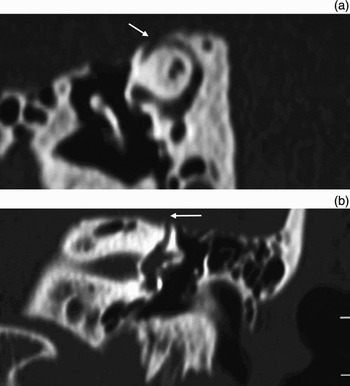
Eventually, high-resolution computed tomography (CT) scans of the temporal bones, without contrast, demonstrated a dehiscent superior semicircular canal on the left side (Figure 1), with no contralateral dehiscence. Electronystagmography with vestibular assessment and caloric testing was normal. Vestibular evoked myogenic potential testing revealed absent potentials on the contralateral side but robust potentials at 70 dB on the ipsilateral (left) side, supporting a diagnosis of superior canal dehiscence syndrome.
Pre-operative high resolution computed tomography scans of left temporal bone, demonstrating superior canal dehiscence syndrome. The reformatted images in the (a) plane of the superior semicircular canal and (b) coronal plane clearly demonstrate a bony dehiscence of the superior canal (arrow).

A middle fossa craniotomy revealed a 2×1 mm defect over the superior semicircular canal. This was resurfaced with hydroxyapatite cement (Bone Source Classic; Stryker, Freiberg, Germany) and temporalis fascia.
Recovery from surgery was uncomplicated, with a complete resolution of symptoms and no change in hearing thresholds.
Discussion
Superior canal dehiscence syndrome is a rare condition the exact prevalence of which remains unknown. Cadaveric studies have shown the superior semicircular canal to be dehiscent in 0.5 per cent of the population, with another 1.4 per cent of the population having only a very thin (<0.1 mm) bony covering.Reference Carey, Minor and Nager2 Otosclerosis is transmitted in an autosomal dominant fashion, with variable penetrance. Despite being seen in 2.5–10 per cent of Caucasian temporal bones in cadaveric studies, it has a clinical prevalence of approximately 0.3 per cent.Reference Declau, van Spaendonck, Timmermans, Michaels, Liang and Qiu3 With this in mind, it is unsurprising that the two conditions can coexist.
Therefore, it is likely that, in this patient with a dehiscent superior semicircular canal, the patient's right ear otosclerosis protected him from developing symptomatic superior canal dehiscence syndrome, by fixing the oval window. Successful stapes surgery created a physiological third window, allowing symptoms of superior canal dehiscence syndrome to develop.
The symptoms of superior canal dehiscence syndrome are usually vestibular, in the form of vertigo or oscillopsia, but can be purely audiological, e.g. hyperacusis or conductive hearing loss.Reference Banerjee, Whyte and Atlas4 Standard audiovestibular examination can be unremarkable; however, fistula testing may reveal a vertical and torsional nystagmus with slow waves directed away from the affected labyrinth. Similar eye movements can be evoked by sound. Audiometric testing may demonstrate a mild to moderate, low frequency, conductive hearing loss.Reference Minor5 Vestibular tests are often normal, although caloric testing may reveal a canal paresis. Vestibular evoked myogenic potential testing assesses the sacculo-collic pathway's response to sound. These potentials are absent in patients with otosclerosis and other conductive hearing losses, and are absent in the majority of patients following stapes surgery.Reference Stapleton and Mills6 In patients with superior canal dehiscence syndrome, robust low threshold responses are very supportive of the diagnosis.Reference Welgampola and Colebatch7 Helical CT scanning of the temporal bones, with reformatting in the plane of the superior semicircular canal, confirms the diagnosis. Computed tomography scans, although extremely sensitive, may have a positive predictive value of only 50–57 per cent in standard CT protocols that use 1 mm collimation protocols. Reducing collimation widths to 0.5 per cent in CT protocols increases the likelihood of demonstrating bone, increasing the positive predictive value to 93 per cent.Reference Belden, Weg, Minor and Zinreich8
In otosclerosis, the commonest site of bony deposition is at the anterior oval window, causing stapes footplate fixation and a conductive hearing loss. A low frequency conductive hearing loss can also be seen in superior canal dehiscence syndrome, and indeed can be the only presenting feature of this syndrome.Reference Mikulec, McKenna, Ramsey, Rosowski, Herrmann and Rauch9 This loss can mimic otosclerosis, and the diagnosis of superior canal dehiscence syndrome should be considered during stapedectomy surgery when a mobile footplate is identified.Reference Merchant, Rosowski and McKenna10 However, in the current case stapes fixation was present, with the anticipated good audiometric outcome post-stapedotomy.
Mild but transient imbalance is relatively common following stapes surgery, being seen in approximately 5–27 per cent of patients.Reference Kujala, Aalto and Hirvonen11 Persistent vertigo and late onset vertigo is much rarer, the commonest causes being prosthesis malposition and perilymphatic fistula. Perilymphatic fistula, characterised by fluctuating sensorineural hearing loss and vertigo, is said to occur in up to 3 per cent of patients after stapedectomy.Reference Atacan, Sennaroglu, Genc and Kaya12, Reference Albera, Canale, Lacilla, Cavalot and Ferrero13 This incidence is almost certainly lower following newer stapedotomy techniques. In cases of persistent vertigo, CT scans of the temporal bone may reveal a prosthesis that is dislocated or too long, or show air within the vestibule indicating a possible perilymphatic fistula.Reference Pickuth, Brandt, Berghaus, Spielmann and Heywang-Köbrunner14 In the current case, with persistent imbalance a minor feature and audiological symptoms predominating, a high resolution CT scan was not initially requested.
• Superior canal dehiscence syndrome is rare, but the anatomical prevalence of dehiscence of the superior semicircular canal is higher, at 0.5 per cent
• Dehiscence creates a ‘third window’ into the inner ear, causing vestibular, and to a lesser extent auditory, symptoms
• Low frequency conductive hearing loss can be the only presenting feature of superior canal dehiscence syndrome, which may mimic otosclerosis
• Superior semicircular canal dehiscence and otosclerosis can coexist; surgical treatment of otosclerosis can unmask latent superior canal dehiscence syndrome by creating a third window at the level of the stapes prosthesis
Since the condition of superior canal dehiscence syndrome has been described, the senior author has seen six cases, four of which presented in an atypical manner. Such atypical presentations emphasise the need for a high index of clinical suspicion for superior canal dehiscence syndrome. Specifically, this syndrome should be considered in any patient with persistent audiovestibular symptoms following stapes surgery. In this setting, high resolution CT scanning of the temporal bone should be requested at an early stage to confirm the diagnosis.
Conclusion
The diagnosis of superior canal dehiscence syndrome should be considered in patients with persistent audiovestibular symptoms following stapes surgery. High resolution CT of the temporal bone and vestibular evoked myogenic potential testing, if available, are the investigations of choice in confirming the diagnosis.