. It is found that the TOF effect has a profound influence on the kinetic energy distribution of implanted ions and subsequent energy deposition process into the target. The HIPIB of mixed H
1. INTRODUCTION
Intense laser and particle beams are highly useful tools in materials modification (Krasa et al., Reference Krasa, Velyhan, Jungwirth, Krousky, Laska, Rohlena, Pfeifer and Ullschmied2009; Li et al., Reference Li, Liu, Cheng, Xu, Ge and Wen2009; Renk et al., Reference Renk, Mann and Torres2008; Stasic et al., Reference Stasic, Gakovic, Krmpot, Pavlovic, Trtica and Jelenkovic2009; Trtica et al., Reference Trtica, Radak, Gakovic, Milovanovic, Batani and Desai2009). Here we discuss the application of high-intensity pulsed ion beam (HIPIB) technique, which has been developed during the last two decades, for surface modification, material synthesis, and high heat flux testing of materials for nuclear fusion devices as well as high-energy density physics research (Rej et al., Reference Rej, Davis, Olson, Remnev, Zakoutaev, Ryzhkov, Struts, Isakov, Shulov, Nochevnaya, Stinnett, Neau, Yatsui and Jiang1997; Renk et al., Reference Renk, Provencio, Prasad, Shlapakovski, Petrov, Yatsui, Jiang and Suematsu2004, Reference Renk, Mann and Torres2008; Linke et al., Reference Linke, Escourbiac, Mazul, Nygren, Rodig, Schlosser and Suzuki2007; Tahir et al., Reference Tahir, Hoffmann, Kozyreva, Shutov, Maruhn, Neuner, Tauschwitz, Spiller and Bock2000). This pulsed ion beam technology is characterized by extremely high-density energy (>1 J/cm2) deposition into target materials within short pulse of not more than 1 microsecond duration, as delivered by implanted ions typically at high current density of 1–103 A/cm2. Significant and coupled thermal and dynamic effects are thus induced, including ultra-high rate heating, melting, ablation and/or evaporation of irradiated materials followed by ultra-fast cooling, and strong compressive wave generation and propagating into the target under the thermal shock and the ablation process (Remnev et al., Reference Remnev, Isakov, Opekounov, Matvienko, Ryzhkov, Struts, Grushin, Zakoutayev, Potyomkin, Tarbokov, Pushkaryov, Kutuzov and Ovsyannikov1999; Renk et al., Reference Renk, Provencio, Prasad, Shlapakovski, Petrov, Yatsui, Jiang and Suematsu2004). The resultant micro- and/or macro-structural changes in the irradiated materials are responsible for the improvement of surface properties such as hardness, fatigue resistance, corrosion and wear resistance, or contrarily, the deterioration of the properties with surface roughening, cracking or exfoliation etc depending on the energy density of HIPIB irradiation. Numerical methods have been employed as a powerful and indispensable way to explore and understand the beam-material interactions, since the thermal-dynamic process is ultimately dependent on the ion energy deposition process otherwise hardly observed experimentally for such a very fast high-density energy deposition of nanosecond to microsecond range. As reported in many ion-matter interaction studies so far, a fixed initial energy of ions can be determined and the subsequent energy deposition (energy loss) of the implanted ions thus be established for various target materials, e.g., as done in TRIM code (Ziegler et al., Reference Ziegler, Biersack and Littmark1996). These methods and database could be introduced for exploring the HIPIB-material interactions, however, a number of assumptions have been made in previous studies, e.g., assuming all ions are the same kinetic energy (i.e., a constant accelerating voltage over the entire time-dependent ion current density) (Akamatsu et al., Reference Akamatsu, Ikeda, Azuma, Fujiwara and Yatsuzuka2001) or connecting ion energy with ion density by assigning a certain time delay between the experimentally measured waveforms of accelerating voltage and ion current (Le et al., Reference Le, Yan, Zhao, Han, Wang and Xue2000). The calculation will be obviously simplified by these assumptions; nevertheless, some important time-dependent characteristics of ion distribution and energy deposition may be excluded unexpectedly due to the simplifications of the numerical models. In this study, the time-of-flight (TOF) of ions was found to be of significance for the energy deposition process, where the TOF, i.e., the period of ion flight from the exit of ion source to the target surface, is determined by the accelerating voltage and ion charge (ion kinetic energy), the ion mass and the ion flight distance. During a pulsed ion beam irradiation of hundred ns duration onto a target material, the time-dependent ion implantation with an ion energy spectrum differed considerably from the initial output of ion source due to TOF of ions, and this process can not be included in the simplified models mentioned above. Numerical simulation of energy deposition by HIPIB of mixed hydrogen and carbon ions with TOF ion energy spectrum was subsequently performed based on experimental observation of time-dependent accelerating voltage and ion current density. The evolution of energy deposition process due to the TOF of ions was discussed with respect to its influence on the thermal and dynamic effect of materials under HIPIB irradiation, for exploring the beam-material interaction.
2. HIPIB-TARGET INTERACTION UNDER UNIPOLAR-PULSE MODE
2.1. HIPIB Characteristics of Unipolar-Pulse Mode
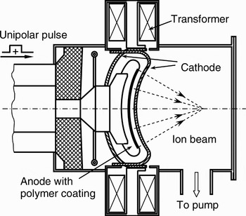
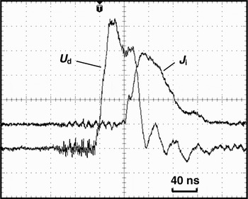
HIPIB is generated from a magnetically insulated ion diode (MID) on TEMP-6 HIPIB apparatus operated in a unipolar-pulse mode (Zhu et al., Reference Zhu, Lei, Dong and Ma2003). The pulsed power system of the apparatus consists of a Marx generator and a double coaxial pulse-forming line, generating a high-voltage pulse of 200–400 kV supplied to the MID of external-magnetic field type for ion beam generation. Figure 1 shows a schematic structure of the external-magnetic field MID operated in the unipolar-pulse mode. A polyethylene coated stainless steel anode is used and confined in a closed stainless steel cathode where the cathode is separated into two parts with a forked connection symmetrically powered by a pulsed current source to generate insulating magnetic field in the anode-cathode gap of MID, and the front part of the cathode has an array of silts for ion beam extraction. A positive high-voltage pulse supplied to the MID causes surface breakdown of the polyethylene, generating dense anode plasma, and simultaneously the ions are accelerated and extracted from the formed plasma. The focusing of the ion beam can be achieved by the cylindrical configuration of MID with curvature radii of anode and cathode being 15 and 14 cm, respectively. The typical waveforms of diode voltage (U d) and ion current density (J i) are given in Figure 2, respectively. The waveform of diode voltage has a Gaussian-like form with pulse duration of 80 ns and could be divided into two periods in relation to the ion beam generation process, in which the first peak of about 380 kV corresponds to surface breakdown of the polyethylene for anode plasma formation, and then the ions were extracted from the formed plasma boundary by the following portion of the diode voltage with a maximal value around 350 kV. The ion beam with a composition of about 70%H+ + 30%C+ is generated and the ion current density measured by Faraday cup has a maximum value of 300 A/cm2 with 140-ns pulse duration at 15 cm downstream from the MID cathode surface.
Schematic drawing of the external-magnetic field MID on TEMP-6 HIPIB apparatus operated in unipolar-pulse mode.

Typical waveforms of diode voltage (U d) and ion current density (J i), with a peak value of about 380 kV and 300 A/cm2, respectively.

2.2. Ion Kinetic Energy Distribution with TOF
According to the HIPIB generation mechanism of the external magnetic field MID in unipolar mode (Zhu et al., Reference Zhu, Lei, Dong and Ma2003) and the experimental observation of diode voltage, the unipolar pulse is treated as a cut-off profile of Gaussian line for ion acceleration from the dense plasma which is formed, following the surface breakdown. The density variation of anode plasma is ignored during the ion extraction by the pulse. This assumption is reasonably based on the fact that the density of anode plasma grows very rapidly due to the surface breakdown at first peak of diode voltage, and the decrease in the plasma density due to its expansion to the cathode could be compensated by sustainable plasma proliferation by electron bombardment under magnetic insulation. Accordingly, the Gaussian profile of the accelerating voltage can be expressed with modified pulse duration of 60 ns

where t U0 is the time point corresponding to the maximum value of accelerating voltage, and t U the pulse duration, A, B, and C are the constants determined by the shape of the accelerating voltage, respectively. The kinetic energy E 0 of ions just extracted out of the MID can be given as E 0 = qU, where q is the ion charge. Then the TOF of ions t TOF is determined by

where d is the ion drift distance in the vacuum and m i is the ion mass. Consequently, the time-dependent kinetic energy distribution E s of ions impinging the target surface is calculated in connection with E 0 by the t TOF of respective ions. With sufficient sampling of ions, the corresponding ion current density at the target surface is numerically derived from the ion acceleration and transportation process.
2.3. Energy deposition of HIPIB irradiation with TOF
The total deposited energy profile in depth during a HIPIB pulse into a Ti target is calculated by
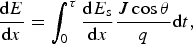
where dE s /dx is the energy loss of the implanted ions with a kinetic energy E s at the target surface, obtained using TRIM code (Ziegler et al., Reference Ziegler, Biersack and Littmark1996), which has been employed as a versatile tool for simulation of ion-matter interaction with a resultant target temperature well below 10 eV order (Tahir et al., Reference Tahir, Hoffmann, Kozyreva, Shutov, Maruhn, Neuner, Tauschwitz, Spiller and Bock2000; Le et al., Reference Le, Yan, Zhao, Han, Wang and Xue2000; Wu et al., Reference Wu, Liu, Zhu and Lei2008), θ is the incident angle of ion beam, and τ is the duration of ion current density. In the calculation, θ = 0° was adopted for one-dimensional model of the impinging ion beam normal to the titanium target. Moreover, for ion beam irradiation with mixed composition of m%M+ + n%N+ + …, the deposited energy is obtained according to the fraction of each type of ions in the mixed ion beam, i.e.

3. RESULTS AND DISCUSSION
3.1. Effect of TOF on Ion Kinetic Energy Distribution at Target Surface
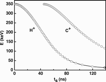
Figure 3 shows the time-dependent profile of kinetic energy of ions bombarding the target surface located 15 cm downstream from the MID, where the arrival of the first ion at the target surface is set as the zero point of time axis. The ion energy distribution profile has a pulse form with a fast rising front up to the peak value of 350 keV followed by a slower decreasing tail extended to more than 100 ns. It is clearly shown that, for each type of ion species, the higher the ion kinetic energy (higher accelerating voltage) was, the earlier the ions arrived at the target surface. This particular feature of ion energy distribution correlates to the respective ion flight period that depends on the kinetic energy of each ions obtained as extracted from the MID by the applied diode voltage. As a result, the varied TOF for ions at the widespread kinetic energies prolonged the duration of ion bombarding of the target surface, more than two times that of the original accelerating voltage in this case. Furthermore, the ion kinetic energy distribution profile presented two separate peaks for ion beam of mixed H+ and C+, i.e., first arrival of C+ was delayed 45 ns with respect to that of H+ at the kinetic energy of 350 keV, which is determined by Eq. (2) with the different ion mass. Consequently, a complex distribution of the ion energy occurred from the first arrival of C+ at the target surface, leading to simultaneous ion energy deposition with two kinds of ions at different kinetic energies.
The time-dependent profile of ion kinetic energy at the target surface locating 15 cm downstream from the MID, where the zero point of time axis is set according with the moment of first arrival of H+ at the target surface.

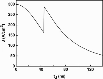
Utilizing the TOF of ions and the experimental data of ion current density obtained by Faraday cup, one can correlate the ion current density to the kinetic energy of ions at the target surface. A typical waveform of ion current density derived from TOF of ions is shown in Figure 4, with a peak value of 300 A/cm2 at the target surface. The ion current density has a similar pulse form to that of ion kinetic energy distribution given in Figure 3, except for a different relative intensity ratio between H+ and C+ peaks determined by the ion beam composition.
Typical waveform of ion current density derived according to TOF of ions at a drifting distance of 15 cm and a peak value of 300 A/cm2.

3.2. Evolution of Energy Deposition by HIPIB of Mixed Ions
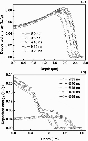
Figure 5 shows the energy deposition profile by a single ion into Ti target at the typical moments with 5 ns interval during a pulse of HIPIB irradiation with composition of 70%H+ + 30%C+. In general, the energy deposition proceeded with a decreasing tendency during the pulse of ion beam-surface interaction, for either single H+ within the first 45 ns (Fig. 5a) or mixed H+ and C+ during the following period (Fig. 5b). The energy deposition profile of H+ presents a feature of peaking at a depth close to the ion range, whereas that of C+ shows a maximum value at the outermost surface, which is mainly attributed to the difference in Bragg peak position for light and heavy ions (Le et al., Reference Le, Yan, Zhao, Han, Wang and Xue2000; Tahir et al., Reference Tahir, Hoffmann, Kozyreva, Shutov, Maruhn, Neuner, Tauschwitz, Spiller and Bock2000). Within the first 45 ns, most energy is delivered by H+ changed from a maximal deposition depth of about 2.6 µm with the highest peak energy density of 0.08 kJ/g to a shallower ion range of about 1.3 µm with a lower peak value about 0.04 kJ/g. As bombardment of C+ onto the target surface started from 45 ns, the energy was deposited by mixed H+ and C+ and developed from a maximal depth of about 1.3 µm to the top surface zone, where the deposited energy per ion was normalized as a sum of 70% energy of H+ and 30% of C+ according to the ion beam composition (Fig. 5b). Note that the deposition profile of the “mixed” ion has a peak value at the outermost surface different from that of H+ peaking at a depth near the ion range. The phenomenon indicates that the energy delivered by C+ implantation constitutes a major portion of deposited energy in the phase of the mixed ions irradiation where the H+ of lower kinetic energy has a comparable ion range with the C+ of higher kinetic energy. Consequently, there was a sharp increase in deposited energy density as C+ arrived at the target surface, resulting in a peak energy density of around 0.23 kJ/g, almost three times of the maximal value of single H+ energy deposition phase.
The energy deposition profile per ion into Ti target at the different periods during a pulse of HIPIB irradiation with 70%H+ + 30%C+: (a) 0–20 ns and (b) 35–55 ns, respectively, where the mixed ions reached the target surface from 45 ns on.

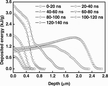
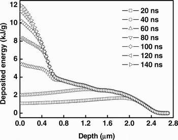
The evolution of HIPIB energy deposition of 70%H+ + 30%C+ with a peak ion current density of 300 A/cm2 is shown in Figure 6, where the profiles of deposited energy were integrated for each 20 ns period up to 140 ns, respectively. It is clear that the HIPIB irradiation led to energy deposition in the target material with a shallowing of depth and decreasing of energy density process during a pulse. Moreover, the mixed ion beam may result in an energy deposition profile of separated peaks with delayed heavier ions due to TOF effect, and the peak values of the deposited energy profile not only depend on the ion species and its kinetic energy, but also on the ratio among the different ions. The cumulative energy deposited at the different time point during a pulse is correspondingly given in Figure 7. The total energy profile of a full pulse has an obvious transient point at a depth of around 0.6 µm between 40 and 60 ns, by which the profile of energy deposition firstly extended to a depth more than 2 µm with a high energy density plateau within 3–4 kJ/g at the top 0.7 µm depth, and then subsequent energy deposition concentrated at the top surface layer developing from a depth of 0.7 µm to the outermost surface with a final accumulative energy peak of around 12 kJ/g. It is evident that the transient point is a sign of the major energy contribution shifting from H+ to C+ irradiation from 45 ns on, being an important characteristic of HIPIB irradiation of mixed ion species.
The evolution of HIPIB energy deposition of 70%H+ + 30%C+ with a peak ion current density of 300 A/cm2, where the profiles of deposited energy were integrated for each 20 ns period up to 140 ns, respectively.

The cumulative energy profiles at the different time points during a pulse of 140 ns.

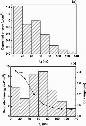
Figure 8 shows the deposited energy fluence and specific energy during a pulse under HIPIB irradiation with mixed H+ and C+. The energy fluence at each 20 ns periods (corresponding to Fig. 6) is integrated to clearly present the evolution of energy deposited in different periods of a HIPIB pulse, which can be regarded as effective energy fluence of HIPIB absorbed by the target material (Fig. 8a). The effective energy fluence had the highest value of about 1.45 J/cm2 by H+ irradiation during the first 20 ns, and then decreased to a value about 0.9 J/cm2 along with the reduction in ion current density of H+. The value was increased again at 40–60 ns period to a value of about 1.0 J/cm2, which is attributed to the arrival of peak current of C+ during mixed ion beam irradiation phase. After 60 ns, the effective energy fluence decreased continuously to a value of 0.05 J/cm2 at the last 20 ns period where the total effective energy fluence is about 4.3 J/cm2 of the 140 ns pulse. It should be pointed out that the specific energy deposited in the target material may have a considerably different evolution taking into account the ion range of respective ions (Fig. 8b), where the ion range is defined as the depth containing 90% of the total deposited energy in each 20 ns period. It is noted that the maximal value of the specific energy density occurred within 40–80 ns when the C+ current density was at relatively high level (Fig. 4), other than at the first 20 ns with the highest effective energy fluence (Fig. 8a), due to a continuously decreased ion range in a pulse.
The deposited energy fluence (a) and specific energy (b) during a pulse under HIPIB irradiation with 70%H+ + 30%C+, where the ion range is defined as the depth of 90% total energy deposited in the respective 20 ns period.

3.3. Energy Deposition Process during HIPIB Irradiation
Investigation on the energy deposition process of HIPIB is crucial for fully understanding the beam-material interactions, where the energy deposition and its rate is the origin of significant thermal and dynamic effects induced by HIPIB irradiation onto target materials. For this purpose, the energy deposition of ions has been intensively studied as the heat source for numerical simulation to explore the thermal and dynamic effects under HIPIB irradiation (Davis et al., Reference Davis, Johnston, Olson, Rej, Waganaar, Ruiz, Schmidlapp and Thompson1999; Pogrebnjak et al., Reference Pogrebnjak, Shablya, Sviridenko, Valyaev, Plotnikov and Kylyshkanov1999; Le et al., Reference Le, Yan, Zhao, Han, Wang and Xue2000; Akamatsu et al., Reference Akamatsu, Ikeda, Azuma, Fujiwara and Yatsuzuka2001; Wu et al., Reference Wu, Liu, Zhu and Lei2008). A number of simplifications of the heat source term were usually assumed to facilitate the calculation, such as taking a constant accelerating voltage over the time-dependent ion current pulse (Akamatsu et al., Reference Akamatsu, Ikeda, Azuma, Fujiwara and Yatsuzuka2001), or connecting the ion kinetic energy and ion current at the target surface by assigning a certain delay time between the measured voltage and current (Le et al., Reference Le, Yan, Zhao, Han, Wang and Xue2000). However, as TOF of ions concerned for the pulsed ion beam irradiation in this study, it is revealed that the energy deposition process by HIPIB thus may differ greatly from that of such simplifications or assumptions. According to the present results, there are mainly three specific features for the energy deposition process by HIPIB irradiation.
First of all, a substantial change in the ion kinetic energy distribution was observed at the target surface during HIPIB irradiation, from the applied history of accelerating voltage (i.e., the form of accelerating pulsed voltage), as a major consequence of TOF effect of ions. The higher the ion kinetic energy of ions extracted from the ion source, the shorter their TOF, and thus the earlier the ions arrived at the target surface. As a consequence of the TOF effect, the ion kinetic energy distribution of each ion species arriving at the target surface has a characteristic pulse form of fast-rising front followed by a long tail, and multi peaks will occur accordingly in presence of multiple ion species (Fig. 3). It is indicated that the beam-material interaction has been initiated by the ion impinging of the highest ion kinetic energy and lightest ion mass. This inherent TOF evolution of ion kinetic energy spectrum during HIPIB-material interaction can not be included in the previous assumptions of a constant kinetic energy (Akamatsu et al., Reference Akamatsu, Ikeda, Azuma, Fujiwara and Yatsuzuka2001) or a kinetic energy distribution profile identical to the waveform of accelerating voltage (Le et al., Reference Le, Yan, Zhao, Han, Wang and Xue2000; Wu et al., Reference Wu, Liu, Zhu and Lei2008).
Second, it is found that the TOF of ions led to ion current density of a fast rising front and extended pulse duration of about 140 ns (Fig. 4), more than two times of that of accelerating history of 60 ns. This prolonged pulse form of ion current density is in accordance with the experimental measurement by Faraday cup (Fig. 1), even though the rising front of ion current and the C+ peak were not sharply resolved in the measured waveforms. The slight difference is reasonable since a number of factors may affect the measured result, including the dynamic response and resolution limits of Faraday cup, the specific ion diode structure, and operation principles etc. It has been also observed by other researchers that for HIPIB generation the ion diode operating parameters may affect the pulse form of measured ion current density with either fast or slow rising front (Noonan et al., Reference Noonan, Glidden, Greenly and Hammer1995; Bystritskii et al., Reference Bystritskii, Garate, Rostoker, Song, Vandrie, Anderson, Qerushi, Dettrick, Binderbauer, Walters, Matvienko, Petrov, Shlapakovsky, Polkovnikova and Isakov2004), and unfocused or focused configuration of ion diode may have changed relative ratios among the different ion species (Isakov et al., Reference Isakov, Kolodii, Opekunov, Matvienko, Pechenkin, Remnev and Usov1991; Davis et al., Reference Davis, Johnston, Olson, Rej, Waganaar, Ruiz, Schmidlapp and Thompson1999; Pogrebnjak et al., Reference Pogrebnjak, Shablya, Sviridenko, Valyaev, Plotnikov and Kylyshkanov1999). Note that a fast rising front of measured ion current density were usually observed in case of a high-density anode plasma formed, e.g., pre-produced in the acceleration gap of ion diode prior to the applying of accelerating voltage (Remnev et al., Reference Remnev, Isakov, Opekounov, Kotlyarevsky, Kutuzov, Lopatin, Matvienko, Ovsyannikov, Potyomkin and Tarbokov1997; Ito et al., Reference Ito, Miyake and Masugata2008). This experimental phenomenon may verify our proposed model in this study where dense anode plasma with little variation is assumed as well as ion current density of a fast rising front is experimentally observed. Consequently, a peak-to-peak correlation between the ion current density and the ion kinetic energy can be established with the TOF effect, i.e., a higher kinetic energy correlates to a higher ion current density. The TOF correlation can be partially understood by the HIPIB composition analysis using Thomson parabola spectrometer, where the ion species detected by CR-39 plate (with the ion track pattern on) usually concentrated in the higher ion kinetic energy range (Remnev et al., Reference Remnev, Isakov, Opekounov, Kotlyarevsky, Kutuzov, Lopatin, Matvienko, Ovsyannikov, Potyomkin and Tarbokov1997; Ito et al., Reference Ito, Miyake and Masugata2008), while the direct correlation of the kinetic energy to the time-dependent ion current can not be obtained. The time-dependent ion kinetic energy and current correlation is of great significance since the two parameters determine the power density of ion beam bombarding the target surface and, ultimately affect the evolution of energy absorption and dissipation in the target materials during subsequent energy deposition process. Therefore, the ion kinetic energy and ion current density of pulsed ion beam can be incorporated to explore the energy deposition of ions, whereas the incorporation was unfeasible in the previous studies where the two primary parameters were treated separately and simply overlapped together with or without delay time.
Besides the ion kinetic energy variation and the correlation between kinetic energy and ion current density at the target surface, the ion beam composition also play an important role in the energy deposition process. The different ions arrived at the respective moment as determined by their mass and charge in addition to accelerating voltage, which can result in a discrete concentrated energy deposition. It has been shown that the profiles of the energy deposition at different periods and the total deposited energy have an obvious transition when the C+ arrived at the target surface starting the contribution (Figs. 5–7). The separated peaks occurred with time in the energy deposition profiles is a unique feature of pulsed ion beam irradiation with mixed ions. Moreover, the contribution by C+ to the specific energy deposition is significant as compared to that of H+ in the Ti target, even though only 30% C+ was taken in this case. Subsequent calculation based on Monte Carlo method revealed a shallowing trend of energy deposition during a pulse even though different ion species were presented, which is ascribed to the featured time-dependent ion kinetic energy distribution and ion current density. It is noted that the effective energy fluence (energy deposited per unit area) presented a decreasing tendency for HIPIB irradiation with single or mixed ion species, nevertheless, the evolution of deposited specific energy may changed greatly due to the presence of mixed ion species having various ion ranges at the same kinetic energy.
4. CONCLUSIONS
Energy deposition by HIPIB irradiation onto a Ti target was investigated taking into account the TOF of ions, at a peak accelerating voltage of 350 kV and ion current density of 300 A/cm2 with a ion drift distance of 15 cm from the ion source to the target surface. Monte Carlo method was employed to simulate the energy deposition process by HIPIB of mixed ion species of 70% H+ and 30% C+. Based on the present findings, the main conclusions are drawn as follows: (1) A time-dependent ion kinetic energy spectrum with peak value of 350 keV at the target surface was derived according to the ion TOF, considerably, differing from the original energy spectrum of ions extracted from the ion source, changing from Gaussian peak with pulse duration of 60 ns to a pulse form with a fast rising front and a long tail extended to 140 ns. H+ arrived at the target surface 45 ns earlier than C+ with the same peak energy of 350 keV, on a drift distance of 15 cm in vacuum. (2) The TOF ion energy spectrum of widespread kinetic energies can be correlated to the ion current density, with higher ion kinetic energies concentrating at the higher ion current. Moreover, separate current peaks were observed for ion beam of mixed ion species, which led to two energy deposition phases with concentrated energy deposition at varied depth by single H+ and mixed C+ and H+, respectively. (3) The specific energy deposition process of HIPIB is essentially determined by the TOF ion energy spectrum and correlated ion current at the target surface. It is indicated that TOF effect of ions is significant for exploring HIPIB-matter interactions, resulting in time-dependent energy deposition phenomena not observed in case of uniform ion energy distribution.
ACKNOWLEDGMENTS
The authors would like to thank Prof. G.E. Remnev and Prof. D. Wu for their technical assistance and helpful discussion. This work is supported by National Natural Science Foundation of China (NSFC) under project No. 50701009.