Introduction
Chronic rhinosinusitis and nasal polyposis are common rhinological diagnoses.Reference Lund1, Reference Scadding, Durham, Mirakian, Jones, Drake-Lee and Ryan2 Nasal polyposis is managed medically, or surgically if the disease is refractory to medical treatment.Reference Fokkens, Lund and Mullol3 Pre-operative computed tomography (CT) of the sinuses is frequently performed. This enables the radiologist and the surgeon to assess the degree of sinonasal disease radiologically, and to examine the sinonasal anatomy, which is highly variable between individuals.Reference Kantarci, Karasen, Alper, Onbas, Okur and Karaman4
Both chronic rhinosinusitis and nasal polyposis have effects on the morphology of the nose and sinuses, such as sinonasal bone expansion, erosion and thickening.Reference Lund1, Reference Lund and Lloyd5–Reference Connor, Hussain and Woo12 Changes are thought to be due to the mass effect of the polyps themselves and may also be secondary to chronic inflammation.Reference Liu, Schaefer, Moscatello and Couldwell6, Reference Connor, Hussain and Woo12 There is increasing evidence of an association between chronic rhinosinusitis and osteitis.Reference Videler, Georgalas, Menger, Freling, van Drunen and Fokkens13
This retrospective, radiological analysis of patients with known nasal polyposis aimed to assess the prevalence of bony changes such as sinonasal bone expansion, erosion and thickening. It also aimed to elucidate whether the severity of polyps, assessed using the Lund–Mackay score,Reference Lund and Mackay14 correlates with an increased prevalence of these bony changes. Such a correlation would highlight these changes as complications of nasal polyposis, as well as emphasising the importance of pre-operative CT in stratifying those at risk of polypectomy complications.
It has been reported that changes such as erosion and expansion can mimic both benign and malignant invasive disease radiologically.Reference Lund1, Reference Liu, Schaefer, Moscatello and Couldwell6, Reference Som, Lawson and Lidov8 A high prevalence of these changes would also emphasise the need to histologically assess polypoid material post-operatively, in order to exclude an alternative diagnosis.
Materials and methods
The current study entailed a retrospective analysis of the pre-operative CT scans of 104 patients with endoscopically confirmed nasal polyposis. The CT scans were viewed in 3 mm coronal sections. Bone algorithm reconstructions were viewed as standard. The CT window width parameters were greater than 150 Hounsfield units for soft tissue windows, and greater than 3500 Hounsfield units for bone windows. These patients underwent surgery at the Royal National Throat, Nose and Ear Hospital (London) over a two-year period. Patients who had undergone previous surgery or who had a diagnosis other than nasal polyposis were excluded from the study. The CT scans of an age- and gender-matched control group consisting of 44 patients with no sinonasal disease were also reviewed.
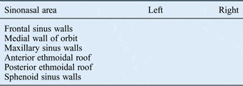
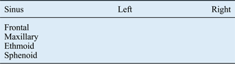
The Lund–Mackay scoreReference Lund and Mackay14 was used to grade the severity of the polyposis bilaterally. Scans were also assessed bilaterally for the presence of: bony expansion and erosion of the frontal sinus walls, medial wall of the orbit, anterior and posterior ethmoidal roofs, maxillary sinus walls, and sphenoid sinus walls; and bony thickening of the frontal, maxillary, ethmoidal and sphenoid sinus walls. Scans were assessed by two clinicians, a consultant radiologist and an otolaryngologist. All scores were agreed upon by both clinicians. The scans were given an overall score for expansion, erosion and thickening, graded out of a total score of 32; the scoring is summarised in Tables I and II.
Sinonasal expansion and bony erosion assessment tool

A score of 1 was given for the presence of expansion, and a score of 1 for the presence of erosion and 0 for its absence on each side. (Expansion and erosion were each scored out of 6 per side, to give a total out of 24.)
Bony thickening of paranasal sinus walls assessment tool
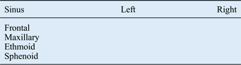
A score of 1 was given for the presence of bony thickening and 0 for its absence. (Bony thickening was scored out of 8.)
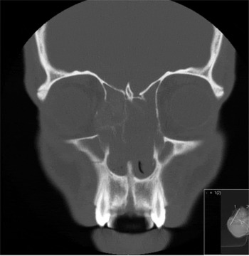
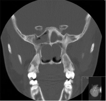
Figures 1 and 2 show examples of sinonasal bony expansion, erosion and thickening.
Coronal computed tomography scan of a nasal polyposis patient, showing bilateral total opacification of the anterior ethmoid sinuses, with local erosion of the right lamina papyracea, expansion of the right anterior ethmoid sinus and erosion of the medial wall of the right orbit.

Coronal computed tomography scan of the sphenoid sinus of a nasal polyposis patient, demonstrating incomplete opacification of the right sphenoid sinus, with sinus expansion and bone erosion, and bone thickening of the left (totally opacified) sphenoid sinus.

Results and analysis
Of the 104 patients, 66 were male and 38 were female. The patients' ages ranged from 11 to 86 years. For the 104 patients, the median Lund–Mackay score was 17 out of 24. For the patients in the control group, the median Lund–Mackay score was 0 out of 24.
Radiological bony changes
Table III shows the findings for radiological bony changes. The mean score for expansion, erosion and thickening was 6.4 out of 32 (range 0–18). Of the 104 patient scans reviewed, 97 (93 per cent) showed evidence of sinonasal expansion, bony erosion or bony thickening. Ninety of the patients (87 per cent) showed evidence of sinonasal expansion, 73 (70 per cent) had sinonasal bony erosion and 61 (59 per cent) had bony thickening of the sinus bones.
Prevalence of bony changes*

* In nasal polyposis patients (total n=104). MWO = medial wall of orbit; AER = anterior ethmoidal roof; PER = posterior ethmoidal roof; FS = frontal sinus; SS = sphenoid sinus; ES = ethmoid sinus; MS = maxillary sinus; B = bilateral; U = unilateral
Bone expansion was seen most frequently in the medial wall of the orbit (78 patients, 75 per cent), followed in order of decreasing frequency by: the maxillary sinus (45 patients, 43 per cent), frontal sinus (20 patients, 19 per cent), anterior ethmoidal roof (8 patients, 8 per cent), the sphenoid sinus (8 patients, 8 per cent) and the posterior ethmoidal roof (5 patients, 5 per cent).
Bone erosion was seen most frequently in the medial walls of the orbits (61 patients, 59 per cent), followed in order of decreasing frequency by: the anterior ethmoidal roof (24 patients, 23 per cent), the sphenoid sinus walls (19 patients, 18 per cent), the maxillary sinus walls (13 patients, 13 per cent), the frontal sinus walls (11 patients, 11 per cent) and the posterior ethmoidal roof (9 patients, 9 per cent).
Bony thickening was seen most frequently in the sphenoid sinus walls (36 patients, 35 per cent) and the maxillary sinus walls (36 patients, 35 per cent). These were followed in order of decreasing frequency by: the frontal sinus walls (28 patients, 27 per cent) and the ethmoidal sinus walls (9 patients, 9 per cent).
No bone expansion, erosion or thickening was seen in the CT scans of the control group.
Statistics
The Spearman's rank correlation coefficient (r s) for the correlation between the Lund–Mackay score and the total score of sinonasal expansion, bony erosion and bony thickening was 0.31 (p < 0.01, two-tailed).
Discussion
Nasal polyposis is a common otorhinolaryngological condition, for which surgery is performed for disease that is refractory to medical treatment.Reference Fokkens, Lund and Mullol3 Sinonasal bone expansion, erosion and thickening secondary to nasal polyposis have all been described in the literature.Reference Lund1, Reference Lund and Lloyd5, Reference Banna, Ewaschuk and Cole7, Reference Som, Lawson and Lidov8, Reference de Vries10, Reference Rejowski, Caldarelli, Campanella and Penn11 However, little has been documented regarding the prevalence of these changes and how they relate to the severity of polyposis.
The results of this study showed that the total prevalence of sinonasal expansion, bony erosion and bony thickening was high (93 per cent) in this group of patients with nasal polyposis. Thus, these changes were a common complication of nasal polyposis in this population. Sinonasal expansion, bony erosion and bony thickening were not observed in the control group.
The bony changes are thought to be due to the mass effect of the polyps themselves and may also be secondary to chronic inflammation.Reference Liu, Schaefer, Moscatello and Couldwell6, Reference Connor, Hussain and Woo12
Woakes' syndrome is a condition of infantile, destructive nasal polyposis. It is associated with broadening of the nose as a result of sinonasal expansion and erosion due to nasal polyps, as well as frontal sinus aplasia, bronchiectasis and dyscrinia. It is thought that the expansile mass effect of the polyps at a young age cause the typical facies in affected patients.
There is increasing evidence of an association between chronic rhinosinusitis and osteitis.Reference Videler, Georgalas, Menger, Freling, van Drunen and Fokkens13 One study showed histological evidence of osteitis in more than 50 per cent of bone specimens from patients undergoing functional endoscopic sinus surgery for chronic rhinosinusitis.Reference Lee, Kennedy, Palmer, Feldman and Chiu15
Nasal polyposis associated with sinonasal bony changes has been shown to present with symptoms such as blindnessReference Rejowski, Caldarelli, Campanella and Penn11 or evidence of intracranial extension,Reference Majithia, Tatla, Sandhu, Saleh and Clarke16 which may complicate surgery. Indeed, hyperostosis in patients with chronic rhinosinusitis may lead to a poorer outcome after surgery.Reference Kim, Dhong, Lee, Chung, Yim and Oh17
In this study, the most frequent change seen was sinonasal bone expansion (90 patients, 87 per cent), followed by bone erosion (73 patients, 70 per cent) and bone thickening (61 patients, 59 per cent). The most commonly expanded and eroded area was the medial wall of the orbit. These frequencies in occurrence were higher than those of a previous investigation that showed ethmoidal expansion in 20 per cent of the patients studied.Reference Lund and Lloyd5 The disparity may be due to the fact that the current study involved CT, whereas the previous study employed plain radiography.
The data of the current study suggest that the medial wall of the orbit is most at risk of damage, both through the disease process and during endoscopic surgery. Bony expansion, erosion and thickening were seen in all areas surveyed.
The most frequently thickened sinus walls were those of the sphenoid and maxillary sinuses (36 patients each, 35 per cent). Bony thickening may complicate sinus surgery as it can make opening of the sinuses and removal of the bony partitions more difficult.
The high prevalence of sinonasal expansion, erosion and thickening emphasises the importance of a pre-operative CT scan in patients with nasal polyposis, which can help to prevent operative intracranial and orbital complications.Reference Kaluskar, Patil and Sharkey18 The findings may also promote the use of intra-operative imaging guidance software, particularly in those with severe bony changes on CT.
Major complications of surgery for nasal polyposis and chronic rhinosinusitis most commonly arise due to the proximity of the nose and sinuses to the orbit and anterior cranial fossa, and due to severe haemorrhage. A national audit on the complications of surgery for chronic rhinosinusitis and nasal polyposis reported that major complications, including orbital or intracranial complications, and severe haemorrhage, occurred in 0.4 per cent of the 3128 patients involved in that study. Minor complications such as moderate haemorrhage occurred in 6.6 per cent of patients. The complication rate was increased in patients with more severe symptoms and those with more opaque sinuses on CT.Reference Hopkins, Browne, Slack, Lund, Topham and Reeves19
In this study, a correlation was found between the severity of polyposis (Lund–Mackay score) and the total expansion, erosion and thickening score (r s = 0.31; p < 0.01). This suggests that it is not only the presence of nasal polyps, but also the severity and extent of polyposis that increases the likelihood of morphological sinonasal changes. These data may help to explain the increased surgical complication rates reported in patients with severe sinonasal disease compared with mild disease.Reference Hopkins, Browne, Slack, Lund, Topham and Reeves19 Georgalas et al. correlated severity of osteitis with Lund–Mackay scores, but this assessment only included bone thickening, and not sinus bone expansion or erosion.Reference Georgalas, Videler, Freling and Fokkens20
A caveat is that the Lund–Mackay score does not differentiate between chronic rhinosinusitis alone and chronic rhinosinusitis with nasal polyposis. However, opacification is often worse in patients with nasal polyposis, and all patients in the current study were endoscopically confirmed to have nasal polyps.
• In this study of nasal polyposis patients, the prevalence of sinonasal expansion, erosion or thickening was 93 per cent
• Disease severity was positively correlated with the prevalence of bony changes
• The findings emphasise the importance of pre-operative imaging and consideration of intra-operative image guidance
• These can aid visualisation of individual morphology and help prevent intra-operative complications
• As bony changes may mimic more invasive disease, the histological assessment of polypoidal material is encouraged
The bony changes surveyed in this study, particularly sinonasal bone expansion and erosion, have been reported by some to mimic more invasive benign disease, including inverted papilloma or even malignant disease such as mucinous adenocarcinoma.Reference Lund1, Reference Liu, Schaefer, Moscatello and Couldwell6, Reference Som, Lawson and Lidov8 Diamantopoulos et al. reviewed the histology results of 2021 patients treated surgically for nasal polyposis, and found that 22 patients (1.1 per cent) had a histological diagnosis that differed from the diagnosis made at the time of surgery, 50 per cent of which showed inverted papilloma.Reference Diamantopoulos, Jones and Lowe21
The high prevalence of bony changes seen in this group of patients with nasal polyposis emphasises the fact that nasal polyps can appear invasive radiologically, which may make differentiation between benign and malignant disease more difficult. Radiological features such as density and location of a mass help the radiologist to distinguish between benign and malignant disease. However, the bone erosion associated with nasal polyps (as seen in this study) and the occasional resemblance to malignancy emphasise the importance of histologically assessing all polypoid material post-operatively.
Conclusion
This study demonstrated a high prevalence of sinonasal bony expansion, erosion and thickening on the pre-operative CT scans of patients with nasal polyposis. The medial wall of the orbit was the most commonly expanded and eroded area, and the walls of the sphenoid sinus and maxillary sinus were the most commonly thickened areas. The severity of nasal polyposis was positively correlated with the observed bony changes. These data emphasise the importance of a pre-operative CT scan in assessing the patient's disease severity and morphology, which can help to prevent operative complications that effect intracranial and orbital areas. Prospective cohort studies are required to further examine whether these bony changes do increase the risk of such operative complications. As these bony changes might mimic a more invasive disease process, histological assessment of polyps post-operatively is important in order to exclude alternative pathology.