Introduction
Dehydroleucodine (DhL), a sesquiterpenic lactone, was isolated and purified from Artemisia douglasiana Besser, a medicinal herb used in Argentina (Giordano et al., Reference Giordano, Guerreiro, Pestchanker, Guzman, Pastor and Guardia1990). It has previously been shown that DhL selectively induces a dose-dependent transient arrest in G2 of both meristematic cells and vascular smooth muscle cells (VSMC) (López et al., Reference Lopez, Giordano and Lopez2002; Cruzado et al., Reference Cruzado, Castro, Fernández, Gomez, Roque, Giordano and Lopez2005). However, the nature of the mechanisms involved in the process is still unknown.
In the meiosis of fully grown amphibian oocytes the transition from the G2 to the M phase is regulated by the maturation promoting factor (MPF), a complex of the cyclin-dependent kinase p34/cdc2 and cyclin B (Lohka et al., 1988; Masui, 1992).
In immature oocytes of Xenopus, Bufo arenarum and starfish there is an inactive complex (pre-MPF) that consists of cyclin B-bound cdc2 phosphorylated on both Thr-161 and Thr-14/Tyr-15. In these species, cdc2 Thr-14/Tyr-15 dephosphorylation is primarily responsible for MPF activation during oocyte maturation (Gautier & Maler, Reference Gautier and Maller1991; Minshul et al., Reference Minshull, Murray, Colman and Hunt1991; Ookata et al., Reference Ookada, Hisanaga, Okano and Kishimoto1992; Galas et al., Reference Galas, Barakat, Dorée and Picard1993; Sánchez Toranzo et al., Reference Sanchez Toranzo, Bonilla, Zelarayán, Oterino and Bühler2006). It has been demonstrated that dephosphorylation of cdc2 Thr-14/Tyr-15 is induced by cdc25 phosphatase (Perdiguero & Nebreda, Reference Perdiguero and Nebreda2004; Dekel, Reference Dekel2005), one of the key events of M-phase reentry.
In G2-arrested Xenopus oocytes, cdc25 is probably maintained inactive by PKA-mediated phosphorylation. When oocyte maturation is initiated by progesterone, cAMP levels are reduced and PKA becomes inactivated. Interestingly, PKA and PP2A are both active in G2-arrested oocytes and become inactivated upon progesterone addition (Duckworth et al., Reference Duckworth, Weaver and Rudeman2002; Schmitt & Nebreda, Reference Schmitt and Nebreda2002).
The injection of only germinal vesicle contents (GV content) from immature Bufo arenarum oocytes is sufficient to cause GVBD in recipient immature oocytes (Sánchez Toranzo et al., Reference Sanchez Toranzo, Bonilla, Zelarayán, Oterino and Bühler2006). This result is similar to that reported for starfish oocytes (Picard & Doree, Reference Picard and Dorée1984; Picard et al., Reference Picard, Labbe, Barakat, Cavadore and Doree1991). In this regard, it has been suggested that a nuclear factor originating from the GV is required for the activation of pre-MPF (Kishimoto et al., Reference Kishimoto, Hirai and Kanatani1981; Picard & Doree, Reference Picard and Dorée1984; Picard et al., Reference Picard, Labbe, Barakat, Cavadore and Doree1991).
In Bufo arenarum and Xenopus oocytes a characteristic of MPF is its ability to undergo auto-catalytic activation in the absence of protein synthesis (Wasserman & Masui, Reference Wasserman and Masui1975; Sánchez Toranzo et al., Reference Sanchez Toranzo, Bonilla, Zelarayán, Oterino and Bühler2006). A small amount of cytoplasm containing active MPF introduced by microinjection into an immature oocyte is enough to activate an amplification loop that promotes the conversion of an inactive complex into its active form.
Bufo arenarum oocytes obtained during the reproductive period (spring–summer) resume meiosis without the need of an exogenous hormonal stimulus if deprived of their enveloping follicle cells, a phenomenon called spontaneous maturation. In this species it is possible to obtain oocytes competent and incompetent to undergo spontaneous maturation according to the seasonal period in which animals are captured (Zelarayán et al., Reference Zelarayán, Oterino and Bühler1995).
As a whole, the model of oocyte maturation in vitro affords four clear situations to start meiosis division: one by deprivation of follicle cells in oocytes competent to undergo spontaneous maturation and three in incompetent oocytes: (i) progesterone stimulation, the induction starts at the upper string of cdc25 activation by inhibiting PKA activity; (ii) GV- content stimulation at the upper string of cdc25 activation, probably by inhibiting PP2A phosphatase activitity; and (iii) a small concentration of active MPF that induces stimulation of the cdc25, the auto-amplification loop of MPF stimulation.
This study was designed to evaluate the effect of DhL on the resumption of meiosis in Bufo arenarum oocytes induced in vitro by the four treatments mentioned above: removal of follicle cells, progesterone stimulation, VG-content injection and injection of a small amount of active MPF.
Materials and Methods
Animals
Adult specimens of Bufo arenarum were collected in the northwestern area of Argentina from May to August (winter animals) and from September to December (summer animals) and kept at 15 °C until use.
Hormones and reagents
All hormones and reagents were purchased from Sigma. Progesterone was dissolved in ethanol and added directly to the culture medium to give a final concentration of 2.5 μM. Dehydroleucodine (DhL) at 93% purity was obtained according to Giordano (Giordano et al., Reference Giordano, Guerreiro, Pestchanker, Guzman, Pastor and Guardia1990, Reference Giordano, Pestchanker, Guerreiro, Saad, Enriz, Rodriguez, Jauregui, Guzman, Maria and Wendel1992). DhL was dissolved in DMSO and various doses were added to the culture medium.
In vitro denuded oocytes culture
Experimental manipulation and culture were performed at room temperature (22–25 °C) in amphibian Ringer solution (AR) (6.6 g NaCl/l, 0.15 g CaCl2/l and 0.15 g KCl/l) containing penicillin G-sodium (30 mg/l) and streptomycin sulphate (50 mg/l), pH 7.4. Denuded full-grown oocytes were obtained according to Lin & Schuetz (1985). Follicle cells were removed by gentle shaking (100 oscillations/min) (Zelarayán et al., Reference Zelarayán, Oterino and Bühler1995).
Randomized samples of 20 oocytes were distributed into separate wells containing 2 ml of AR; the reagents were added (5 μl) directly to the culture medium. Oocyte maturation was assessed 24 h after hormone or reagent addition. Meiosis reinitiation was scored both by the presence of a transient white spot in the animal pole and by the absence of GV after dissection of the oocytes fixed in trichloroacetic acid.
Cytoplasm and germinal vesicles contents
Cytoplasm from mature and immature oocytes was obtained according to Hedeimann's method, modified by Bühler & Petrino (Reference Bühler and Petrino1983). Oocytes matured with progesterone (2.5 μM) or immature oocytes were suspended at the interface of 60% Ficoll (w/v) in Ringer's solution and then centrifuged for 30 min at 2500 g at 4 °C in a Sorval HB4 rotor. After this procedure the oocytes were stratified into four layers: yolk platelets, pigment granules, clear cytoplasm and oil cap. The content of the clear cytoplasm layer was used for microinjections.
To obtain the GV content, fully grown oocytes were isolated manually and pricked at the animal pole with a fine glass pipette. The GVs were squeezed out into AR Tris–HCl (pH 7.4). Forty GVs were packed into a capillary tube with AR to a final volume of 18 μl and centrifuged at 10 000 g for 10 min at 15 °C. The supernatant is referred to hereafter as ‘GV content’.
Microinjections
Denuded fully grown immature oocytes were microinjected with 50 nl of mature cytoplasm, with 50 nl of immature cytoplasm or with 50 nl of the GV content. Following injection, oocytes were placed in AR and scored for GVBD after incubation for 24 h.
Microinjection was performed with a sharpened glass micropipette (outer diameter 40–50 μm) attached to a micromanipulator.
Results and discussion
Effect of DhL on spontaneous and progesterone-induced oocyte maturation
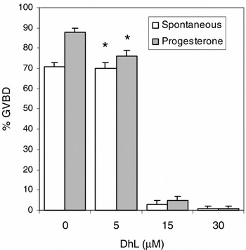
Denuded oocytes competent and incompetent to mature spontaneously were cultured in AR with different doses of DhL (5–30 μM) 60 min before the removal of follicular cells or the addition of progesterone. Oocytes were examined for GVBD after 24 h of culture. Results show that 15 μM of DhL induced inhibition of GVBD in 90% of both types of oocytes (Fig. 1).
Effect of DhL on spontaneous and progesterone-induced oocyte maturation. Denuded oocytes competent or not competent to mature spontaneously were cultured in AR with different doses of DhL (5–30 μM) 60 min before the removal of follicular cells or the addition of progesterone (2.5 μM). GVBD was scored after 24 h of culture. Values are the mean ± SEM (n = 5). Each experiment was performed on a different animal. *Significant difference to 0 μM DhL. p < 0.01 (one-way analysis of variance, ANOVA).

When oocytes that had been incubated previously with progesterone and DhL, like above, were incubated a further 24 h only with progesterone, the number of oocytes with GVBD increased up to 50%. These results indicate that the effect of DhL was reverted if the inhibitor was removed from the incubation medium (data not shown).
Effect of DhL on GVBD in oocytes incompetent to mature spontaneously injected with GV contents
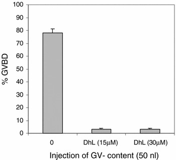
Denuded full-grown oocytes incompetent to mature spontaneously were preincubated with and without 15 μM DhL for 60 min and microinjected with 50 nl of GV content. Injected oocytes were incubated in AR with and without DhL (15 μM) for 16 h before GVBD scoring. Meanwhile, 80% of oocytes incubated without DhL showed GVBD and only 5% matured when they were incubated in 15 μM of DhL (Fig. 2), indicating that DhL inhibited the ability of GV content to induce GVBD.
Effect of DhL on the GVBD in oocytes not competent to mature spontaneously injected with GV content. Denuded oocytes incompetent to mature spontaneously were preincubated with and without 15 μM DhL for 60 min and microinjected with 50 nl of GV content. Injected oocytes were incubated in AR with and without DhL (15 μM) for 16 h before GVBD scoring. Values are the mean ± SEM (n = 4). Each experiment was performed on a different animal.

The GV component responsible for this activity has not been identified yet; however, Picard et al. (Reference Picard and Dorée1991) reported that the effect of the injection of GV content on starfish is similar to the injection of 1 μM okadaic acid. This suggests that the GV contains a factor that inhibits PP2A phosphatase activity in the same way as does okadaic acid.
In Bufo arenarum the results found by Sánchez Toranzo et al. (Reference Sanchez Toranzo, Bonilla, Zelarayán, Oterino and Bühler2006) agree with those of Picard (Reference Picard and Dorée1991) and suggest that the GV content would possess a type 2A phosphatase inhibitor capable of promoting MPF amplification through a loop of unidentified regulatory events that depend on protein synthesis. It is possible that the target of DhL could be the PP2A phosphatase inhibitor.
Effect of DhL on MPF amplification in oocytes unable to mature spontaneously
The effect of DhL on MPF amplification of Bufo arenarum oocytes was tested by microinjection of cytoplasm from mature oocytes. Denuded fully grown oocytes that were unable to mature spontaneously were preincubated with and without DhL for 60 min and microinjected with 50 nl of mature cytoplasm. Injected oocytes were cultured in AR with and without DhL for 24 h and the GVBD were scored.
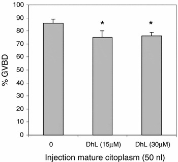
Results showed that DhL at the doses assayed had no effect on MPF amplification and the percentages of GVBD obtained were similar to that of the control oocytes (Fig. 3). Previous results from our laboratory demonstrated that in Bufo arenarum the mechanism of amplification of MPF by injection of mature cytoplasm requires the presence of an active cdc25 phosphatase (Sánchez Toranzo et al., Reference Picard, Labbe, Barakat, Cavadore and Doree2006). On the basis of the results shown in this paper, we can infer that DhL inhibited GVBD in the earliest events of cdc25 activation, suggesting that the target of DhL is a specific event in the early step of the activation cascade reaction of cdc25. These results have encouraged us to search for the target of DhL.
Effect of DhL on MPF amplification in oocytes incompetent to mature spontaneously. Denuded oocytes incompetent to mature spontaneously were preincubated with and without DhL for 60 min and microinjected with 50 nl of mature cytoplasm. Injected oocytes were cultured in AR with and without DhL for 24 h and GVBD were scored. Values are the mean ± SEM (n = 4). Each experiment was performed on a different animal. *Significant difference to 0 μM DhL. p < 0.01 (one-way analysis of variance, ANOVA).

Acknowledgements
This work was supported by a grant from the Science Council of the National University of Tucumán (CIUNT) and the National Council of Scientific and Technological Research (CONICET).