Introduction
Anomalous single coronary artery originating from the pulmonary artery (ASCAPA) is an exceptionally rare and life-threatening congenital coronary anomaly. Reference Ochoa-Ramirez, Valdez-Garza and Reyes-Gonzalez1 Due to the limited number of reported cases, its exact incidence remains unknown. Reference Shetty, Thareen, Ramaiah, Narayan, Das and Chandrashekariah2 However, artery originating from the pulmonary artery poses a significant diagnostic and therapeutic challenge because of its nonspecific presentation and rapid clinical deterioration if left untreated. Given the high surgical risk and potential for poor outcomes, early recognition and preoperative stabilisation are essential. This case report presents a unique instance of artery originating from the pulmonary artery in an infant with advanced endocardial fibroelastosis and severe left ventricular dysfunction, emphasising the importance of early diagnosis and a tailored extracorporeal membrane oxygenation-based management strategy.
Case presentation
The patient was a term male infant born via normal spontaneous vaginal delivery with a birth weight of 3200 g. At presentation, he was 2 months and 3 days old, weighing 4800 g. The family reported that the infant had been asymptomatic until approximately one week prior to admission, when they noticed tachypnoea and respiratory distress, prompting them to seek care at a paediatric emergency department. The patient underwent cardiopulmonary resuscitation at an outside facility, after which he was intubated and admitted to the ICU. Chest radiography revealed cardiomegaly and signs of heart failure, leading to referral to our paediatric cardiology department for further evaluation and management.
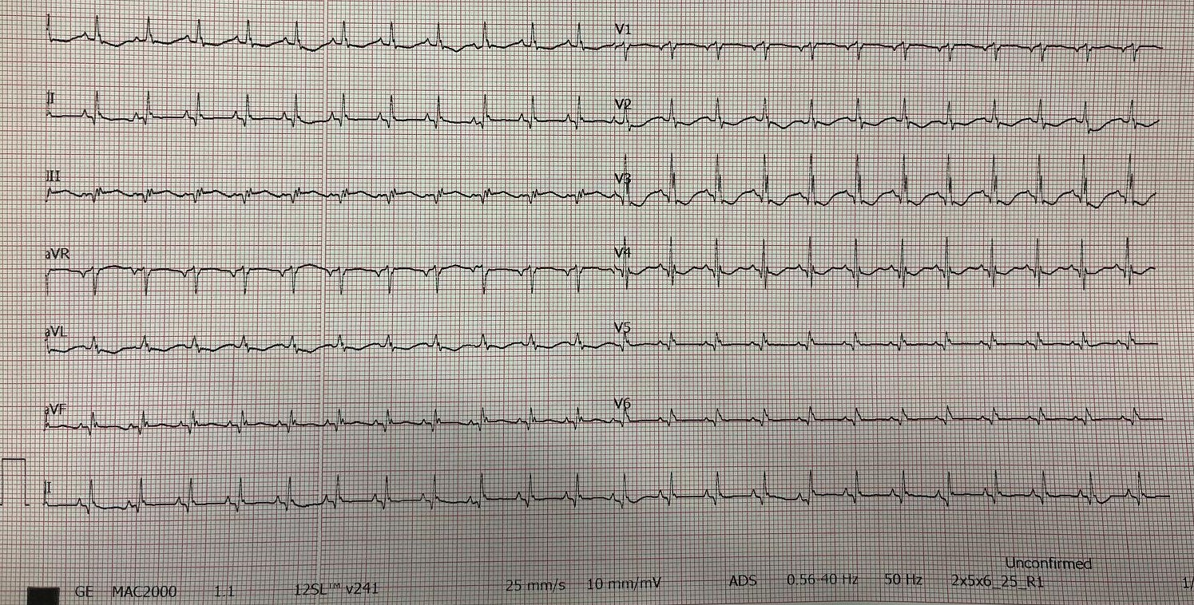
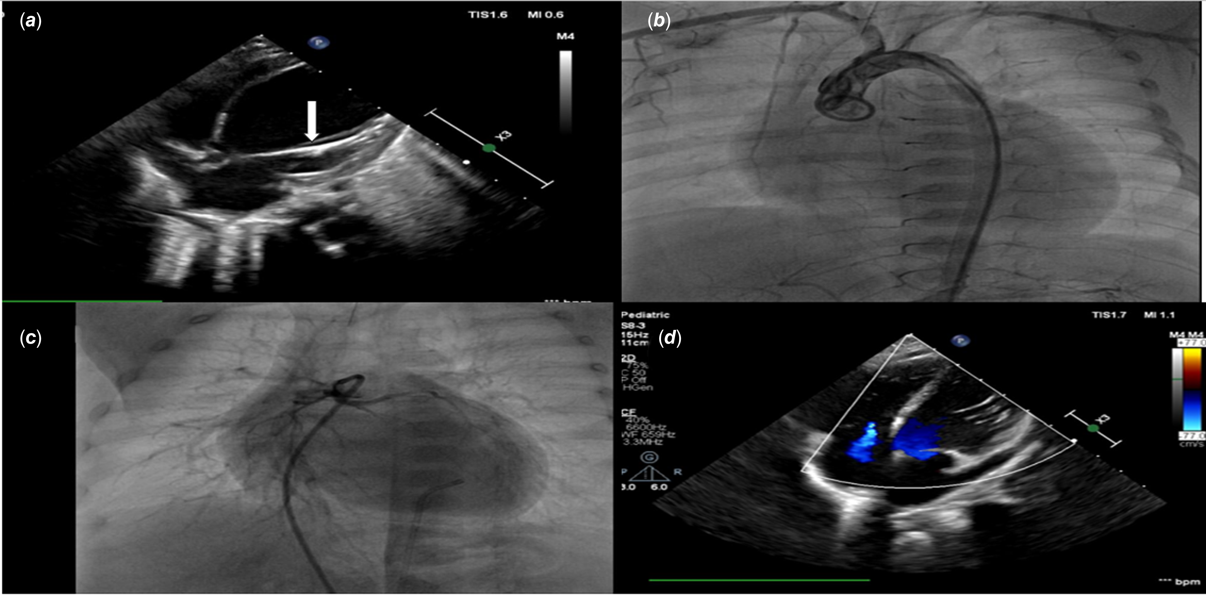
On physical examination, oxygen saturation was 97% (while intubated with FiO2 40%), heart rate was 150 beats per minute, and blood pressure was 60/30 mmHg. Twelve-lead electrocardiography revealed diminished RS voltages and relatively prominent Q waves in leads D3 and aVF. Additionally, decreased R wave voltages and proportionally increased Q waves were noted in leads V5 and V6 (Figure 1). Echocardiography demonstrated significantly impaired left ventricular function (ejection fraction: 28%), increased echogenicity of the papillary muscles suggestive of endocardial fibroelastosis (Figure 2a), and a patent ductus arteriosus.
Decreased rs voltages in D3 and aVF, with relatively increased q waves by ratio. Additionally, decreased R wave voltages and relatively increased q waves by ratio are observed in leads V5 and V6.

(a) Prolonged reduced coronary perfusion leads to fibrosis and elastic tissue proliferation in the endocardium of the left ventricle, resulting in endocardial fibroelastosis (EFE) (arrow). A functionally impaired, spherical, and dilated left ventricle (LV) secondary to EFE has been demonstrated. (b) Aortic root injection was performed during angiography, and no coronary artery arising from the aorta was visualised, supporting the diagnosis of ASCAPA. (c) Pulmonary artery root injection was performed during angiography, and a single coronary artery originating from the pulmonary artery was observed, supporting the diagnosis of ASCAPA. (d) Postoperative follow-up echocardiography, compared to the preoperative echocardiogram (Figure 2a), showed regression of the spherical morphology of the left ventricle and improvement in its function. ASCAPA, artery originating from the pulmonary artery.

The presence of left ventricular dysfunction and endocardial fibroelastosis raised suspicion for an anomalous left coronary artery originating from the pulmonary artery. To confirm the diagnosis, cardiac catheterisation and angiography were performed (Supplementary Video). Aortic root injection revealed no coronary arteries arising from the aorta (Figure 2b). Pulmonary artery injection demonstrated a single anomalous coronary artery originating from the pulmonary artery, confirming the diagnosis of artery originating from the pulmonary artery (Figure 2c).
Following angiography, the patient’s blood pressure dropped despite inotropic support, and desaturation developed. Therefore, veno-arterial extracorporeal membrane oxygenation (V-A extracorporeal membrane oxygenation) was initiated. Given the patient’s clinical deterioration, stabilisation with extracorporeal membrane oxygenation was considered more appropriate than proceeding directly to surgery.
Prior to corrective surgery, we employed a modified extracorporeal membrane oxygenation strategy. In conventional V-A extracorporeal membrane oxygenation, venous blood from the right heart is diverted through cannulas into the extracorporeal membrane oxygenation circuit, thereby reducing flow to the pulmonary artery. In the context of a single coronary artery originating from the pulmonary artery, this reduction in pulmonary arterial flow could compromise coronary perfusion, resulting in coronary steal.
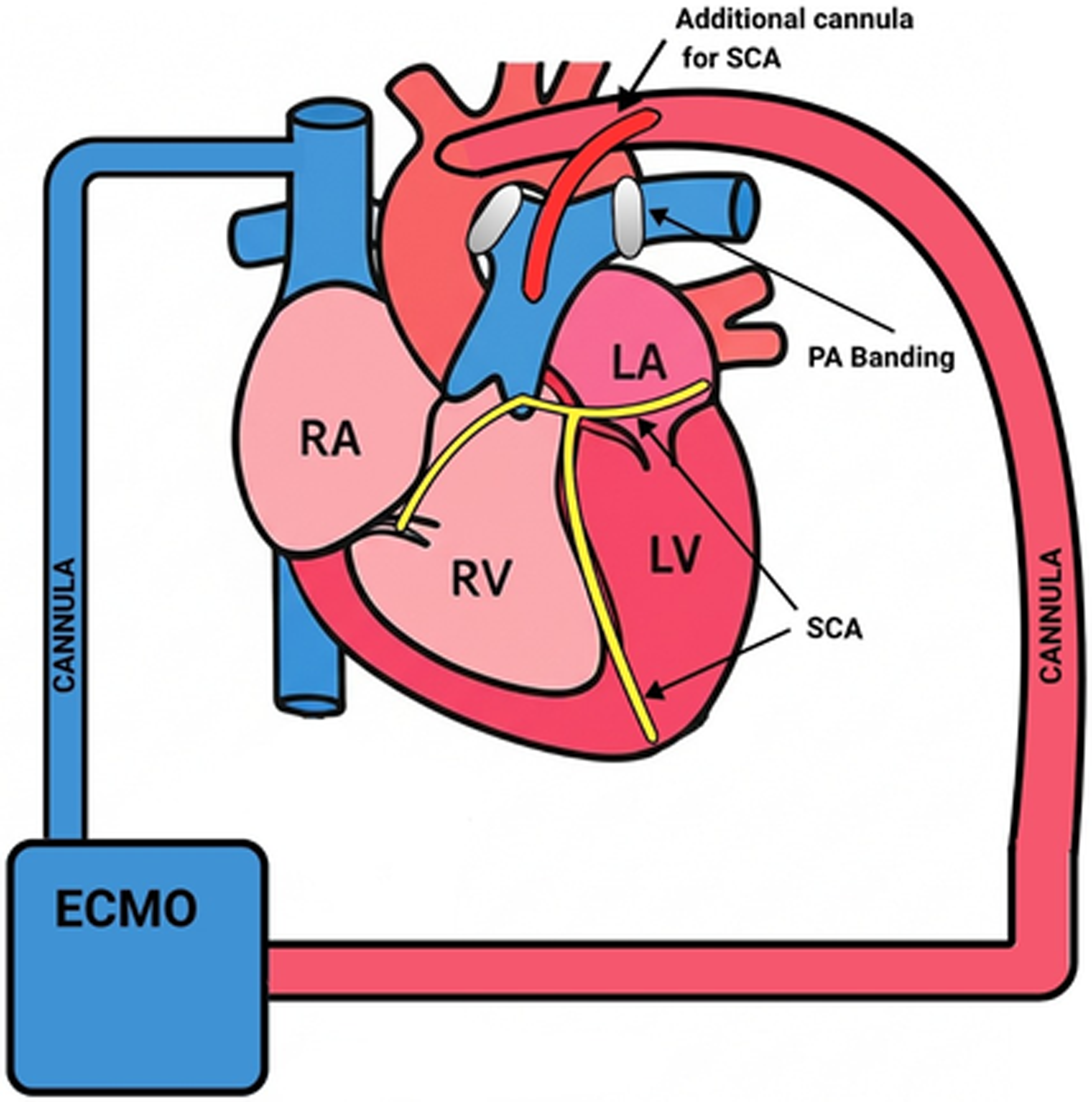
Cannulas were placed to drain blood from the right atrium to the oxygenator and then return it to the aorta. Additionally, a smaller cannula was connected to the circuit between the oxygenator and the aortic return and directed into the main pulmonary artery to ensure perfusion of the coronary artery originating from it. To prevent runoff of oxygenated blood into the peripheral pulmonary arteries and avoid coronary steal, the distal branches of the pulmonary arteries were banded (Figure 3).
Visual representation of the ECMO technique applied to the patient. ECMO, extracorporeal membrane oxygenation.

After achieving haemodynamic stabilisation, the patient was taken to surgery. Intraoperative findings confirmed the diagnosis: both the left main coronary artery and the right coronary artery originated from a single ostium located in the left (posterior) sinus of the pulmonary artery. Following reimplantation of the single coronary artery into the aorta, intraoperative epicardial echocardiography revealed poor contractile function, and the patient was transferred to the ICU with postoperative extracorporeal membrane oxygenation support.
He was monitored under high-dose inotropic and extracorporeal membrane oxygenation support. Extracorporeal membrane oxygenation was successfully weaned on postoperative day 11, and the patient was discharged on day 22. At present, the patient is 2 years old and remains under outpatient follow-up with mildly reduced left ventricular function (ejection fraction: 52%) and moderate tricuspid regurgitation (Figure 2d).
Discussion
Artery originating from the pulmonary artery is among the rarest coronary anomalies and is often diagnosed postmortem; its preoperative identification is exceptionally uncommon. Reference Guenther, Chen, Gustafson, Ing, Brothers and Raff3 Although the true incidence remains unknown, artery originating from the pulmonary artery is significantly rarer than the more commonly described anomalous left coronary artery from the pulmonary artery. Reference Shetty, Thareen, Ramaiah, Narayan, Das and Chandrashekariah2
Typical signs and symptoms may include respiratory distress, feeding intolerance, unexplained irritability, and crying during feeding, with clinical manifestations generally emerging between the second and fourth months of life. Chest radiographs frequently show cardiomegaly, while echocardiography often reveals mitral regurgitation and ventricular dysfunction. Electrocardiographic findings typically indicate myocardial ischaemia and infarction. Reference Shetty, Thareen, Ramaiah, Narayan, Das and Chandrashekariah2
Due to the rarity of artery originating from the pulmonary artery and the limited number of surviving patients, robust evidence-based recommendations for long-term follow-up are lacking. Coronary artery stenosis may develop even after successful surgical repair. Left ventricular recovery is a gradual process, particularly in infants with severely impaired left ventricle function at diagnosis. Lifelong cardiology follow-up—including clinical history, electrocardiogram, and echocardiographic assessment—is essential for the early detection of potential coronary complications. Reference Hoashi, Hirsch, Bove and Ohye4
The differential diagnosis of left ventricular dysfunction is broad and includes myocarditis, metabolic disorders (e.g., mitochondrial diseases and fatty acid oxidation defects), structural heart diseases (e.g., critical aortic stenosis, and coarctation), cardiomyopathies (including noncompaction and dilated types), arrhythmia-induced cardiomyopathy, and coronary anomalies. Reference King, Kingery and Casey5 These differential diagnoses were thoroughly considered during the patient’s initial evaluation. However, specific findings—such as significantly reduced left ventricle function, increased echogenicity of the papillary muscles, and unclear coronary origins—pointed strongly towards a coronary anomaly.
In infants presenting with signs of ischaemia and left ventricle dysfunction early in life, artery originating from the pulmonary artery is more frequently considered. Reference Shetty, Thareen, Ramaiah, Narayan, Das and Chandrashekariah2 Based on our prior experience, this diagnosis was initially suspected in our patient as well, given the echocardiographic findings and clinical status.
For haemodynamically stable patients, our first-line imaging modality for coronary evaluation is coronary computed tomography angiography. However, due to the patient’s instability, urgent and definitive diagnosis was required, making direct cardiac catheterisation the preferred method. In our institution, catheterisation has successfully clarified the diagnosis in similar clinical scenarios. Thus, catheterisation was performed and unexpectedly revealed artery originating from the pulmonary artery.
In cases of a single coronary artery from the pulmonary artery, coronary perfusion is directly dependent on pulmonary artery pressure. The normal postnatal decline in pulmonary vascular resistance decreases perfusion pressure, leading to ischaemia and dysfunction. Reference Abdelmohsen, Alkhushi, Bahaidarah, Shihata and Jamjoom6 Congenital defects such as ventricular septal defect, aortopulmonary window, truncus arteriosus, and patent ductus arteriosus may elevate pulmonary artery pressure and oxygen content, potentially delaying artery originating from the pulmonary artery symptoms. Reference Guenther, Chen, Gustafson, Ing, Brothers and Raff3,Reference Heusch, Quagebeur, Paulus, Krogmann and Bourgeois7 In our patient, the presence of a patent ductus arteriosus likely contributed to the delayed symptom onset and relatively favourable prognosis.
At presentation, pulmonary artery pressure had physiologically declined, and the patient’s condition rapidly worsened post-catheterisation. Under such conditions, immediate surgery would likely have been fatal. Thus, extracorporeal membrane oxygenation support was initiated, and bilateral pulmonary artery banding was performed to maintain adequate coronary perfusion. The patient tolerated surgery well and was discharged, supporting the feasibility and benefit of this extracorporeal membrane oxygenation strategy.
Conclusion
This case highlights not only the diagnostic complexity of artery originating from the pulmonary artery but also the potential life-saving role of a tailored extracorporeal membrane oxygenation strategy during the preoperative period. Given the rarity and severity of this condition, early and accurate diagnosis is paramount to reduce mortality risk. Cardiac catheterisation remains a vital tool for definitive diagnosis when non-invasive imaging is limited by clinical instability. Moreover, our experience suggests that individualised extracorporeal membrane oxygenation approaches can provide critical haemodynamic support and optimise surgical outcomes in infants with severe ventricular dysfunction due to artery originating from the pulmonary artery.
Supplementary material
The supplementary material for this article can be found at https://doi.org/10.1017/S1047951125101406