Introduction
Innominate artery compression syndrome, also known as brachiocephalic artery compression syndrome, is an infrequent yet significant cause of tracheal obstruction in the paediatric population. Reference Wenig and Abramson1 This syndrome can present with a spectrum of respiratory symptoms, often leading to misdiagnosis due to its rarity and nonspecific presentation. Understanding this syndrome’s pathophysiology and clinical presentation is crucial for timely diagnosis and treatment, thereby preventing potential long-term respiratory complications.
Case presentation
A 5-month-old male infant began experiencing intermittent episodes of stridor and apnoea at 3 months of age. These episodes were particularly pronounced when the infant was lying flat or during episodes of excitement or crying. Each apneic episode lasted approximately 10–15 s, followed by gasping and wheezing that would continue for up to an hour. Notably, there were no signs of cyanosis or other constitutional symptoms, which initially led to considerations of more common conditions such as gastroesophageal reflux or upper respiratory infections.
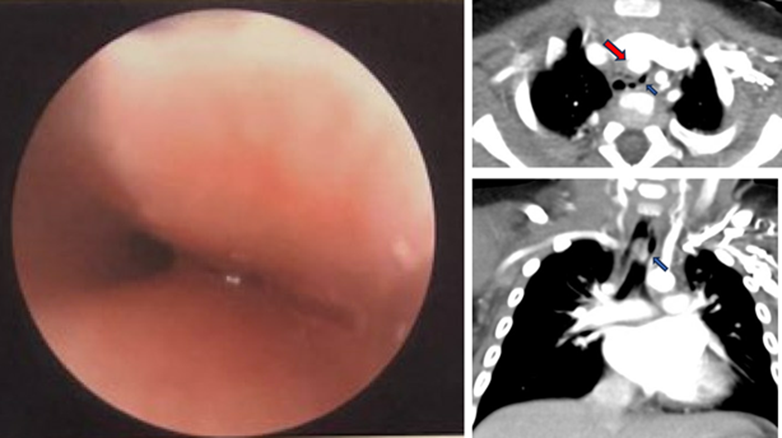
Upon referral to a paediatric otolaryngology and pulmonology team, a series of diagnostic evaluations were conducted. Flexible and rigid bronchoscopy revealed moderate laryngomalacia and severe tracheomalacia, characterised by a pulsatile anterior collapse of the mid-trachea. These findings prompted further imaging studies. An echocardiogram was obtained and did not show any abnormality. A CT angiogram of the chest detailed a left aortic arch with a normal branching pattern; however, the innominate artery was noted to be prominent and aberrantly positioned (distal and posterior), exerting significant anterior compression on the trachea. The tracheal lumen was severely narrowed in the affected region, confirming the diagnosis of innominate artery compression syndrome (Figure 1).
A; Bronchoscopy of the mid trachea revealing pulsatile compression of the anterior wall. B and C; CT angiogram of the chest showing compression of the trachea (blue arrow) by the brachiocephalic artery (red arrow).

Given the severity of the anatomical anomaly and its impact on the airway, surgical intervention was deemed necessary. The procedure involved relocating the innominate artery proximally and anteriorly on the ascending aorta to alleviate the compression. Intraoperative findings confirmed the preoperative imaging, and subsequent intraoperative bronchoscopy post-relocation demonstrated a significant improvement in tracheal patency.
The infant’s postoperative course was uneventful, with a notable resolution of respiratory symptoms. Follow-up over 24 months post-surgery showed the patient to be asymptomatic, with normal growth and developmental milestones, underscoring the success of the intervention.
Discussion
Innominate artery compression syndrome represents an incomplete vascular ring, which presents significant diagnostic challenges due to its rarity and the nonspecific nature of its symptoms. This condition, affecting the paediatric population, often presents with respiratory symptoms that can easily be mistaken for more common paediatric issues such as asthma, gastroesophageal reflux, or respiratory infections. The misdiagnosis or delayed diagnosis of innominate artery compression syndrome can lead to prolonged periods of discomfort, though the compression is thought to decrease with age as some patients outgrow the narrowing. Reference Donuru, Kandula and Madueme2
This case report highlights the critical nature of interdisciplinary collaboration in diagnosing and managing complex vascular anomalies. The involvement of paediatric pulmonology, otolaryngology, radiology, and cardiothoracic surgery was pivotal in successfully resolving this case. This multidisciplinary approach facilitated a precise diagnosis and enabled a successful surgical intervention, underscoring the value of comprehensive care teams in handling such intricate cases. Moreover, this case emphasises the importance of advanced non-invasive imaging techniques over angiography in diagnosing innominate artery compression syndrome. While physical examination and standard imaging may suggest the presence of a tracheal compression, detailed imaging modalities such as CT angiography are crucial for visualising the vascular anatomy and confirming the diagnosis. Such precise imaging guides surgical planning and can significantly affect the outcome, as demonstrated by the successful surgical relocation of the innominate artery in this infant.
The abnormal distal and posterior takeoff of the innominate artery from the aortic arch is thought to cause tracheal compression, which occurs as the artery courses from the left chest across the midline to the right arm. Since these findings are found in a large number of infants and innominate artery compression syndrome is rare, the cause is thought to be likely multifactorial with additional factors such as anterior crowding of mediastinum and tracheomalacia contributing to the genesis of symptoms. Reference McKiernan and Lee4 Surgical intervention (aortopexy, aortoinnominopexy or innominate arteriopexy) is indicated in patients with severe symptoms, complications, frequent exacerbations (occurring monthly), short symptom-free periods, >50% tracheal compression, and worsening symptoms. Whereas those with mild symptoms, long symptom-free periods, and <50% tracheal compression can be managed conservatively. Reference Corcione, Borrelli and Radice3,Reference McKiernan and Lee4,Reference Gardella, Girosi and Rossi5 In conclusion, the discussion of this patient provides an opportunity to reflect on the potential for underdiagnosis or misdiagnosis of innominate artery compression syndrome in the paediatric population. It serves as a reminder for healthcare professionals to consider vascular causes when presented with atypical respiratory symptoms and to pursue further diagnostic evaluation when initial assessments do not conclusively explain the symptoms.
Data availability
Majority of data associated with this study are provided in text and figures. Additional data will be made available upon request.
Financial support
This research received no specific grant from any funding agency, commercial, or not-for-profit sectors.
Competing interests
All authors have reported that they have no relationships relevant to the contents of this paper to disclose.
Ethical standard
Informed written consent was obtained from the patient or their parents as applicable, as per West Virginia University IRB guidelines.