Introduction
The term ‘mucocele’ was first used by Rollet in 1896.Reference Rollet1 The histological components of this lesion were first described by Onodi in 1901. A mucocele is an epithelium-lined, mucus-filled sac located within a paranasal sinus. It is capable of expansion by bone resorption and new bone formation.Reference Canalis, Zajtchuk and Jenkins2 This dynamic process distinguishes a mucocele from a blocked sinus with trapped mucus. Although the commonest cause of mucocele development is previous surgery, the process may be initiated by chronic inflammation, benign tumours or trauma.
Mucoceles progress slowly, and symptoms and signs are the result of their progressive expansion. Symptoms vary from mild (e.g. nasal obstruction) to severe (e.g. diplopia and impaired vision). Mucoceles can invade the skull base and cause severe problems.
The uncinate process plays a role in paranasal sinus drainage and pathology; it is also a key landmark during endoscopic sinus surgery.
The presence of a pneumatised uncinate process (uncinate bulla) can cause narrowing of the middle meatus, which may lead to obstruction of drainage through the infundibulum.Reference Isobe, Murakami and Kataura3
In this report, we present a case of a mucocele of a pneumatised uncinate process which presented as recurrent sinusitis.
Case report
A 43-year-old woman was referred to our ENT department suffering from persistent symptoms of sinusitis. These were predominantly left-sided and remained unresolved despite courses of steroids and antibiotics prescribed by the patient's general practitioner.
The patient was seen in our department a month after the initial referral. Upon examination, she was noted to have what appeared to be an enlarged left ethmoidal bulla.
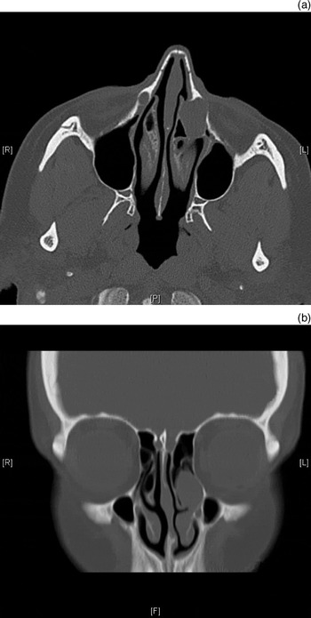
Due to this finding and the unilateral nature of her symptoms, a computed tomography (CT) scan was performed (Figure 1). This showed a 1.5 cm diameter, soft tissue mass lying in the fossa of the lacrimal sac, extending down the course of the left nasolacrimal duct and protruding into the ethmoidal air cells and the middle and upper meati. The CT also suggested a lacrimal gland mass, although there were no symptoms of epiphora.
(a) Axial and (b) coronal computed tomography scans of the sinuses, showing left uncinate mucocele. R = right; L = left; P = posterior; F = inferior

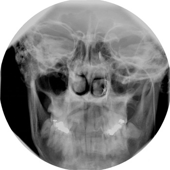
Further investigation with dacryocystography showed no definite filling defect within the lacrimal sac (Figure 2).
Left dacryocystogram showing flow of contrast into the nasal cavity.

Due to the inconclusive nature of the radiological findings and the need to exclude sinister pathology, early endoscopic surgery was planned.
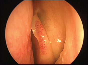
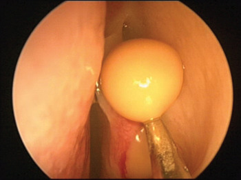
At surgery, a fullness of the left lateral nasal wall was seen in the area of the uncinate process (Figure 3). Incision of the anterior border of the fullness caused an outflow of pus (Figure 4). Further endoscopic visualisation identified the cavity as an uncinate bulla. A diagnosis of mucocele of a left uncinate bulla was made. The mucocele was decompressed and an uncinectomy performed.
Endoscopic view of intact uncinate mucocele.

Endoscopic view showing incision of uncinate mucocele.

Discussion
The current literature reports a high frequency of anatomical variation of facial bone pneumatisation. However, as yet there is no agreed theory of explanation. During research into the present case, we performed a literature search for articles referring to the anatomy of the uncinate process and its anatomical variations. No reports of uncinate mucocele could be found.
The uncinate process is a thin, boomerang-shaped bone structure that projects from the ethmoid bone to the ethmoid process of the inferior concha.
• There is a high frequency of anatomical variation of the paranasal sinuses
• Pneumatisation of the uncinate process may impair sinus drainage
• A mucocele of an uncinate bulla is a rare, previously unreported finding
Between the 10th and 12th week of intrauterine development, invagination of the mucus of the middle meatus occurs and the primary maxillary sinus is formed.Reference Arredondo de Arreola, López Serna, de Hoyos Parra and Arreola Salinas4 At this stage, the uncinate process and bulla ethmoidalis delineate a narrow groove (the hiatus semilunaris). In rare circumstances, as development continues pneumatisation can occur where there is an extension of an air cell into the uncinate process, most frequently in the anterosuperior region.Reference Isobe, Murakami and Kataura3 The prevalence of this anatomical variation has been reported as 0.4–5 per cent.Reference Kantarci, Karasen, Alper, Onbas, Okur and Karaman5 Such an uncinate bulla (as it could also be termed) can cause narrowing of the infundibulum and impaired sinus drainage.Reference Scribano, Ascenti, Loria, Cascio and Gaeta6 This functional blockage seems to correspond to an area of contact between the uncinate bulla and the middle turbinate. Such a blockage can lead to recurrent sinusitis, as in our case.
This case report furthers our knowledge of this anatomically diverse region, and brings to light a new and clinically relevant pathology.