The Melody® transcatheter pulmonary valve; Medtronic, Minneapolis, MN, United States of America, has been used for “off-label” implantations in the mitral position of infants and children too small to receive available traditional prosthetic valves.Reference Quiñonez, Breitbart, Tworetsky, Lock, Marshall and Emani 1 – Reference Trezzi, Cetrano, Albano and Carotti 4 This approach theoretically allows for further percutaneous balloon dilation of the valve to accommodate somatic growth. However, reports of subsequent dilations are limited. We present a 15-month-old child undergoing further percutaneous expansion of a modified Melody valve in the mitral position after initial implantation in early infancy who experienced an unusual complication.
Case report
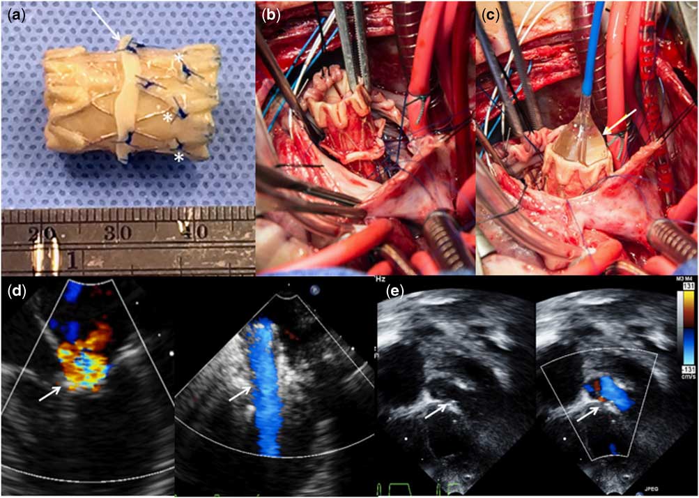
A 4.1 kg 5-month-old female infant underwent repair of a complete atrioventricular canal defect with severe congenital left-sided atrioventricular valve regurgitation. She was noted to have deficiency of the left-sided component of the superior bridging leaflet, and despite repair with commissuroplasty, postoperative transesophageal echocardiography still demonstrated severe insufficiency. The patient’s left atrioventricular valve annulus size of only 10 mm made her unsuitable for traditional atrioventricular valve replacement. Three days after initial repair, she underwent surgical implantation of a melody valve in the mitral position. The valve was implanted via atriotomy using a 12 mm balloon and sutured to the annulus using a pericardial cuff. A small fenestration was left in the atrial septal patch for future access and further dilation. On transesophageal echocardiography, the valve was unobstructed with a 10 mm inner diameter, trace regurgitation, and the left ventricular outflow tract was unobstructed (Fig 1a–e). By 15 months of age and 8.4 kg, echocardiography showed stable valve size, no insufficiency, and a mean gradient of 18–20 mmHg.
( a ) Modified Melody valve prepared for implantation. The terminal crowns were folded back on both ends to lie flat against the stent and the anterior folded crowns were stitched down to prevent protrusion into the left ventricular outflow tract (LVOT) upon further expansion (*). A pericardial cuff is sewn into place to secure the valve onto the mitral annulus (arrow). ( b ) The valve is implanted, secured onto the annulus and the distal end stitched to the posterior left ventricular wall to avoid LVOT obstruction, and ( c ) dilated using a 12 mm balloon (arrow). ( d ) Before Melody valve implantation, there is severe mitral valve insufficiency on transesophageal echocardiography (left, arrow). After implantation, the valve (right, arrow) spans mid-atrium to mid-ventricle, measures 10 mm in internal diameter, and has unobstructed laminar inflow and trivial insufficiency. ( e ) No obstruction of the LVOT (arrow) is noted on subsequent transthoracic echocardiography.

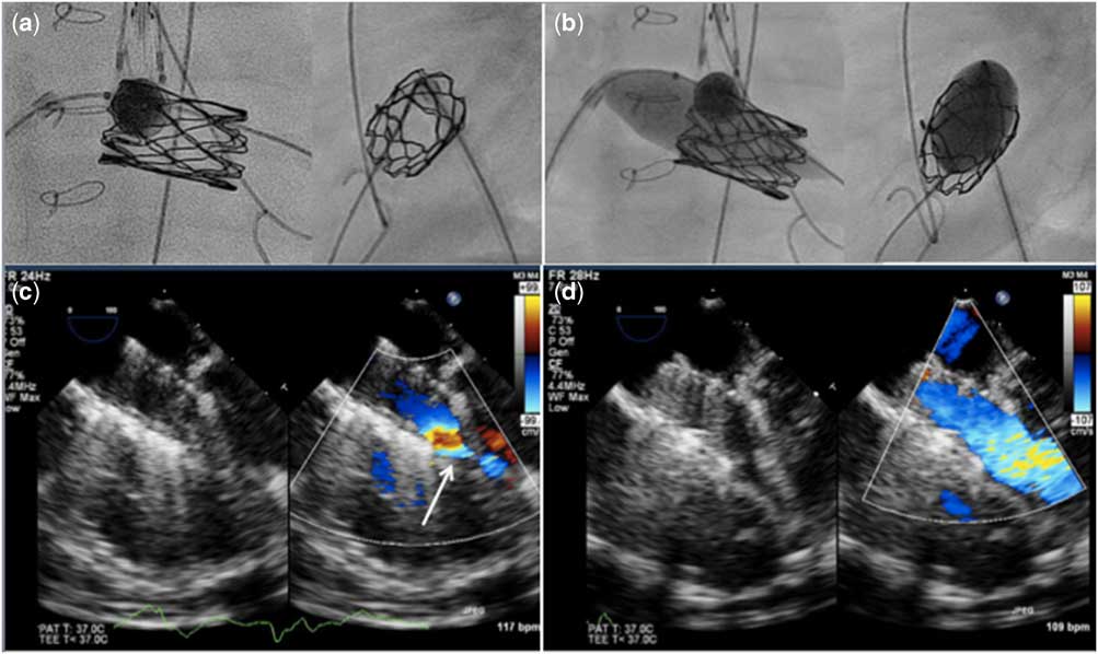
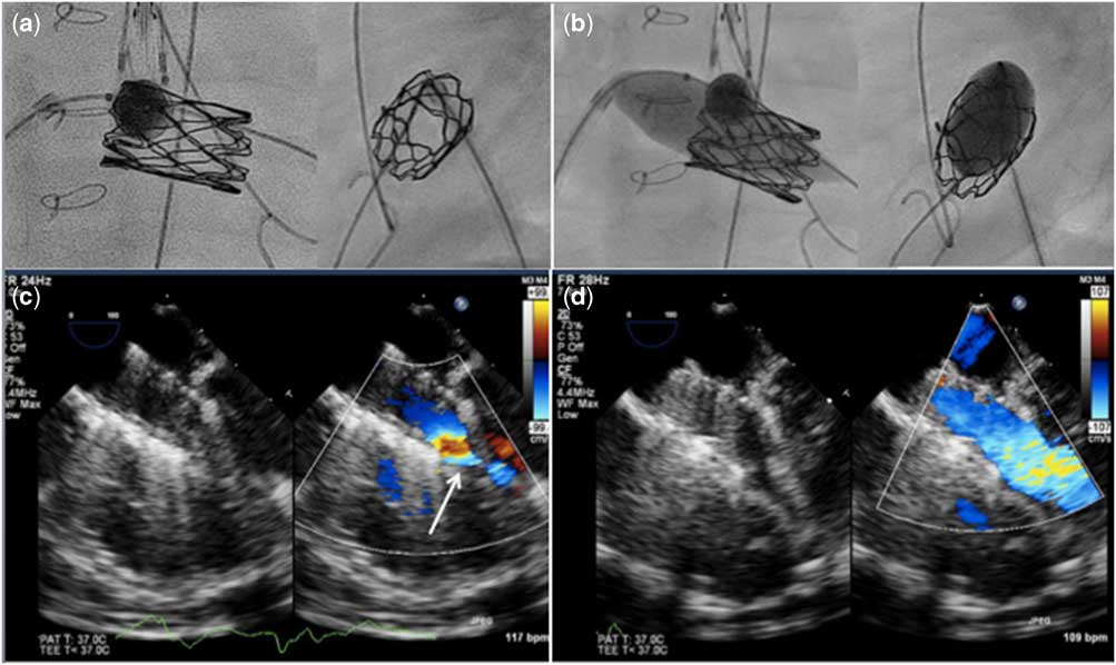
She was brought to the catheterisation laboratory, where transesophageal echocardiography showed spontaneous closure of the atrial septal fenestration. A transseptal puncture was performed and a 6 French long sheath was placed in the left atrium. Mean transmitral gradient was 19 mmHg. There was a peak gradient of 15 mmHg in the left ventricular outflow tract where the stent protruded slightly. An initial left atrial angiogram was performed to evaluate the posterior atrioventricular groove to assess proximity of the left circumflex artery to the Melody valve. The valve was crossed from the left atrium using a tip-deflecting wire and a 4 F angled glide catheter; Terumo Interventional Systems, Somerset, NJ, United States of America, and a 0.035″ wire was positioned in the left ventricular apex. An additional wire was positioned retrograde across the left ventricular outflow tract for balloon dilation in the event of outflow obstruction following Melody valve dilation. A 14 × 20 mm Atlas Gold balloon; Bard, Tempe, AZ, United States of America, was dilated to 20 atm in the valve. During inflation, the balloon slipped slightly back towards the atrium, preferentially expanding the proximal portion of the stented valve (Fig 2a,b). Upon balloon deflation, the patient developed complete heart block and became hypotensive. Sinus rhythm resumed shortly thereafter, but the patient remained hypotensive and bradycardic. The echo revealed poor left ventricular systolic function, no increase in left ventricular outflow tract obstruction, and a dilated right ventricle presumably because of acute pulmonary hypertension. The distal portion of the valve stent was distorted and narrowed. Color Doppler of the inflow at the distal end of the stent appeared turbulent (Fig 2c). The balloon was used again to further dilate the entire length of the stent valve. The patient’s blood pressure rapidly recovered. Her function and right ventricular distension normalised and echo demonstrated unobstructed laminar flow through the Melody valve (Fig 2d). The inner diameter of the valve had increased to 12 mm by echo. The mean mitral valve gradient improved to 9 mmHg. The left ventricular outflow tract peak gradient remained unchanged. The patient recovered without incident and was discharged the following day. She has not required reintervention in the 14 months since.
( a ) Before intervention, the Melody valve appears uniformly cylindrical in the AP (left) and lateral (right) views. ( b ) With preferential expansion of the proximal portion, there is deformation of the distal portion, more notable in the lateral projection (right). ( c ) Transesophageal echo (TEE) with color Doppler shows narrowing and restricted flow exiting the stent (arrow). ( d ) After uniform dilation of the stent, TEE shows unobstructed laminar flow.
Discussion
The Melody valve provides a viable mitral valve replacement option for infants in whom prosthetic valves are not feasible or associated with poor outcomes. Although the valve’s longevity in this position is unknown, its theoretical advantage is that it can be further expanded by percutaneous balloon dilation as the patient grows, thereby potentially reducing reoperations. Reports describing subsequent percutaneous dilations of the Melody valve in the mitral position in small patients are limited,Reference Quiñonez, Breitbart, Tworetsky, Lock, Marshall and Emani 1 , Reference Frigiola, Pluchinotta and Saracino 2 and these reports do not detail the techniques and considerations for reintervention. Our case supports the reasonable short-term durability of the Melody valve in the mitral position and the feasibility of further dilation to accommodate growth.
The atrial septum should be fenestrated at the time of implantation to facilitate future percutaneous dilations, but operators should be prepared for a transseptal puncture in a small patient in the event that the fenestration closes spontaneously. The septal fenestration should be made with enough distance from the Melody valve to allow room for the wire and balloon to smoothly curve into the valve orifice without an acute turn. In addition, operators must anticipate complications, including acute left ventricular outflow tract obstruction, acute mitral regurgitation, compression of the circumflex branch of the left coronary artery, and deformation of the valve leading to acute mitral valve obstruction. It is advised to have three pressure transducers for this procedure: a retrograde catheter in the left ventricle, a transseptal sheath to monitor the left atrial pressure, and an additional arterial monitoring angiocatheter so that the left atrium, left ventricle, and arterial pressure can be simultaneously assessed to evaluate the residual mitral gradient and the outflow gradient. Furthermore, this allows rapid wire positioning and balloon expansion of the left ventricular outflow tract if obstruction occurs.
We observed two previously unreported complications – acute obstruction of the distal end of the Melody valve because of stent deformation following dilation of the proximal portion of the valve as well as transient complete heart block. In contrast to dilating a stent in a vessel or conduit, a stent anchored only at the single fulcrum of the atrioventricular valve annulus may flatten at one end if the other end is preferentially dilated. Operators should also be aware of the location of the atrial septum in relation to the balloon so that the transseptal puncture site is not dilated with a large balloon. Use of the transesophageal echocardiography throughout this procedure is helpful in evaluating possible sources of haemodynamic instability and for rapid assessment of the patency of the valve after further dilation.
Acknowledgements
Dr Jay Pruetz and Dr Cheryl Takao also played critical roles in the care of this patients and the planning of the procedures. The authors were supported by the Children’s Hospital Los Angeles Heart Ambassadors and the Hearst Foundations.
Financial Support
This research received no specific grant from any funding agency, or from commercial or not-for-profit sectors.
Disclosures/Conflicts of Interest
None.