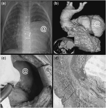
A 3-year-old girl presented with progressive chest tightness and decreased appetite for 5 days preceding her admission. She was diagnosed with extreme tetralogy of Fallot with pulmonary atresia. She underwent right ventricular outflow tract patch correction and neo-pulmonary trunk reconstruction 40 days previously and was discharged after 20 days. Plain chest radiography showed an abnormal bulging mass-like shadow at the upper left cardiac border (@ in Fig a). Emergent computed tomography (Light-Speed 16-slice; GE Healthcare) scan disclosed a 4 × 5 centimetre lobulated pseudo-aneurysm from the trans-annular right ventricular outflow tract patch (@ in Fig b). Under the impression of leaked right ventricular outflow tract patch with pseudo-aneurysm formation, she underwent emergent aneurysmectomy. The surgical finding confirmed this huge pseudo-aneurysm (@ in Fig c) and the resected specimen by Masson's Trichrome stain (Fig d) revealed no myocardial or vascular wall pathologically with full thickness of collagenous component of the fibrotic tissue (light grey) mixed with petechial haemorrhage (dark grey).
Figure 1