Introduction
Histological analysis is a routine technique applied to evaluate the quality of tissues and cells. This procedure can be used to evaluate pathophysiological changes with the help of optic instruments, to access biochemical changes using immunohistochemistry, or even in tissue proteomic studies (Longuespée et al., Reference Longuespée, Fiéron, Pottier, Quesada-Calvo, Meuwis, Baiwir, Smargiasso, Mazzucchelli, De Pauw-Gillet, Delvenne and De Pauw2014). Classical histology is often applied to evaluate ovarian tissue because it allows the analysis of morphological, morphometrical, and cell density changes.
This classic procedure consists of tissue fixation followed by processing, that is dehydration, diaphanization, infiltration, inclusion in paraffin, sectioning, and microscopic analysis. The main variation within this process is the type of fixative used and time of fixation. When ovarian tissue is submitted to immunohistochemical analysis, it is preferable that the tissue is fixed in neutral-buffered formalin up to 48 h before embedding in paraffin (Grazul-Bilska et al., Reference Grazul-Bilska, Borowicz, Reynolds and Redmer2013; Rowley et al., Reference Rowley, Rubenstein, Manuel, Johnson, Surgnier, Kapitsinou, Duncan and Pritchard2020), but other fixatives are also used such as Carnoy (Grazul-Bilska et al., Reference Grazul-Bilska, Borowicz, Reynolds and Redmer2013; Rowley et al., Reference Rowley, Rubenstein, Manuel, Johnson, Surgnier, Kapitsinou, Duncan and Pritchard2020), Davidsons’s solution (Rowley et al., Reference Rowley, Rubenstein, Manuel, Johnson, Surgnier, Kapitsinou, Duncan and Pritchard2020), or paraformaldehyde (Castilho et al., Reference Castilho, Dalanezi, Franchi, Price, Ferreira, Trevisol and Buratini2019). The incubation period for these fixatives varies in time from 6 h (Grazul-Bilska et al., Reference Grazul-Bilska, Borowicz, Reynolds and Redmer2013), 12 h (Santos et al., Reference Santos, Rodrigues, Costa, Silva, Matos, Lucci, Bao, van den Hurk and Figueiredo2006), 24 h (Brito et al., Reference Brito, Brito, Silva, Rodrigues, Figueiredo, Domingues and Santos2018; Rowley et al., Reference Rowley, Rubenstein, Manuel, Johnson, Surgnier, Kapitsinou, Duncan and Pritchard2020), and 48 h (Grazul-Bilska et al., Reference Grazul-Bilska, Borowicz, Reynolds and Redmer2013). This variation depends on the tissue size (whole ovary, half or fragments). In addition to the labelling purpose, the tissue is fixed with these chemicals with the main goal to study the morphological integrity. For instance, classical histology is helpful to evaluate the quality of the ovarian tissue and its follicles.
In the present study we fixed ovarian fragments for 12 h or 24 h in neutral-buffered formalin (NBF), Carnoy’s solution (CAR), Davidsons’s solution (DAV), or paraformaldehyde (PFA) for further analysis of their effect on the morphology of ovine preantral follicles.
Materials and Methods
Chemicals
All chemicals and media used in this study were purchased from the Sigma Chemical Company (St. Louis, MO, USA).
Ovarian tissue collection
Ovaries (n = 10) from adult mixed breed sheep (n = 5) were obtained at a local slaughterhouse. The ovaries were trimmed of adhering tissue, washed in 70% alcohol and then, twice in Minimum Essential Medium (MEM) containing HEPES. The ovaries were put into tubes containing 20 ml of MEM HEPES and transported to the laboratory within 1 h in a thermos flask filled with water at 20 °C.
Experimental design
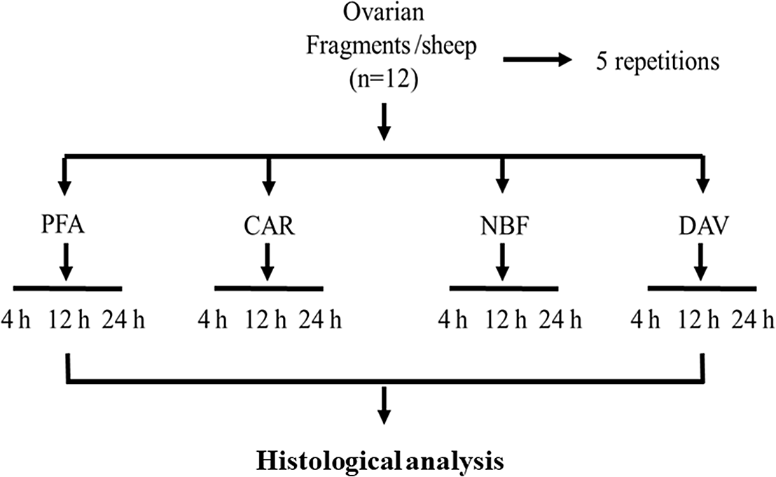
In the laboratory, each ovarian pair was divided into 12 fragments of 3 × 3 × 1 mm, which were randomly distributed in the treatments, as described in Figure 1. In brief, each ovarian fragment was fixed in paraformaldehyde (PFA), Carnoy’s solution (CAR), neutral buffered formalin (NBF) or Davidson’s solution (DAV), for 12 or 24 h. After this fixation time, each fragment was prepared for histological analysis. The main composition of each tested fixative is given in Table 1.
Experimental design to test different fixatives and fixation periods.

Composition of the test fixatives

Histological analysis
After fixation, ovarian fragments were dehydrated in ethanol, clarified with xylene and embedded in paraffin wax. Serial sections (5 μm) of ovarian tissue were cut and every fifth section was mounted on glass slides and stained with periodic acid Schiff (PAS)–haematoxylin. All sections were examined using a light microscope (Leica) at a magnification of ×40.
The analyzed preantral follicles were classified into primordial (oocyte surrounded by a layer of squamous granulosa cells), primary (oocyte surrounded by a layer of cubic granulosa cells), or secondary (oocyte surrounded by two or more layers of cubic granulosa cells) (Silva et al., Reference Silva, Brito, Sa, Silva, Ferreira, Silva, Guedes, Rodrigues, Santos and Figueiredo2019).
To avoid counting a follicle more than once, preantral follicles were counted only in the sections where their oocyte nucleus was observed. Follicular quality was evaluated based on the morphological integrity of the oocyte, the granulosa cells and the basement membrane (Santos et al., Reference Santos, Rodrigues, Costa, Silva, Matos, Lucci, Bao, van den Hurk and Figueiredo2006).
The density of primordial, primary, and secondary follicles in the ovarian fragments was calculated as the total number of each follicular class divided by the total evaluated tissue volume and expressed as the number of follicles/mm3 of evaluated ovarian tissue (Fransolet et al., Reference Fransolet, Henry, Labied, Masereel, Blacher, Noël, Foidart, Nisolle and Munaut2015).
Ovarian stroma density was evaluated by calculating the stromal cell per 100 m2. For each treatment, 10 fields per slide were assessed and the mean number of stromal cell per field was calculated (Faustino et al., Reference Faustino, Santos, Silva, Pinto, Celestino, Campello, Figueiredo and Rodrigues2010).
Statistical analysis
The ovarian fragments were randomly assigned to treatment conditions before each experiment. Statistical analysis was conducted using GenStat statistical software (GenStat for Windows 20th Edition, VSN International, Hemel, Hempstead, UK; https://www.vsni.co.uk/downloads/genstat/). Data were compared with analysis of variance (ANOVA). The null hypothesis was that there was no treatment effect on the response parameter. Treatment means were compared according to Fisher’s least significant difference (LSD) (for a two-sided test and P ≤ 0.05) after a significant treatment effect was confirmed by ANOVA. Data are presented as mean ± standard error of the mean (SEM). Given the factorial design, the statistical model used to analyze the data was:
 $${\rm{Y}} = \mu + {\rm{bloc}}{{\rm{k}}_{\rm{i}}} + {\rm{Fixativ}}{{\rm{e}}_{\rm{j}}} + {\rm{Exposur}}{{\rm{e}}_{\rm{k}}} + {\rm{Fixative}} \times {\rm{Exposur}}{{\rm{e}}_{{\rm{jk}}}} + {{\rm{e}}_{{\rm{ijk}}}}$$
in which:
Y = Response parameter
µ = General mean
Blocki = Effect of block (i = 1…5)
Fixativej = Effect of fixative (j = 1…4)
Exposurek = Effect of exposure time (k = 1, 2)
Fixative × Exposurejk = Effect of the interactions between fixative and exposure time
Errorijk = Error term
Values with P ≤ 0.05 were considered to be statistically significant.
Results and Discussion
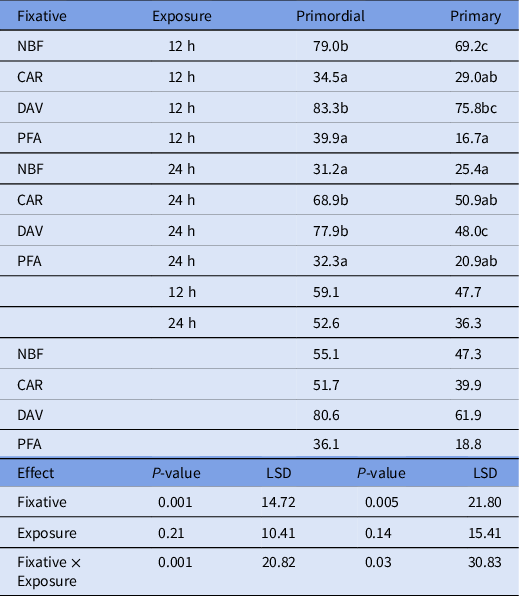
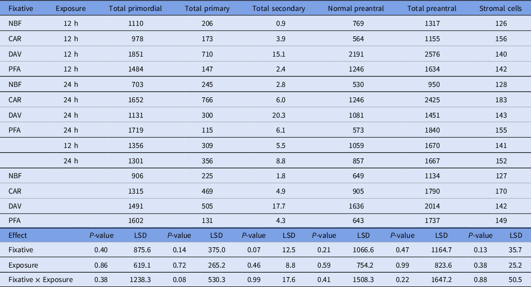
There was a significant interaction between tested fixatives and exposure period on the percentages of morphologically normal primordial and primary follicles (Table 2). Due to the huge variation in the number of secondary follicles, no statistical analysis was performed with this follicular class. The percentages of morphologically normal primordial and primary follicles were significantly the highest when ovarian tissue was fixed for 12 h in NBF or DAV, or when fixed for 24 h in CAR and DAV (Table 2). Preantral follicles are classified as degenerated if the cytoplasm is retracted or present vacuolization, if the granulosa cells are not connected to the oocyte, or some signals of pyknosis are present. Paraformaldehyde can cause morphological changes and does not always fix the samples completely (Melan, Reference Melan1994). Furthermore, Richter et al. (Reference Richter, Revelo, Seitz, Helm, Sarkar, Saleeb, D’Este, Eberle, Wagner, Vogl, Lazaro, Richter, Coy-Vergara, Coceano, Boyden, Duncan, Hell, Lauterbach, Lehnart, Moser, Outeiro, Rehling, Schwappach, Testa, Zapiec and Rizzol2018) demonstrated that PFA6 fixation is associated with the formation of membrane blebs and vacuoles and a general change in the cell morphology. This may explain the misleading classification of the primordial and primary follicles as degenerated in the present study. To avoid these artefacts, usually paraformaldehyde is combined with other compounds such as glutaraldehyde. However, this is not an advantage for studies on follicular morphology, which require more simple methods. NBF is a low-cost fixative that penetrates tissues well (Perry et al., Reference Perry, Chung, Ylaya, Choi, Simpson, Matsumoto, Smith and Hewitt2016). However, attention must be given to the fixation period, which may affect cell morphology as observed in the present study where the percentage of normal follicles decreased to almost the half when increasing fixation time from 12 h to 24 h. Studies on the effects of NBF fixation time are related to the labelling of biomolecules (Chung et al., Reference Chung, Song, Ylaya, Sears, Choi, Cho, Rosenberg and Hewitt2018). This is of high importance when fixing tissues with already a high rate of degenerated follicles such as feline ovaries, that is already c. 63–79% in healthy ovaries (Jewgenow, Reference Jewgenow1998; Lima et al., Reference Lima, Silva, Santos, Sales, Evangelista, Figueiredo and Silva2006; Brito et al., Reference Brito, Domingues, Rodrigues, Silva, Alves, Wu, Francisco, Barroso Neto, Freire, Figueiredo, Pieczarka and Santos2020). Recently, Hamouzouva et al. (Reference Hamouzova, Cizek, Bartoskova and Novotny2020) demonstrated that feline and canine ovaries are well preserved in Carnoy’s solution. Another advantage of Carnoy’s solution is that this fixative increases the eosinophilic staining in ovarian tissue (Rowley et al., Reference Rowley, Rubenstein, Manuel, Johnson, Surgnier, Kapitsinou, Duncan and Pritchard2020). In the present study, the Carnoy solution was efficient to preserve follicular morphology after 24 h fixation. Similarly, Bufalo et al. (Reference Bufalo, Gonzalez, Silva, Lindquist, Bergamo, Costa, Marinho and Seneda2016) showed that Carnoy was able to preserve the morphology of equine preantral follicles when the ovarian tissue was fixed for 24 h. Davidson’s solution was able to preserve follicular morphology regardless of the fixation period. Naumowicz et al. (Reference Naumowicz, Pajdak, Szarek, Schulz, Terech-Majewska and Felsmann2018) demonstrated that Davidson’s solution was more suitable than NBF for preserving fish ovaries, because the last one resulted in shrunken and misshaped oocytes. These former authors reported the negative effects of formalin and alcohol on the preservation of the oocyte morphology. Both Carnoy and Davidson’s solutions contain alcohol, but at very different proportions, being much lower in Davidson’s solution. When evaluating follicular and stromal cells density, no significant differences were observed (Table 3). This suggests that fixative or fixation period cannot affect the counting of follicular and stromal structures and confirms that different fixatives may confound the observer when classifying follicular morphology, but do not interfere in cell counting.
Effects of fixatives and time of exposure on the mean percentages of morphologically normal primordial and primary follicles

a,b,cValues followed by a different letter within a column differ significantly (P ≤ 0.05).
Effects of fixatives and time of exposure on the density of preantral follicles and stromal cells

In conclusion, when fixing ovarian fragments for the morphological analysis of primordial follicles it is indicated to use NBF with an exposure time of 12 h, Carnoy’s solution with exposure for 24 h, or Davidson’s solution for 12 or 24 h. The use of a proper protocol is crucial to avoid misleading data when evaluating the morphology of preantral follicles submitted to toxicity tests of in vitro culture.
Conflict of interest
All the authors have no conflict interest.
Ethical approval
This study was approved and performed under the guidelines of the Ethics Committee for Animal Use of the State University of Ceará (project no. 4156483/2018).
Financial support
This research received no specific grant from any funding agency, commercial or not-for-profit sectors,