Hypercalcaemia in patients with primary hyperparathyroidism can have numerous adverse effects on multiple organ systems including the cardiac system, where it can precipitate conduct disorders such as ventricular arrhythmia.Reference Surawicz 1 , Reference Surawicz 2
Case report
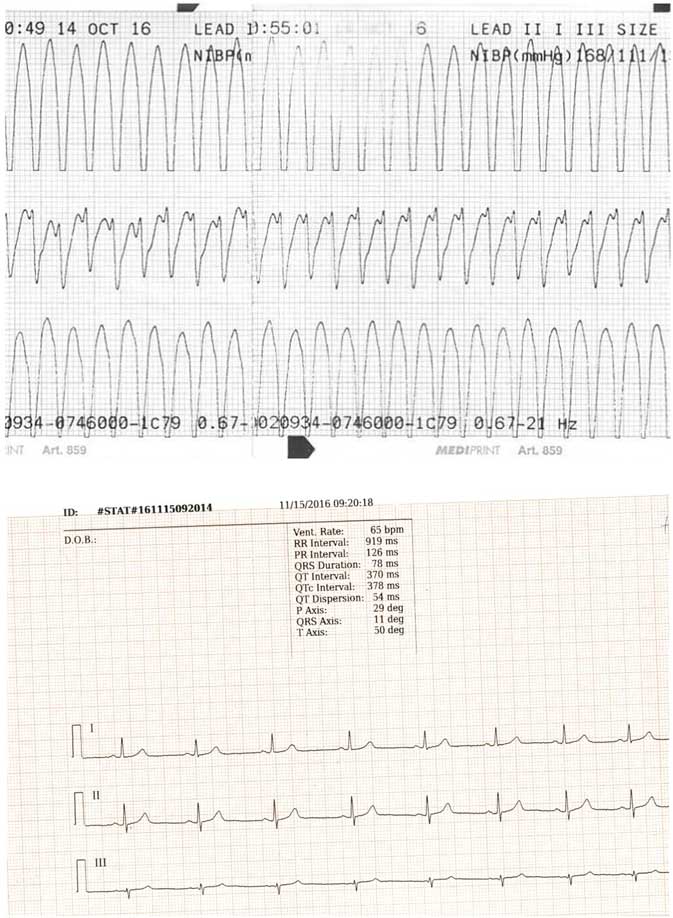
A previously healthy 16-year-old boy with a positive family history of cardiovascular diseases was sent to the emergency room with complaints of palpitation, chest tightness, and dizziness after physical activity; the patient’s mother has hypertension, whereas the father had a heart attack after which a stent was implanted in the coronary artery. Cardiac monitoring at arrival showed a wide QRS complex tachycardia with a ventricular rate of 253 beats per minute with preserved pulse. Blood pressure was 150/80 mmHg. Adenosine was administered intravenously three times at 6 mg, with additional two doses of 12 mg, in order to stop the attack of tachycardia but without any success. Sinus rhythm was restored with intravenous application of 450 mg of amiodarone. Considering that our patient weighed 60 kg, the administered dose was 7.5 mg/kgReference Helmy, Herre and Garwood 3 (Fig 1).
( a ) A three-lead electrocardiogram obtained in the emergency room showed classic monomorphic tachycardia with wide QRS and QRS duration of 160 ms, QS in II and III, and indeterminate axis. ( b ) Normal three-lead electrocardiogram of the patient after surgical treatment.

Laboratory findings at admission showed elevated concentrations of total (14.2 mg/dl, ref. 8.4–10.3 mg/dl) and ionised calcium (6.3 mg/dl; ref. 4.4–5.4 mg/dl), with low phosphate level at 2.38 mg/dl (3.1–4.7 mg/dl) and high parathyroid hormone level (173 pg/ml; 10–55 pg/ml). Other laboratory findings including concentrations of sodium, potassium, and magnesium were normal. By performing genetic testing on the patient, we excluded the presence of the most common gene mutations related with MEN 1 and MEN 2b. These panels identify about 90% of patients with either MEN 1 and MEN 2B. Taking this into account, follow-up by an endocrinologist is needed.
As laboratory findings indicated primary hyperparathyroidism without any other organ symptoms except ventricular tachycardia, treatment with hyperhydration and loop diuretics was immediately initiated according to the protocol for severe hypercalcaemia. By definition, severe hypercalcaemia is defined as ionised serum Ca level >5.8 mg/dl; the highest measured value in our patient was 6.3 mg/dlReference Bilezikian 4 . Despite the latter treatment, calcium level continued to increase up to 15.6 mg/dl, so bisphosphonate (pamidronate) was included. After 3 days of pamidronate therapy, total calcium level declined to 11.2 mg/dl.
After treating for hypercalcaemia, 12-lead electrocardiogram showed normal sinus rhythm (67/min), normal PR (128 ms), QRS complex (78 ms), and QTc (340 ms) interval. Echocardiography showed normal, healthy heart without structural abnormalities. Telemetry monitoring performed on the patient showed no conduct disorder.
Neck ultrasound was performed, which showed two hypoechoic masses: one located in the right inferior portion of the thyroid gland measuring about 1×0.5×0.8 cm and another in the thymus measuring about 0.9×0.7×0.5 cm. Cytological examination revealed a high concentration of parathyroid hormone in the obtained sample (181 pg/ml), whereas Technetium-99m (99mTc) sestamibi scintigraphy showed hyperfunctional parathyroid tissue.
Despite scintigraphy findings, and based on the intraoperative findings after inspection of all four parathyroid glands on the 21st admission day, the patient was referred for parathyroidectomy of the lower left parathyroid gland and partial thymectomy. Pathological findings showed hyperplasia of the parathyroid gland and confirmed the diagnosis of primary hyperparathyroidism. The post-operative course was uneventful, and serum calcium level returned to normal, with total calcium level at 9.5 mg/dl.
During the post-operative period, we performed an electrophysiological study during which programmed stimulation could not induce any ventricular arrythmia, even after infusion of isoprotenol. Therefore, we came to a conclusion that normalisation of serum calcium level after surgery had a curative effect on arrhythmia as well.Reference Chang, Chen and Tai 5 During the 6-month follow-up period, no ventricular tachycardia occurred. Control electrocardiogram and echocardiography were normal.
Considering that there were no subjective or clinical symptoms, there was no need to perform another electrophysiological testing.
Discussion
In conclusion, ventricular tachycardia as a presenting symptom of primary hyperparathyroidism has not been reported in such a young patient so far in medical literature. Serum calcium levels should be routinely measured in previously healthy children presenting with ventricular tachycardia. One might hypothesise that primary hyperparathyroidism was the cause of this calcium-dependent arrhythmia. Therefore, after successful treatment of the underlying disorder and normalization of serum calcium levels, ventricular tachycardia was not inducible during the electrophysiological study. As it was not in the best interest of the patient to perform the electrophysiological study before normalization of calcium levels, and that the electrocardiography performed in the emergency room was three-lead, in which we cannot identify any characteristic sign of hypercalcaemia such as J wave (Osborn), which is considered to be mostly identified in precordial leads, diagnosis of verapamil-sensitive ventricular tachycardia remains a hypothesis.Reference Tchou, Tarakji and Kanj 6
Acknowledgements
The authors are grateful to Dr Nataša Rojnić Putarek for help in reporting this case.
Financial Support
This research received no specific grant from any funding agency or from commercial or not-for-profit sectors.
Conflicts of Interest
None.