Friedreich’s ataxia is an autosomal recessive disorder characterised by progressive nervous system damage and ataxia. Other manifestations of Friedreich’s ataxia include cardiomyopathy, scoliosis, and diabetes.Reference Harding 1 , Reference Schulz, Boesch and Bürk 2 More than 60% of patients with Friedreich’s ataxia may develop hypertrophic cardiomyopathy.Reference Durr, Cossee and Agid 3 , Reference Kipps, Alexander and Colan 4 Attempts to identify treatments to prevent and manage hypertrophic cardiomyopathy in these patients have been limited.Reference Hausse, Aggoun and Bonnet 5 , Reference Kearney, Orrell and Fahey 7 This article details the first reported case of Friedreich’s ataxia-associated hypertrophic cardiomyopathy successfully managed with septal myectomy.
Case report
Our patient was diagnosed with Friedreich’s ataxia at 4 years of age following a period of progressive ataxia. As part of the initial workup, an echocardiogram was performed, which demonstrated hypertrophic cardiomyopathy. Follow-up echocardiograms showed worsening left ventricular outflow tract obstruction, and he was referred to our centre. His echocardiogram at that time demonstrated a peak left ventricular outflow tract gradient of 80 mmHg and moderate septal wall hypertrophy. During the time of this evaluation, the patient was treated with propranolol (0.7 mg/day).
Following presentation to our institution, preoperative echocardiography showed severe hypertrophic cardiomyopathy (septal thickness of 16 mm) with a peak left ventricular outflow tract gradient of 104 mmHg and mitral valve systolic anterior motion. In addition, moderate mitral valve regurgitation and borderline left atrial enlargement were observed. The right ventricle had normal size and systolic function.
The following day, an extended left ventricular septal myectomy was performed with resection of an accessory mitral chord. The mitral papillary muscles were noted to be highly hypertrophied (up to 1.5 cm in diameter). The septal myectomy was performed starting at the nadir of the right coronary cusp and extending leftward towards the free wall. There was a large and thickened accessory cord from the papillary muscle of the mitral valve to the posterior edge of the free wall and this was resected. The myectomy was then extended towards the edge of the mitral valve. The cross-clamp time for this procedure was 83 minutes, and total bypass time was 91 minutes. Postoperative trans-oesophageal echocardiography demonstrated no evidence of left ventricular outflow tract obstruction and trivial mitral regurgitation. Mild dynamic mid-ventricular obstruction was noted at the level of the mid-papillary muscles with a maximum gradient of 40 mmHg; this was secondary to the hypertrophied papillary muscles and did not cause outlet obstruction. Postoperatively, the patient required an esmolol infusion for heart rate and blood pressure control. Transthoracic echocardiography was performed on day 11, which demonstrated no evidence of left ventricular outflow tract obstruction. The patient was discharged on postoperative day 11. His hospitalisation was prolonged due to pleural effusion requiring chest tube placement on postoperative day 2. The chest tube was subsequently removed on postoperative day 9.
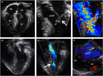
The patient had close outpatient surveillance following surgery. It is now 7 years since the surgery, and serial echocardiograms of the patient continue to demonstrate no significant outflow tract obstruction and only trivial mitral regurgitation (Fig 1). He currently takes no cardiac medications.
Still-frame images from the patient’s preoperative ( a – c ) and postoperative ( d – f ) echocardiograms. ( a ) Preoperative four-chamber view demonstrating prominent septal hypertrophy. ( b ) Systolic anterior motion of the anterior mitral leaflet into the left ventricular outflow tract (arrow). ( c ) Doppler imaging demonstrating increased velocity through the left ventricular outflow tract (peak gradient 80 mmHg). ( d ) Postoperative four-chamber view showing the site of the septal myectomy and a widely patent left ventricular outflow tract. ( e ) Doppler imaging demonstrating decreased flow acceleration through the left ventricular outflow tract and mild dynamic mid-ventricular obstruction at the level of the papillary muscles (arrow). ( f ) Parasternal long-axis view showing a widely patent left ventricular outflow tract. LA=left atrium; LV=left ventricle; LVOT=left ventricular outflow tract; RA=right atrium; RV=right ventricle; *ventricular septum.

Discussion
This case report is the first to describe the use of septal myectomy in the management of hypertrophic cardiomyopathy with left ventricular outflow tract obstruction in Friedreich’s ataxia.
Kipps et alReference Kipps, Alexander and Colan 4 in 2009 sought to describe the natural progression of cardiomyopathy in patients with Friedreich’s ataxia. In this study, patients initially demonstrated left ventricular hypertrophy on echocardiogram, with a portion then progressing to systolic dysfunction at an average age of 22 years. In all, 12 of the 23 patients (52%) with Friedreich’s ataxia and hypertrophic cardiomyopathy developed systolic dysfunction. A portion of the patients in this study received coenzyme Q or idebenone during their follow-up, with some demonstrating improvement or at least stabilisation of their disease; however, this was not a targeted outcome evaluated in this study. Despite early promising evidence,Reference Hausse, Aggoun and Bonnet 5 a recent study published on the use of idebenone did not show clinically relevant improvement in left ventricular hypertrophy or systolic function over a 6-month follow-up period.Reference Lagedrost, Sutton and Cohen 6
At present, there are other anti-oxidant treatments that are undergoing evaluation of their use in patients with Friedreich’s ataxia; however, a recent Cochrane review identified no other treatments that have shown significant benefits for patients with Friedreich’s ataxia-associated hypertrophic cardiomyopathy.Reference Kearney, Orrell and Fahey 7
The use of septal myectomy as a technique for the management of patients with hypertrophic cardiomyopathy and left ventricular outflow tract obstruction has been in practice for almost 50 years.Reference Maron and Maron 8 This procedure reliably relieves outflow tract obstruction, thereby normalising left ventricular pressures and decreasing the risk of progressive heart failure.Reference Maron, McKenna and Danielson 9 It is generally indicated in patients with significant symptoms due to left ventricular outflow tract obstruction, such as fatigue, exertional chest pain, and palpitations, which are refractory to pharmacologic therapy. Usually the maximal instantaneous gradient is at least 50 mmHg across the left ventricular outflow tract.Reference Gersh, Maron and Bonow 10
Our patient had symptoms of fatigue and palpitations before surgery with a preoperative left ventricular outflow tract gradient of 104 mmHg secondary to systolic anterior motion of the mitral valve. Despite treatment with propranolol, symptoms and echocardiographic findings did not improve. Therefore, he met the outlined criteria to undergo a septal myectomy, which provided relief of obstruction and improvement in symptoms. Given the progressive nature of cardiomyopathy in Friedreich’s ataxia, it might be a concern that the hypertrophy and obstruction would recur; however, this patient did not demonstrate any recurrence over a 7-year follow-up period. In addition, this patient has not developed systolic dysfunction during this follow-up period. This suggests that myectomy may be a viable option for the management of select Friedreich’s ataxia patients who would otherwise meet the standard criteria for septal myectomy. Myectomy should not be withheld if standard hypertrophic cardiomyopathy indications are present in patients with Friedreich’s ataxia.
It should be noted that there has been sustained improvement in the degree of mitral regurgitation following septal myectomy. Although speculative, it is possible that an increasing degree of mitral regurgitation related to left ventricular outflow tract obstruction, over time, could have accelerated his degree of systolic dysfunction and symptomatology related to his hypertrophic cardiomyopathy. Preventing ventricular dysfunction related to mitral regurgitation or residual obstruction may have specific importance to patients with Friedreich’s ataxia.
Conclusion
This case report suggests that septal myectomy may be an effective and appropriate choice of intervention in specifically selected patients with Friedreich’s ataxia and hypertrophic cardiomyopathy, who also demonstrate left ventricular outflow tract obstruction. Further research is needed to describe the long-term outcomes for patients who undergo this procedure.
Acknowledgements
The authors thank Dr. Ralitza Garvilova and Dr. Frank Cetta for reviewing this manuscript before publication.
Financial Support
This research received no specific grant from any funding agency, commercial or not-for-profit sectors.
Conflicts of Interest
None.
Ethical Standards
The authors assert that all procedures contributing to this work comply with the ethical standards of the relevant national guidelines on human experimentation in the United States of America and with the Helsinki Declaration of 1975, as revised in 2008, and has been approved by the institutional committees of Mayo Clinic.