Paediatric liver transplantation has greatly changed the outcomes of chronic liver diseases in children. With the average long-term survival rate of 80% at 10–15 years post transplant,Reference Soltys, Mazariegos, Squires, Sindhi and Anand 1 – Reference Bucuvalas 3 there is a paradigm shift in the emphasis of post-transplant care. Besides ensuring long-term graft survival, the goal of therapy focuses on monitoring for long-term systemic complications of transplantation and the potential impact on the young person’s quality of life.Reference Ng and Otley 4
Reports from adult studies have associated orthotopic liver transplantation with early postoperative cardiovascular complications, as well as long-term cardiovascular risks.Reference Fussner, Heimbach and Fan 5 This can be attributed to the significant haemodynamic changes during the surgical procedure of transplantation itself, as well as the associated diabetogenic side effects of current immunosuppressants. Metabolic syndrome was prevalent in 17% of the cohort of paediatric liver transplant recipients.Reference Baskar, George and Eghtesad 6 It seems logical that paediatric liver transplant recipients would similarly be at risk of accelerated atherosclerotic disease. Varying cardiovascular complications such as hypertrophic cardiomyopathyReference Atkison, Joubert and Barron 7 and congestive cardiac failure have been reported in the paediatric population, implicating the possible cardiotoxic effects of tacrolimus. In addition, de novo cardiovascular aortic structural anomalies were previously reported.Reference Kotb, Abd El Satar and Badr 8 We hypothesise that the haemodynamic stress of liver transplant, coupled with the microvascular injury and metabolic side effects from chronic calcineurin inhibitor use, may induce permanent structural abnormalities in previously normal cardiac anatomy. We aim to describe the de novo cardiovascular abnormalities that occurred in otherwise asymptomatic paediatric liver transplant recipients, detected during routine post-transplant monitoring.
Patients and methods
The data of paediatric recipients who received liver allografts between 1991 and 2014 at the National University Hospital, Singapore, were retrospectively reviewed, specifically looking at cardiovascular outcomes. Ethics approval was obtained from our local institutional review board.
Pre-transplant and post-transplant echocardiographic screening protocol
All paediatric liver transplant candidates undergo cardiac echocardiography as part of a standardised pre-transplant evaluation. Post-liver transplant echocardiograms are also performed on an yearly or at least 2-yearly basis for all transplant recipients, as part of a standardised echocardiographic surveillance protocol by cardiologists. Echocardiographic measurements of cardiac structure, including left ventricular diameter, interventricular septum, aortic sinus size, and ascending aorta size, were quantified in terms of their Z-scores.Reference Pettersen, Du, Skeens and Humes 9 , Reference Gautier, Detaint and Fermanian 10 We defined significant aortic dilation when the Z-score of at least one of the aortic dimensions was greater than 2.00 – that is, aortic size greater than 2 standard deviations from the reference means.Reference Gautier, Detaint and Fermanian 10 To account for inter-observer variation in echocardiographic findings of aortic dilation, at least two serial echocardiograms by two independent cardiologists confirming a aortic sinus and/or ascending aorta size of more than Z-score +2.00 were required for the diagnosis. Extrapolating from data on aortic dilation in children with bicuspid aortic valves,Reference Fernandes, Khairy and Graham 11 we considered aortic dilation as mild for Z-scores between 2 and 3.99, moderate for Z-scores between 4 and 5.99, and severe for Z-scores ⩾ 6.
Risk factors for de novo cardiovascular anomalies
Pre-transplant and peri-transplant risk factors
We first examined possible influences of pre-transplant factors – age at transplant, ethnicity, pre-transplant diagnosis, and pre-transplant weight. We also examined the incidence of abnormal pre-transplant echocardiographic findings and whether these worsen or reverse after transplantation. We subsequently described key perioperative indices that may predispose transplant recipient to cardiovascular insult: operative time, intraoperative inotrope use, estimated intraoperative blood loss, blood product transfusion requirements, and immediate post-transplant renal function/glomerular filtration rate, as a marker of intraoperative visceral ischaemic injury. The revised Schwartz equation was used to estimate glomerular filtration rate in children and adolescents between the ages of 1 and 18 yearsReference Schwartz and Work 12 , Reference Staples, LeBlond, Watkins, Wong and Brandt 13 and the Chronic Kidney Disease Epidemiology equation used in adults >18 years of age.Reference Stevens, Claybon and Schmid 14 We also examined the incidence of transient postoperative hypertension, as defined by intra-arterial blood pressure and/or automated sphygmomanometer measurements above the 95th age- and gender-specific blood pressure centiles for height, and hypertension that occurred in the immediate postoperative period while on high-dose steroids, and resolved within the first 3 months of transplant without need of anti-hypertensive agents.Reference Flynn, Kaelber and Baker-Smith 15
Post-transplant risk factors
We thereafter compared the incidences of traditional cardiovascular risk factors extrapolated from adult literature during long-term follow-up – hypertension, hypercholesterolaemia, and post-transplant obesity. With regard to hypertension, we examined the incidence of hypertension that occurred or persisted beyond the first 3 months of transplant, to distinguish from transient postoperative hypertension. We also followed up on renal function/glomerular filtration rate at 5 years and 10 years post transplant to assess the correlation with decline in renal function with onset of cardiac anomalies. We had defined hypertension as serial clinic blood pressure readings on separate occasions, measuring above age- and gender-specific 95th centiles for height. The blood pressure measurements were obtained by means of an automated sphygmomanometer at our paediatric clinic, with the age-appropriate cuff size. The 95th centiles were obtained from blood pressure tables described in the 2017 American Academy of Pediatrics Clinical Practice Guideline for Screening and Management of High Blood Pressure in Children and Adolescents.Reference Flynn, Kaelber and Baker-Smith 15 The 24-hour ambulatory blood pressure monitoring was not routinely used to diagnose hypertension amongst our paediatric transplant recipients. Hypercholesterolaemia was defined as a fasting serum cholesterol level above 5.2 mmol/L; 16 post-transplant obesity was defined by measurements on the Singapore Health Promotion Board body mass index percentile charts,Reference Lee, Biddle and Chan 17 specific for age and gender: acceptable weights lying between 5th and 90th percentile, overweight children between 90th and 97th percentile, and severely overweight (obesity equivalent) children having weights above the 97th percentile.
We attempted to define the extent of immunosuppression, as measured by immunosuppressant trough levels, time to first acute cellular rejection, and the total number of biopsy-proven acute cellular rejection episodes within the first post-transplant year and in the entire post-transplant period up to the date of data analysis. These latter factors would invariably expose the recipient to heightened immunosuppression, and we aimed to correlate them with the incidence of cardiac injury. We also retrospectively analysed our medication records of all transplant recipients for the type of induction immunosuppression and long-term maintenance immunosuppression. Our immunosuppression protocol for liver transplant recipients involves a single pulsed dose of intravenous methylprednisolone at 10 mg/kg at graft reperfusion: an initial prednisolone dose of 2 mg/kg/day followed by a rapid wean over the first post-transplant month to 0.5 mg/kg/day and thereafter gradually weaning off steroids completely by the end of the first post-transplant year. Certain transplant recipients may require a second course of steroids or a prolonged steroid dose beyond the first post-transplant year, owing to a variety of factors such as late-onset graft rejection, de novo or recurrence of autoimmune hepatitis in the graft, or post-transplant lymphoproliferative disease requiring tacrolimus withdrawal. We correlated the proportion of patients requiring a second or extended course of steroids with the onset of de novo structural cardiovascular anomalies, as well as metabolic outcomes – hypertension and obesity/being overweight. Excluding the initial 5 years (1991–1995) of our centre’s transplant experience when cyclosporine was used, tacrolimus has remained our centre’s drug of choice for maintenance immunosuppression since 1996. Cyclosporine is only used after this period for recipients intolerant to tacrolimus. The starting dose of tacrolimus at 0.1–0.2 mg/kg/day is commenced on the first post-transplant day; the dose is titrated to maintain target tacrolimus trough levels measured by whole-blood microparticle whole enzyme immunoassay. Target trough levels are 8–12 μg/L for the first 3 months post transplant, 6–8 μg/L for the consecutive quarter, and 4–6 μg/L beyond the first 6 months. We also examined for any correlation between cardiovascular outcomes and the use of mycophenolate mofetil, which is a common adjunct immunosuppressant used as a tacrolimus-sparing agent in patients at risk of, or currently experiencing, tacrolimus toxicity.
Last, we looked at long-term graft function and tolerance. All transplant recipients undergo an annual screen of their graft function as part of a post-transplant screening protocol; this is defined by graft transaminase levels (serum alanine aminotransferase and serum aspartate aminotransferase), serum gamma-glutamyl transferase and total serum bilirubin levels, and an ultrasound Doppler of the transplanted liver vessels. We compared the mean alanine aminotransferase, aspartate aminotransferase, gamma-glutamyl transferase, and bilirubin levels between affected recipients and non-affected recipients; we also compared the presence of sonographically measured splenomegaly and/or varices at 5 years and 10 years post transplant, the latter factor acting as a marker of portal hypertension from long-standing graft fibrosis and/or graft vascular obstruction.
All statistical analyses were performed by STATA/SE Edition 13.1 – a univariate and multivariate logistic regression analysis was performed for potential risk factor variables. Continuous variables were compared across affected and unaffected cases by the Student’s t-test and categorical variables were compared by Pearson’s χ2 test/Fisher’s exact.
Results
Of the 96 paediatric liver transplants in 90 children (Table 1), biliary atresia accounted for the largest proportion of pre-transplant diagnoses (64.8%), and grafts were predominantly from living donors (70.8%). The re-transplant rate was 6.3%.
Characteristics of the transplant cohort.

Recipients with biliary atresia and Alagille’s syndrome accounted for the majority (75.3%) of abnormal pre-transplant echocardiograms; in fact, all recipients with Alagille’s syndrome had an abnormal pre-transplant echocardiogram, with pulmonary artery stenosis in 50.0% of recipients with Alagille’s syndrome, pulmonary artery stenosis and ventricular septal defect in 25.0%, and ventricular septal defect alone in 25.0% of recipients. Only one of the six recipients with Alagille’s syndrome experienced de novo aortopathy – the remaining recipients had stable non-progression in pulmonary stenosis or ventricular septal defect with no sequelae of post-transplant cardiovascular complications.
In all, 23.5% of our transplant recipients (Table 1) had an abnormal pre-transplant echocardiogram – pulmonary artery stenosis (5.9%), typically in association with Alagille’s Syndrome, and dilated ventricles/cardiac chambers (5.9%) were the two most common pre-transplant cardiac abnormalities seen. Atrial and ventricular septal defects were the next most common cardiac lesions (5.9% collectively). None of these cardiac findings were associated with reduced systolic or diastolic function. On post-transplant echocardiographic follow-up, abnormal pre-transplant echocardiographic features, namely dilated ventricles and cardiac chambers, ventricular/septal hypertrophy, and intrapulmonary shunting in hepatopulmonary syndrome, had resolved upon the first annual screening post-transplant echocardiogram.
De novo aortopathy
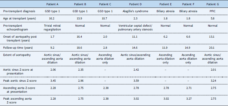
We identified 7/90 (7.8%) recipients (Table 2) with new-onset aortopathy, presenting with aortic root dilation and/or ascending aorta dilation not noted previously in their pre-transplant echocardiograms. All of them had mild aortic dilation, which stayed fairly constant on longitudinal follow-up up to 20 years post transplant. None of the recipients had a pre-existing connective tissue disorder or aortic valvular disorder, which would predispose them to aortopathy: five of the affected recipients had completely normal pre-transplant echocardiograms, whereas two recipients had trivial mitral regurgitation and small ventricular septal defect/pulmonary artery stenosis, respectively. The median time to onset of aortopathy was 6.4 (1.7–16.4) years post transplant: two recipients with glycogen storage disease type I had early onset of aortopathy within 2 years post transplant.
Characteristics and risk profile of seven patients with de novo aortopathy.

GSD=glycogen storage disease
Risk factor analysis for aortopathy
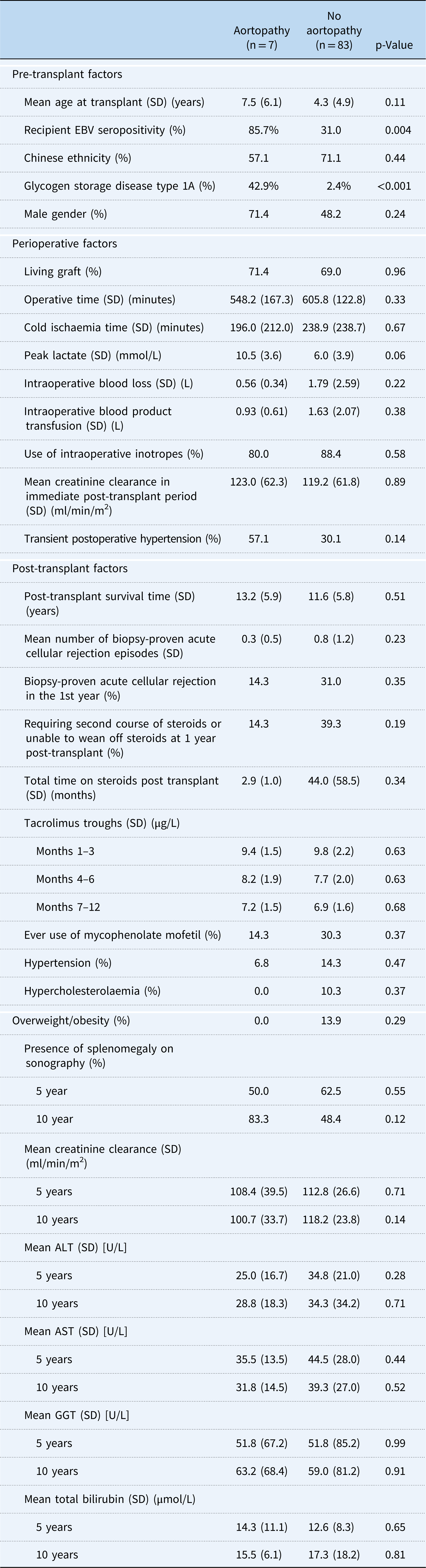
Traditional risk factors for cardiovascular morbidity and mortality in the adult transplant recipient – hypertension, hyperlipidaemia, and obesity – did not show a significant association nor trend to significance among recipients with aortopathy. These cardiovascular risk factors were in contrast to the higher prevalence among recipients without aortopathy. The overall incidence of transient postoperative hypertension was high at 32.1%, of which there was a non-significant trend for recipients with aortopathy to have a higher incidence (57.2 versus 30.1%, p=0.15) (Table 3). The overall incidence of hypertension, which occurred or persisted beyond the first 3 months of transplant, was 7.3%, with the median time to hypertension occurring at 5.3 (3.1–12.4) years post transplant; the overall incidence of post-transplant hypercholesterolaemia was 9.2%, occurring at a median time of 2.9 (0.0–8.5) years post transplant. The 11.9% of the recipients were at least overweight at one point in their post-transplant follow-up; 6.6% of recipients met the predefined criteria for obesity. The odds of obesity significantly increased with a longer post-transplant survival time (odds ratio [OR] 1.23 [95% confidence intervals (CI): 1.05–1.44, p=0.011]). There was a non-significant trend towards having slightly higher rates of post-transplant obesity and being overweight among recipients who required a second course of steroids and/or prolonged course of steroids beyond the first post-transplant year – 16.0% overweight in those requiring repeat course or prolonged steroids versus 9.8% in those weaned off steroids successfully, p=0.451. There was no such trend to association between repeat steroid dose and hypertension – 8.7% hypertension with repeat steroid course versus 9.8% in those weaned off steroids, p=0.889.
Comparison of risk factors in the aortopathy and no-aortopathy cohort.

ALT=alanine aminotransferase; AST=serum aspartate aminotransferase; EBV=Epstein-Barr virus; GGT=gamma-glutamyl transferase
Pre-transplant and perioperative factors
Glycogen storage disease type I (42.9 versus 2.4%, p<0.001) and recipient Epstein-Barr virus seropositivity – defined by a positive Epstein-Barr virus viral capsid IgG titre before transplant (85.7 versus 31.0%, p=0.004) – were the two most significant associations for aortopathy on univariate analysis; an older mean age at transplant and a higher intraoperative peak lactate showed a trend towards significance among recipients with aortopathy (Table 3). However, on multivariate analysis, only glycogen storage disease type I remained as the single most significant risk factor (OR 51.3 [95% CI: 1.1–2498.1, p=0.047]). Other operative indices that could impose haemodynamic stress to the recipient, particularly the total operative time, the use of intraoperative inotropes, the amount of blood loss, and total volume of blood product transfusion, did not show any significant relationship with aortopathy. Considering immediate post-transplant renal function as a surrogate marker for end-organ ischaemic insult, recipients with aortopathy in fact had a slightly higher mean calculated glomerular filtration rate compared with unaffected recipients (123.0 in aortopathy versus 119.2 in unaffected ml/min/m2, p=0.89).
Post-transplant factors
There appeared to be a non-significant trend for recipients with aortopathy to have slightly lower glomerular filtration rate compared with unaffected recipients at the 10th post-transplant anniversary – 100.7 affected versus 118.2 ml/min/m2 unaffected, p=0.14. As glycogen storage disease type I was significantly associated with aortopathy in our cohort, the finding of a lower glomerular filtration rate among recipients with aortopathy could have been explained by glycogen storage disease type I patients generally having progressively lower, albeit non-significant, glomerular filtration rates by their 10th year of post-transplant follow-up (mean glomerular filtration rate 94.3 ml/min/m2 in glycogen storage disease versus 120 ml/min/m2 in non-glycogen storage disease, p=0.12) (Table 3).
Factors that could subject a recipient to increased immunosuppression – the number of biopsy-proven acute cellular rejection episodes within the first post-transplant year, as well as within the entire post-transplant period to the date of data analysis, steroid exposure time, tacrolimus trough levels, and prior or current use of adjunct mycophenolate in the post-transplant year – were not significantly associated with the onset of aortopathy. In all, one out of the seven recipients with aortopathy had early acute cellular rejection occurring 0.4 months post transplant versus a mean time to rejection of 2.2 months in those without aortopathy. In fact, there appears to be a trend towards recipients with aortopathy having a lower mean number of biopsy-proven acute cellular rejection episodes (0.3 versus 0.8, p=0.23), lower steroid exposure time (2.9 versus 44.0 months, p=0.35), lower requirements for repeat steroid course (14.3 versus 39.3%, p=0.19), and lower exposure to adjunct mycophenolate (14.3 versus 30.3%, p=0.37). These findings support that over-immunosuppression was not a significant associative factor.
Graft function measured in terms of total bilirubin levels, serum alanine aminotransferase /aspartate aminotransferase transaminase levels, and gamma-glutamyl transferase levels at the 5th and 10th post-transplant anniversary were also similar between both groups, although we did note a non-significant trend towards recipients with aortopathy having persistent splenomegaly on sonography on their 10th post-transplant anniversary (83.3 versus 48.4%, p=0.12).
Discussion
Liver transplantation can be indeed seen as a double-edged sword with our study describing de novo cardiac anomalies, particularly in vulnerable recipients with an underlying predisposition to vasculopathies. From the perspective of cardiovascular benefits in our cohort, liver transplantation reversed echocardiographic findings of dilated cardiac chambers and ventricular hypertrophy, as well as resolved intrapulmonary shunting associated with hepatopulmonary syndrome within the first post-transplant year. The former findings of cardiac dilation and hypertrophy, in association with cardiac dysfunction, have been described as cirrhotic cardiomyopathy, an entity thought to be consequent to the hyperdynamic circulation in end-stage liver disease.Reference Moller and Henriksen 18 Prior studies on cirrhotic cardiomyopathy in adults have consistently showed improvement in cardiac performance after transplant,Reference Zardi, Zardi, Chin, Sonnino, Dobrina and Abbate 19 with post-transplant cardiac dysfunction lasting between a few days and a period of 6 monthsReference Henderson, Mackay and Hooks 20 The strengths of the study were that we had a routine echocardiogram protocol for all transplant recipients commencing from the pre-transplant phase – we were thus able to monitor for improvement or deterioration in underlying cardiac lesions, as well as detect de novo cardiac structural pathology, which would have been otherwise asymptomatic. The novelty of this study relies on the identification of a subgroup of paediatric transplant recipients with de novo aortopathy, and the analysis of possible associative risk factors.
Prior literature on paediatric aortic dilation has been reported in the context of connective tissue disorders, such as Marfan’s syndrome, and congenital cardiac defects, particularly bicuspid aortic valve and systemic hypertension.Reference Zarate, Sellars, Lepard, Carlo, Tang and Collins 21 Our data on de novo aortopathy mirrored certain aspects reported in prior studies of aortic dilation in children with bicuspid aortic valve – ascending aorta dilation was the most common,Reference Zarate, Sellars, Lepard, Carlo, Tang and Collins 21 and the extent of dilation stayed fairly constant during longitudinal follow-up.Reference Fernandes, Khairy and Graham 11 Prevalence of aortic dilation of 2.8% is reported among a cohort of 142 hypertensive children.Reference Gupta-Malhotra, Devereux, Dave, Bell, Portman and Milewicz 22 A novel finding of aortic dilation in 30.9% of a paediatric cohort with end-stage renal disease compared with an incidence of 2.3% in healthy children is reported, although this cohort was a heterogenous cohort with both children on dialysis and children who were kidney transplant recipients.Reference Kaddourah, Uthup and Madueme 23 On the basis of the same working definitions of hypertension as those of our study, Kaddourah et alReference Kaddourah, Uthup and Madueme 23 reported significantly higher incidences of uncontrolled hypertension, although mean blood pressure values were actually comparable between the two groups, and glomerular diseases among those with aortic dilation, compared with those without.Reference Kaddourah, Uthup and Madueme 23 Similar to the data within our study, Kaddourah et alReference Kaddourah, Uthup and Madueme 23 did not find any significant influence of dyslipidaemia on the incidence of aortic dilation.
We did observe differences in the local prevalence of typical cardiovascular risk factors, with that reported in prior cohort studies. The prevalence of hypertension among our transplant recipients with at least a 5-year follow-up (7.1%) was substantially lower than the rates (20.7%) reported by a recent large North-American cohort study within the Studies of Pediatric Liver Transplantation (SPLIT) registry using the same case definition.Reference McLin, Anand, Daniels, Yin and Alonso 24 Ethnic variations in individual susceptibility to hypertension,Reference Sorof, Lai, Turner, Poffenbarger and Portman 25 as well as very low cyclosporine usage as maintenance immunosuppression in our cohort (4.4 versus 28% in SPLIT cohort), could explain this observation. The prevalence of dyslipidaemia among those with at least 5-year survival was comparable to that from the SPLIT registry – 7% hypercholesterolaemia and 10% hypertriglyceridaemia;Reference Ng, Fecteau and Shepherd 26 we also reported similar incidences of being overweight (11.9%) but a lower incidence of obesity (6.6%) when compared with United Network for Organ Sharing post-transplant obesity data on a cohort of similar follow-up time (16% overweight and 13% obese).Reference Perito, Glidden, Roberts and Rosenthal 27 These latter differences could be explained by ethnic and genetic differences, and the variations between different growth centile charts used.
In the multivariate analysis, our study suggests that glycogen storage disease type I is the single most significant risk factor in the onset of aortopathy. We also noted that both recipients who had developed early-onset aortopathy within 2 years of their transplant had glycogen storage disease. Cardiac manifestations in glycogen storage disease type I have not been described in medical literature,Reference Kishnani, Austin and Abdenur 28 even in prior transplant cohorts of recipients with glycogen storage disease.Reference Iyer, Chen and Wang 29 , Reference Maheshwari, Rankin, Segev and Thuluvath 30 Previously published studies have, however, described the possible increased risk for vascular dysfunction in non-transplanted glycogen storage disease type I patients, evident by increased carotid intima–media thickness and augmentation indices in glycogen storage disease patients versus controls.Reference Yekeler, Dursun and Emeksiz 31 , Reference Bernier, Correia, Haller, Theriaque, Shuster and Weinstein 32 It is uncertain whether the dyslipidaemia in these patients was indeed the main factor contributing to vascular dysfunction, as there are conflicting data on the actual impact of glycogen storage disease-associated hyperlipidaemia on endothelial function.Reference Lee, Celermajer, Robinson, McCarthy, Betteridge and Leonard 33 Glycogen storage disease type I patients with de novo aortopathy in our cohort had resolution of their underlying metabolic milieu – dyslipidaemia, hyperuricaemia, and lactic acidosis – after transplant, which may suggest that additional factors, either related to glycogen storage disease or liver transplant itself, may have contributed to underlying endothelial dysfunction and the onset of aortopathy.
In our study, only one recipient with de novo aortopathy had underlying Alagille’s syndrome, which has the closest association with cardiovascular diseases of all the pre-transplant diagnoses. This child had a pre-existing ventricular septal defect and pulmonary artery stenosis, which could be rarely associated with aortic dilation even in the absence of bicuspid aortic valve.Reference Zarate, Sellars, Lepard, Carlo, Tang and Collins 21 The associated cardiac lesions of pulmonary stenosis and ventricular septal defect observed in our subgroup of recipients with Alagille’s syndrome remained stable post transplant without complications of cardiac failure; these lesions were not expected to reverse or improve with liver transplantation owing to underlying mutations in the JAG1 gene affecting vital cell signalling receptors in organ development.Reference Li, Krantz and Deng 34 We also did not expect these cardiac lesions to deteriorate post transplant, as the lesions in our cohort were mild in severity without consequence to cardiac function – that is, pulmonary stenosis not requiring valvotomy or balloon dilatation and small ventricular defect not requiring pre-transplant cardiac surgery. There are data to support that even severe pulmonary stenosis in Alagille’s is not a contraindication to liver transplantation.Reference Ozcay, Varan, Tokel, Cetin, Dalgic and Haberal 35 The fact that we had reported de novo aortopathy in seven recipients with a wide spectrum of pre-transplant diagnoses suggests that there may be other factors that predispose to vasculopathy besides the diagnosis alone. For instance, we had observed a non-significant trend towards higher incidence of transient postoperative hypertension and higher incidence of splenomegaly 10 years post transplant among recipients with aortopathy. Persistent splenomegaly reflects long-standing portal hypertension, and elevated portal pressures with increased portosystemic shunting may contribute to added haemodynamic stress through a hyperkinetic circulatory state.Reference Al-Hamoudi 36 These factors may cumulatively increase the risk of aortopathy.
Another possible factor to explain this novel finding of de novo aortopathy among our transplant recipients in a predominantly Chinese population would be the effects of ethnicity. Turkbey et al reported a large multi-ethnic adult study of ascending aorta dimensions defined on cardiac MRI; adjusting for age, blood pressure, anthropometry, and cardiovascular comorbidities, the Chinese participants had a significantly higher rate of increase in ascending aorta diameter with age than in Caucasians. Chinese participants also had a larger ascending aorta luminal diameter (1.5 mm larger, p<0.001), whereas African Americans had a smaller ascending aorta diameter (−0.5 mm, p<0.001) at baseline compared with Caucasian participants.Reference Turkbey, Jain and Johnson 37 These findings support an ethnic and possible genetic basis to the novel findings of de novo aortopathy that we observe.
Acquired mitochondrial dysfunction, which is well associated with cardiovascular and endothelial dysfunction,Reference Rosca and Hoppel 38 – Reference Tang, Luo, Chen and Liu 40 may also be a plausible aetiologic factor in de novo structural cardiovascular anomalies. Farah et alReference Farah, Sinha and Wu 41 reported the presence of hepatic mitochondrial dysfunction in mouse models of glycogen storage disease 1A, the latter factor being strongly associated with de novo aortopathy in our cohort. Tacrolimus, the main maintenance immunosuppressant in liver transplant recipients, has been associated with reduced oxygen consumption in rat cell models, and reduction in synthesis of mitochondria DNA-encoded proteins;Reference Palacin, Coto and Llobet 42 tacrolimus is also postulated to cause new-onset diabetes after transplant by impairing mitochondrial calcium uptake and leading to pancreatic beta-cell dysfunction.Reference Lombardi, Trimarco, Iaccarino and Santulli 43 Interestingly, a Taiwanese group observed recovery in mitochondria number and antioxidant enzyme activity in a cohort of children with biliary atresia who underwent liver transplantation.Reference Chen, Tang and Huang 44 This supports the double-edged sword effect that we observed in our study – liver transplantation had the positive effect of reversing pre-transplant cardiac anomalies such as ventricular/septal hypertrophy, cardiac dilatation, and cirrhotic cardiomyopathy; yet, in certain susceptible transplant recipients, de novo cardiovascular anomalies were observed. Further studies are required to elucidate the molecular mechanisms underlying these observed findings.
There is limited literature documenting de novo structural cardiac anomalies in children – Kotb et alReference Kotb, Abd El Satar and Badr 8 had reported pulmonary hypertension, increased cardiac mass, and a case of de novo aortic stenosis in a cohort of 25 paediatric liver transplant recipients; hypertrophic cardiomyopathy has been described in association with tacrolimus in paediatric liver transplant recipients, requiring a dose reduction or switch to cyclosporine;Reference Atkison, Joubert and Barron 7 Chang et alReference Chang, McDiarmid, Alejos, Drant and Klitzner 45 reported two cases of transient asymmetric septal hypertrophy detected in hospitalised paediatric liver transplant recipients. We did not report any cases of post-transplant pulmonary hypertension nor increased cardiac mass nor cardiac hypertrophy: a postulated reason could be that tacrolimus-associated ventricular hypertrophy is an acute and transient phenomenon occurring within weeks of transplantation as described in Chang’sReference Chang, McDiarmid, Alejos, Drant and Klitzner 45 and Atkison’sReference Atkison, Joubert and Barron 7 reported cohorts, and our centre only commences protocolised echocardiographic screening from the first post-transplant anniversary. Another reason could be genetic polymorphisms accounting for variation between different transplant populations in terms of susceptibility to tacrolimus-induced cardiotoxicity and cardiac hypertrophy.Reference Poirier, Nicaud and McDonagh 46
The way forward would be to prospectively investigate for endothelial dysfunction in liver transplant recipients from the pre-transplant phase to the post-transplant period, particularly in those inherently at higher risk for worse cardiovascular outcomes such as Alagille’s syndrome, glycogen storage disease, and certain inborn errors of metabolism – for example, mitochondrial hepatopathies and fatty acid oxidation disorders. Endothelial injury with release of vasoactive factors and an increase in overall arterial stiffness and reduced arterial compliance could be contributing factors to the pathogenesis of aortopathy. Endothelin-1, a potent vasoconstrictor, has been implicated in the pathogenetic mechanisms in hepatorenal syndrome and hepatopulmonary syndrome. Typically, levels of endothelin-1 decrease after liver transplantation, with corresponding improvement in renal function. At the same time, transient elevations in endothelin-1 levels in the early post-transplant period may contribute to systemic hypertension,Reference Neal, Brown, Wilkinson and Alexander 47 whereas persistently elevated endothelin-1 levels in the later post-transplant period have been associated with the use of calcineurin inhibitors and nephrotoxicity.Reference Textor, Taler, Canzanello, Schwartz and Augustine 48 In addition, endothelin-1 has been demonstrated to increase central aortic systolic pressure and arterial stiffness,Reference Vuurmans, Boer and Koomans 49 and arterial stiffness has been established as a determinant of progressive aortic dilation in patients with Marfan syndrome or after correction of congenital cardiac defects.Reference Chen, Wong, Wong and Cheung 50 , Reference Nollen, Groenink, Tijssen, Van Der Wall and Mulder 51 Shalev et alReference Shalev, Nir and Granot 52 demonstrated a strong correlation between endothelin-1 levels and B-type natriuretic peptide – a biomarker for left ventricular volume overload – levels (r=0.79, p<0.05) among a small cohort of paediatric liver transplant recipients with normal echocardiographic cardiac function; mean endothelin-1 levels and B-type natriuretic peptide levels also tended to be higher in post-liver transplant recipients versus healthy controls.Reference Shalev, Nir and Granot 52 These findings suggest that elevation of certain biomarkers related to endothelial damage and cardiac dysfunction may precede the onset of gross cardiac structural abnormalities, and could be potentially used as adjunctive measures of endothelial dysfunction along with standard echocardiographic surveillance.
The small sample size of affected recipients with aortopathy limited the ability to achieve statistical significance in risk factor analysis. Similar to the limitations of prior paediatric liver transplant cohort studies on hypertension, we did not perform 24-hour ambulatory blood pressure monitoring for all recipients, which could unmask nocturnal hypertension. In addition, we did not review other cardiac variables such as shortening fraction, left ventricular wall thickness in diastole, and septal thickness in diastole, as the focus of our study was to specifically look at the aortic dimensions rather than all aspects of cardiac abnormalities.
Conclusions
Liver transplant is a double-edged sword that reverses certain cardiopulmonary complications of end-stage liver disease, but may induce de novo structural cardiac injury in the form of aortic dilation. Transplant recipients with glycogen storage disease type I may be at a higher risk for aortopathy. Our study supports the need for long-term cardiac echocardiographic surveillance in otherwise asymptomatic paediatric liver transplant recipients.
Acknowledgements
The authors thank Dr Dimple Rajgor for her assistance in editing, formatting, reviewing, and submission of the manuscript for publication. The authors also thank Maria Janelle Liwanag for her assistance in collating the data for the transplant cohort.
Financial Support
This research received no specific grant from any funding agency in the public, commercial, or not-for-profit sectors.
Conflicts of Interest
None.
Ethical Standards
The authors assert that all procedures contributing to this work comply with the ethical standards of the relevant national guidelines on human experimentation, SGGCP, and with the Helsinki Declaration of 1975, as revised in 2008, and has been approved by the institutional committees, NHG DSRB.