Introduction
The genus Melaspilea Nyl. (Melaspileaceae W. Watson) is in general characterized by lirelliform apothecioid ascomata, an exciple composed of several cell layers and brown 1-septate ascospores, and it comprises c. 80 saprotrophic, lichenized and lichenicolous species (Ryan & Nimis Reference Ryan, Nimis, Nash, Ryan, Diederich, Gries and Bungartz2004). At present, its systematic position is uncertain and it is tentatively placed in the Arthoniales Henssen ex D. Hawksw. & O. E. Erikss. (Lumbsch & Huhndorf Reference Lumbsch and Huhndorf2010).
The genus as currently circumscribed is heterogeneous and in need of a modern revision. The generic type, M. arthonioides (Fée) Nyl., is a corticolous, lichenized species (Coppins et al. Reference Coppins, James and Hawksworth1992) and most likely unrelated to the lichenicolous members of the genus; according to Sanderson et al. (Reference Sanderson, Hawksworth, Aptroot, Smith, Aptroot, Coppins, Fletcher, Gilbert, James and Wolseley2009), it appears to be close to Buelliella Fink. Eleven obligately lichenicolous Melaspilea species have been accepted so far (Nylander Reference Nylander1868; Tuckerman Reference Tuckerman1872; Müller Reference Müller1888, Reference Müller1895; Vouaux Reference Vouaux1913; Hawksworth Reference Hawksworth1982; Clauzade et al. Reference Clauzade, Diederich and Roux1989; Aptroot et al. Reference Aptroot, Diederich, Sérusiaux and Sipman1997; Lawrey & Diederich Reference Lawrey and Diederich2003; Sanderson et al. Reference Sanderson, Hawksworth, Aptroot, Smith, Aptroot, Coppins, Fletcher, Gilbert, James and Wolseley2009; Kalb et al. Reference Kalb, Buaruang, Mongkolsuk and Boonpragob2012; Zhurbenko & Zhdanov Reference Zhurbenko and Zhdanov2013). One of them, M. canariensis D. Hawksw., was recently transferred into a newly distinguished genus, Labrocarpon Etayo & Pérez-Ortega (Pérez-Ortega & Etayo Reference Pérez-Ortega and Etayo2010), differing from Melaspilea s. str. mainly by the presence of short hyphae emerging from the inner part of the exciple (periphysoids), and specific ascomata with a slit-like disc and lip-shaped margin (Pérez-Ortega & Etayo Reference Pérez-Ortega and Etayo2010).
During our lichenological studies conducted in Bolivia (e.g. Flakus & Wilk Reference Flakus and Wilk2006; Flakus Reference Flakus2009; Flakus & Kukwa Reference Flakus and Kukwa2012; Kukwa et al. Reference Kukwa, Etayo and Flakus2012; Etayo et al. Reference Etayo, Flakus and Kukwa2013), a further lichenicolous species of Melaspilea s. lat. was discovered in Tucumano-Boliviano vegetation in the tropical Andes and is described in this paper.
Material and Methods
The material is deposited in KRAM, LPB, UGDA, hb. Etayo and hb. Flakus. The morphology and anatomy were examined using NIKON SMZ800 and NIKON ECLIPSE 80i (DIC) microscopes. The anatomy was studied on material mounted in water, KOH solution (K), lactophenol cotton blue (LPCB), and for ascus structure Lugol's iodine solution without (I) or with pretreatment with KOH (K/I). Structure of the exciple was additionally studied in commercial bleach (C) and adding Congo Red. Measurement of ascospores and other structures was made on material mounted in water. Ascospore measurements are presented as: (arithmetic mean—standard deviation)—arithmetic mean—(arithmetic mean + standard deviation), flanked by the minimal and maximal measurements in parentheses, and the length/breadth ratio (l/b) is presented in the same way, followed by the number of measurements (n).
The New Species
Melaspilea tucumana Flakus, Etayo & Kukwa sp. nov.
MycoBank No.: MB 808226
Differs from Labrocarpon canariense by aggregated apothecioid ascomata with a widely exposed disc, and by inducing galls on the host thallus.
Type: Bolivia, Dept. Tarija, Prov. Aniceto Arce, Papachacra, near Papachacra valley, 21°41′14″S, 64°30′19″W, 2050 m, open area with shrubs in Tucumano-Boliviano vegetation, on saxicolous Pertusaria sp., 7 August 2012, A. Flakus 23898 (KRAM—holotype; LPB, UGDA, hb. Etayo, hb. Flakus—isotypes).
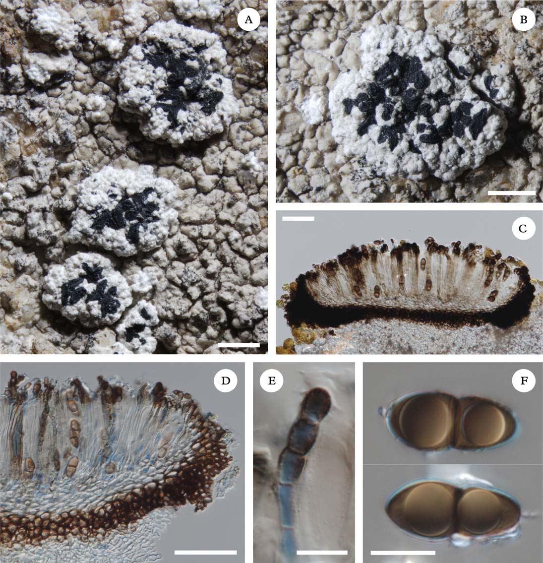
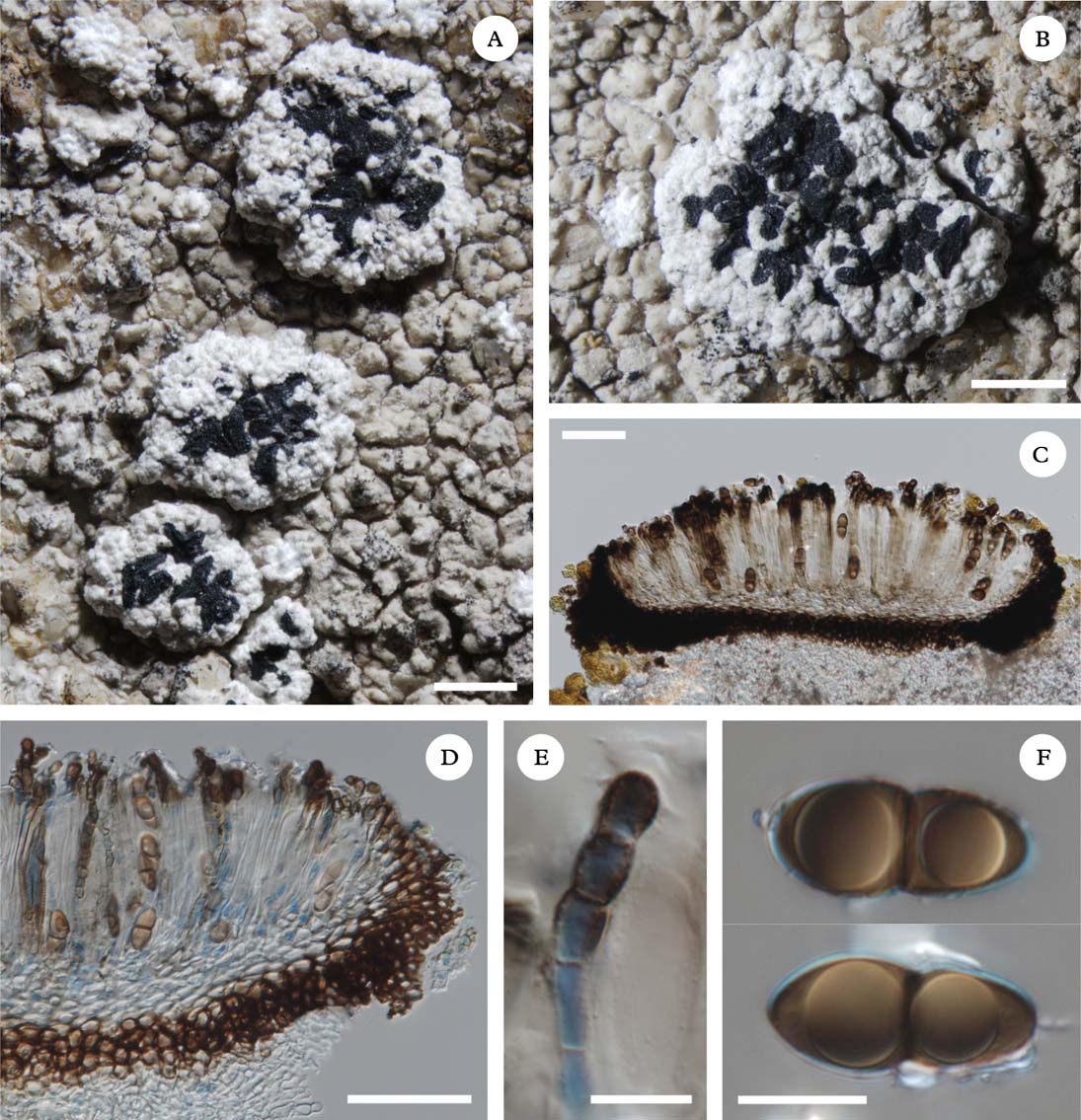
Melaspilea tucumana (holotype). A, habit of fungus galls induced on host thallus; B, single gall with aggregated ascomata; C, section of ascomata (mounted in tap water); D, paraplectenchymatous exciple (mounted in LPCB); E, paraphysis with swollen apical cells (mounted in LPCB); F, mature ascospores (mounted in LPCB). Scales: A & B=1 mm; C & D=50 μm; E & F=10 μm. In colour online.
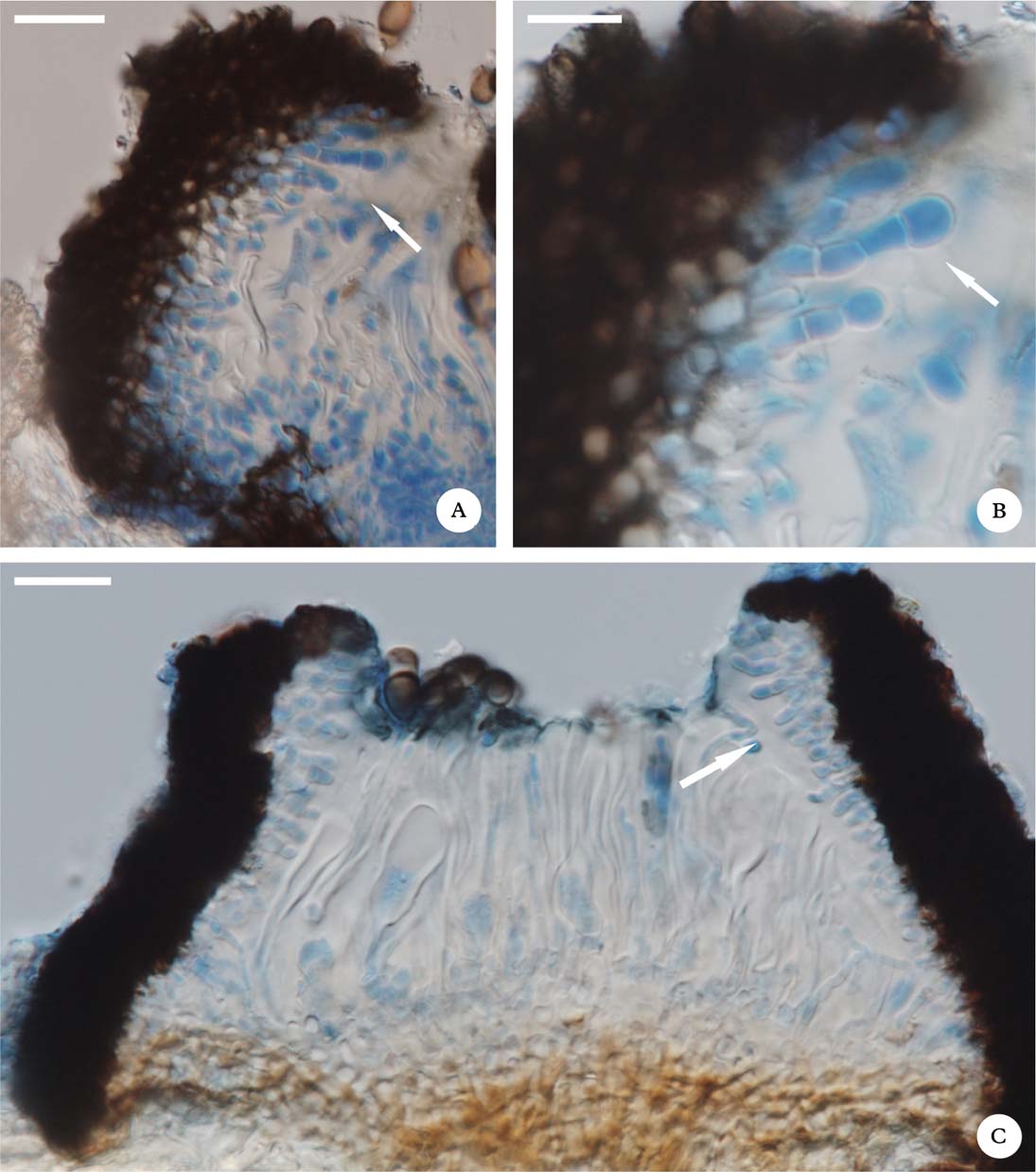
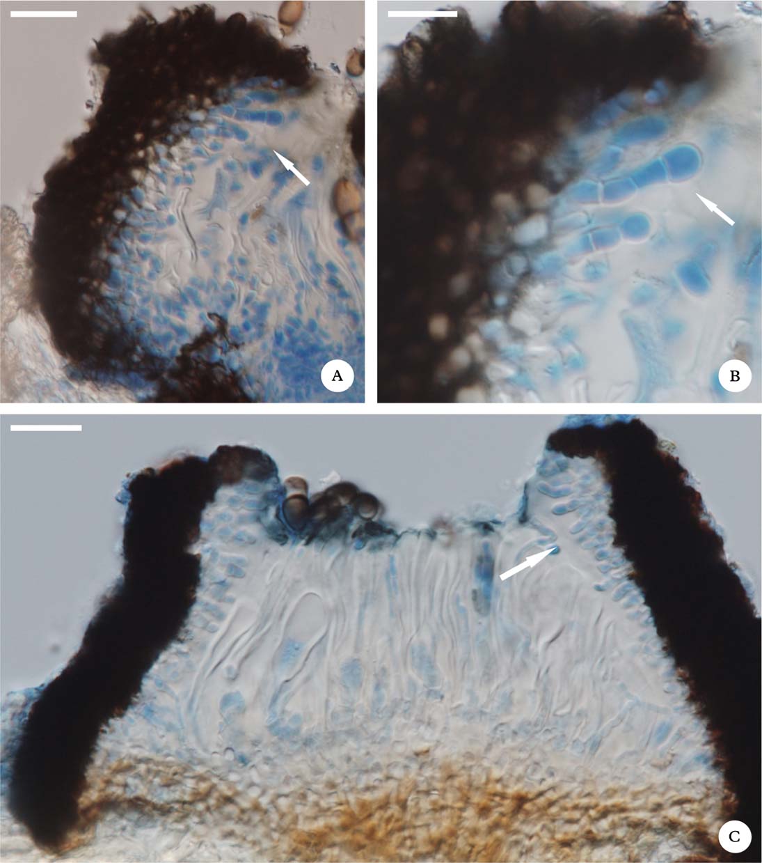
Section of exciple showing periphysoids (mounted in LPCB). A & B, Melaspilea tucumana (holotype); C, Melaspilea sp. (Flakus 24745). Scales: A & C=20 μm; B=10 μm. In colour online.
Ascomata apothecioid, 0·4–1·0×0·2–0·4 mm, black, lirelliform to branched, aggregated to confluent on galls induced on the host thallus, initially perithecioid and immersed in host thallus but apothecioid and superficial when mature. Galls of 1–5 mm diam. Disc widely exposed, flat, concolorous with the margin, black, matt, with a rough surface. Margin black, thick, with a rough surface, prominent when young, later reduced. Exciple paraplectenchymatous, laterally 25–40 µm wide, basally 20–60 µm, continuous below hymenium, composed of cells 3–9 µm diam., dark brown pigmented, K−, N−. Periphysoids 1–3-septate, present in inner part of the exciple. Hymenium hyaline to brownish, 90–120 µm, I+ pale blue, K/I+ pale blue. Subhymenium hyaline, c. 20–30 µm. Epihymenium c. 15–20 µm, dark brown pigmented, K−, N−. Paraphyses sparsely branched to almost simple when mature, septate, constricted at the septa (moniliform), 2–4 µm wide at half height, with 1–3 apical cells strongly swollen (4–10 µm wide) and dark brown pigmented. Asci 4–6-spored, broadly clavate, I+ pale blue, K/I+ pale blue, functionally bitunicate, wall apically strongly thickened, ascus apex without any visible structure (I−, K/I−), 20–25×50–80 µm. Ascospores dark brown, 1-septate (overmature ascospores very rarely 3-septate), strongly constricted at the septa, smooth-walled, without gelatine epispore, lower cell narrower than the upper one, usually with one guttule in each cell, (18·0–)19·8–20·9–22·1(–25·0)×(7·0–)8·4–9·1–9·9(–11·0) µm, l/b=(1·9–)2·1–2·3–2·6(–2·8) µm (n=60).
Conidiomata not observed.
Host
The species causes gall formation on a saxicolous sorediate lichen containing lichexanthone in the thallus cortex and cf. hypothamnolic acid in soralia that apparently represents a so far undescribed, sterile Pertusaria s. lat. species. Thallus of the host: UV+ yellow, K−, P−, C−, K/C−; galls UV+ yellow, K−, P−, C−, K/C−; soralia UV+ ice blue, K+ violet (first K- but after a while K+ violet), P± faintly yellow-orange, C+ red (colour reaction transient), K/C+ red-violet; TLC: lichexanthone, barbatic and cf. hypothamnolic acids.
Etymology
The new species is named after its locus classicus located at higher elevation in the Tucumano-Boliviano vegetation in the Bolivian Andes.
Notes
A careful examination of thin sections of the exciple of M. tucumana (especially when decolouring by C reagent or mounted in LPCB) revealed the presence of 1–3-septate periphysoids in its inner part, previously reported by Pérez-Ortega & Etayo (Reference Pérez-Ortega and Etayo2010) for Labrocarpon canariense (D. Hawksw.) Etayo & Pérez-Ortega. Despite that fact, we decided to place the new species provisionally in the genus Melaspilea s. lat., until the presence or absence of periphysoids has been checked in all lichenicolous species of Melaspilea s. lat., and in the type of the genus. Although the origin of those structures is uncertain, they were observed not only in L. canariense and M. tucumana, but also in another probably undescribed species of Melaspilea s. lat. (Fig. 2C), lichenicolous on corticolous Pyrenulales from Bolivia (Flakus 24745).
Melaspilea tucumana is characterized by aggregated (to confluent when overmature) lirelliform, simple to branched apothecioid ascomata, with widely exposed discs that are located superficially on galls induced on the host thallus. The new species most closely resembles Labrocarpon canariense (Hawksworth Reference Hawksworth1982; Pérez-Ortega & Etayo Reference Pérez-Ortega and Etayo2010). However, L. canariense has comparatively different, regularly dispersed (not aggregated) labriform ascomata with never widely exposed discs (slit-like), it does not cause gall formation on the host thallus, and its exciple is densely inspersed with a dark brown granular pigment (Hawksworth Reference Hawksworth1982; Pérez-Ortega & Etayo Reference Pérez-Ortega and Etayo2010).
Due to host preferences, similar ascomata morphology and gall induction, the new species can also be confused with M. galligena Zhurb. & I. Zhdanov, recently described from Russia (Zhurbenko & Zhdanov Reference Zhurbenko and Zhdanov2013). However, M. galligena can easily be separated by the lack of periphysoids in the inner part of the exciple, larger ascospores (20·7–)22·5–25·9(–30·0)×(10·0–)11·2–13·6(–15·6) µm, angular rounded to elongate and never superficial ascomata, an I− hymenium, and strongly branched paraphyses (Zhurbenko & Zhdanov Reference Zhurbenko and Zhdanov2013).
It is also worth noting that the ascomata of M. tucumana, which are initially perithecioid and immersed in the host thallus, resemble in ontogeny and anatomy those of the genus Buelliella. Such development of ascomata has been noticed also in Melaspilea arthonioides (Fée) Nyl. and M. galligena, respectively by Ryan & Nimis (Reference Ryan, Nimis, Nash, Ryan, Diederich, Gries and Bungartz2004) and Zhurbenko & Zhdanov (Reference Zhurbenko and Zhdanov2013). The relationship of Melaspilea tucumana and those two Melaspilea species to Buelliella needs further investigation, including a molecular framework. It will also allow the identification of its systematic position, which currently seems more appropriate in Dothideomycetes (where Buelliella is currently placed) than Arthoniomycetes.
Distribution and habitat
So far the species is known only from the type locality, where an abundant population on several thalli of the host was observed.
Additional specimens examined. Bolivia: Dept. Tarija: same locality as type, M. Kukwa 10967 (LPB, UGDA, hb. Diederich); ibid., J. Etayo 28152 (LPB, hb. Etayo).
Specimen of Melaspilea sp. studied. Bolivia: Dept. Beni: Prov. Marbán, near road from Camiaco to San Lorenzo, 15°14′49″S, 64°49′11″W, 160 m, lowland Amazon forest surrounded by Los Llanos de Moxos savanna, 2012, A. Flakus 24745 & P. Rodriguez Flakus (LPB).
A key to lichenicolous species of Melaspilea s. lat.
-
1 Ascospores mostly >9 µm broad ... 2
Ascospores mostly <9 µm broad ... 5
-
2(1) Fungus inducing gall formation; ascomata immersed or superficial ... 3
Fungus not inducing gall formation; ascomata superficial ... 4
-
3(2) Ascomata immersed; hymenium I−; ascospores 21·0–30·0×10·0–15·5 µm. On Pertusaria cf. cribellata (Russia) ... M. galligena Zhurb. & I. Zhdanov
Ascomata superficial; hymenium I+ pale blue; ascospores 18–25×7–11 µm. On Pertusaria s. lat. (Bolivia) ... M. tucumana Flakus et al.
-
4(2) Ascomata 0·3–1·5×0·2–0·5 mm; disc slit-like; ascospores 19·5–32·0×9·5–16·0 µm. On Graphis elegans; causing orange-brown decoloration on host thallus (cosmopolitan) ... M. diplasiospora (Nyl.) Müll.Arg.
Ascomata 0·15–0·50×0·1–0·2 mm; disc exposed; ascospores 18–22×9–12 µm. On Verrucaria (France, UK) ... M. leciographoides Vouaux
-
5(1) Ascospores mostly <13 µm long ... 6
Ascospores mostly >13 µm long ... 7
-
6(5) Ascomata up to 0·2 mm long; disc exposed, black; ascospores 10–12×5 µm. On Reimnitzia santensis (Paraguay) ... M. epigena Müll. Arg.
Ascomata 0·3–0·8×0·09–0·15 mm; disc slit-like; ascospores 9–11×3·5–4·5 µm. On Acanthothecis consocians (New Caledonia) ... M. epigraphella (Nyl.) Müll. Arg.
-
7(5) Disc exposed ... 8
Disc slit-like only rarely exposed when old ... 9
-
8(7) Ascomata 0·20–0·25 mm long; disc exposed, black; ascospores 15–23×5–9 µm. On Pyrenula sp. (USA) ... M. cupularis Müll. Arg.
Ascomata 0·2–0·4×0·1–0·2 mm; disc exposed, orange-brown; ascospores 14–17×7–8 µm. On Sarcographa labyrinthica (Thailand) ... M. lakae Brackel & Kalb
-
9(7) Ascomata 0·3–1·1×0·2–0·3 mm, sessile with a broader base; disc slit-like (rarely exposed when old); ascospores 15–17×7·5–9·0 µm. On Anthracothecium australiense and Pyrenula cruenta (Papua New Guinea, USA) ... M. tribulodes (Tuck.) Müll. Arg. (=Encephalographa anthracothecii Diederich)
Ascomata 0·3–0·5×0·1–0·2 mm, with a narrower base (stalk-like extension) immersed in host thallus; disc slit-like ... 10
-
10(9) Ascospores 14–21×6·0–9·5 µm. On Pertusaria (Europe, Macaronesia, Brazil) ... Labrocarpon canariense (D. Hawksw.) Etayo & Pérez-Ortega
Ascospores 10–16×5·0–7·5 µm. On Phaeographis dendritica (Europe, Hawaii, Oceania, USA) ... M. lentiginosa (Lyell ex Leight.) Müll. Arg.
We are greatly indebted to Paul Diederich (Luxembourg) and anonymous reviewers for helpful comments, and also to Rosa I. Meneses Q., the Director of Herbario Nacional de Bolivia, Instituto de Ecología, Universidad Mayor de San Andrés, La Paz, for generous cooperation. This research received support from the National Centre for Research and Development (NCBiR) in Poland, under the LIDER Programme for the years 2010–2013 (no. 92/L–1/09).