Introduction
The rapidly aging population (Armstrong et al., Reference Armstrong2007) will inevitably result in ever increasing demand for memory clinic resources. Resource intensive investigations, such as Magnetic Resonance Imaging (MRI) and Single Photon Emission Computed Tomography (SPECT), should be especially scrutinized. The memory clinic diagnostic process varies significantly internationally. The standard process across Europe can be as extensive as routine use of laboratory testing, cognitive assessment, MRI with dedicated measurement of the medial temporal lobes, SPECT scan, and lumbar puncture (Brækhus et al., Reference Brækhus2011). Some clinicians consider structural imaging integral to the assessment of undiagnosed cognitive impairment (Frisoni et al., Reference Frisoni2010); however, there is a lack of consensus regarding the optimal patient sub-group to refer for more advanced diagnostic imaging.
Advanced imaging should improve diagnostic accuracy, a concept which relies on neuropathological studies to provide the gold standard despite recent questions (Scheltens et al., Reference Scheltens2011). Diagnostic accuracy matters in terms of offering effective treatment (Birks, Reference Birks2006), relevant prognostication (Brodaty et al., Reference Brodaty, Seeher and Gibson2012), and appropriate medico-legal advice (Skene, Reference Skene2002). Imaging may also reduce the misdiagnosis of potentially reversible conditions that present with cognitive impairment. A Danish study (Hejl et al., Reference Hejl, Høgh and Waldemar2002) followed 1,000 consecutively referred patients in a memory service and found that 18 (4%) of the 430 patients diagnosed with dementia had a potentially reversible primary cause identified, which were a combination of normal pressure hydrocephalus (NPH) and space occupying lesions.
There can be significant variation in the results of different diagnostic modalities used to investigate cognitive impairment. Yokoyama et al. (Reference Yokoyama2014) looked at the discrepancies between MRI, SPECT, and amyloid-Positron Emission Tomography (PET) in patients with mild cognitive impairment (MCI) or early Alzheimer's dementia (AD). They found that amyloid deposition was highest in the group with typical findings on MRI and SPECT. For patients with hippocampal atrophy on MRI and atypical SPECT scan findings for AD, amyloid-PET was negative in 60%. In addition to the discrepancies between imaging modalities, interpretation of the imaging findings can also be subject to significant variability (Boelaarts et al., Reference Boelaarts, Scheltens and de Jonghe2014).
Despite their limited availability and potential for significant variation between each modality and in the interpretation of findings within a single modality, the clinical utility of MRI and SPECT scanning has not been prospectively measured in a memory clinic population. The available international evidence indicates that there is significant utility in MRI combined with neuropsychology testing (Meijs et al., Reference Meijs2015), MRI combined with SPECT (Borghesani et al., Reference Borghesani2010), and MRI combined with Computed Tomography (CT) (Chui and Zhang Reference Chui and Zhang1997; Geroldi et al., Reference Geroldi2008), particularly if non- AD is the initial diagnosis. The same studies (Chui and Zhang Reference Chui and Zhang1997; Geroldi et al., Reference Geroldi2008; Borghesani et al., Reference Borghesani2010; Meijs et al., Reference Meijs2015) indicate that there is limited scope for improvement in diagnostic accuracy when AD is the initial expert diagnosis but surgically treatable causes may still be found in a very small number of cases.
Aims
The primary aim of this study is to prospectively measure the clinical utility of MRI and/or SPECT brain scanning in addition to the routine diagnostic workup of patients presenting to a local memory clinic. The secondary aims are to identify variables associated with positive MRI and/or SPECT scan outcomes and to compare baseline characteristics between patients who undergo MRI and/or SPECT scanning and those who receive a consensus diagnosis without MRI and/or SPECT scanning.
Methods
A single center study was conducted over a convenience of 12-month sampling period in a specialist Australian memory clinic. Each patient underwent a minimum of non-contrast CT brain scanning and screening pathology testing prior to their first medical appointment at the clinic. Screening pathology included full blood examination, urea and electrolytes, liver function, thyroid function, vitamin B12, folate, serum calcium, ESR, and syphilis serology. For each patient referred for MRI and/or SPECT scanning, as part of the in clinic diagnostic workup, the primary geriatrician or psychogeriatrician was asked to assign an initial diagnosis, either single or multiple, and an associated level of diagnostic certainty by using a rating scale of 1–3 (1 = high, 2 = intermediate, 3 = low). If an initial diagnosis was not assigned prior to imaging, the patient was excluded from the study as the primary clinician was presumed not to have consented.
The initial diagnosis was then compared with the final consensus diagnosis after any scans or neuropsychology testing had been completed. Demographic information and baseline cognitive testing scores were collected for each patient. The consensus diagnosis, demographic information, and baseline testing scores were also collected for all patients, not referred for MRI and/or SPECT, whose initial medical consult occurred in the same 12-month period. All demographic information was collected from pre-clinic questionnaires routinely filled out by clinicians in consultation with the patient, with reference to the referral letter, prior to the first medical review.
The scans were reported by the local radiology service and the results of each scan were categorized based on their impact on the diagnostic process. Following routine clinical practice, no attempt was made to standardize radiologist reporting or limit the radiology service to a single radiologist for all studies. The diagnosis from any neuropsychology testing performed was also recorded. Neuropsychology assessments were conducted by the two experienced neuropsychologists attached to the memory clinic.
The positive scan outcome categories used were (1) initial diagnosis changed consistent with scan, (2) initial diagnosis confirmed, (3) potentially reversible cause identified. The negative scan outcome categories used were (1) normal or not suggestive of diagnosis, (2) initial diagnosis confirmed or changed through other means. Diagnostic change included if a single initial diagnosis became a mixed consensus diagnosis or vice versa.
Ethics approval was obtained from the local low risk ethics committee.
Statistical analysis
All data were de-identified prior to analysis. F-tests were used to determine the variances for the two-tiered T tests conducted to compare the means of continuous variables and χ 2 statistics were calculated to compare proportions for dichotomous variables. Fisher’s exact test was used for dichotomous variables if there were insufficient expected numbers (<5). Significance was set at p < 0.05. Broad variables, including an initial AD diagnosis or an initial dementia, of any type, diagnosis, as well as the completion of neuropsychology testing and the pre-test level of diagnostic certainty were investigated for their association with a positive scan outcome.
The Number Needed to Test (NNT) for one positive outcome, with 95% CI, was calculated for MRI only, SPECT only, and combined MRI and SPECT testing. The NNT was calculated using the following formula: 1/((positive outcomes after scans/total outcomes after scans) – (positive outcomes before scans/total outcomes before scans)) (Geroldi et al., Reference Geroldi2008). To calculate the NNT, all patients referred for MRI and/or SPECT were assumed to have an unconfirmed diagnosis or negative outcome prior to scanning, which would have remained unchanged without the scan/s. For the MRI only NNT, all patients who underwent SPECT only or MRI and SPECT were excluded from analysis. For the SPECT only NNT, all patients who underwent MRI only or MRI and SPECT were excluded from analysis. For the MRI and SPECT NNT, all patients who underwent MRI only or SPECT only were excluded from analysis.
Results
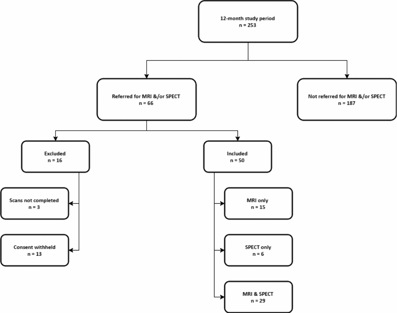
During the 12-month study period, 66 patients (26%) were referred for MRI and/or SPECT scanning out of a total of 253 patients included in the study. Patient flow through the study is depicted in Figure 1 above.
Study patient flow chart.

Demographic and pre-scan cognitive testing comparison
The included scanned patients were significantly younger (mean 69.4 ± 9.2 vs. 76.5 ± 10.2, p < 0.001), more educated (mean on 1–6 range, 2.9 ± 1.3 vs. 2.3 ± 1.0, p = 0.01) and more likely to have a prior head injury (20% vs. 8%, χ 2 = 6.29, p = 0.01).
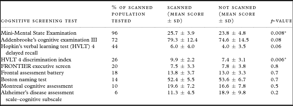
They scanned patients had significantly higher baseline Mini-Mental State Examination scores (mean 25.7 ± 3.9 vs. 23.8 ± 4.8, p = 0.008) and Hopkin's verbal learning test (HVLT) 4 discrimination index scores (mean 9.9 ± 2.2 vs. 7.4 ± 3.1, p = 0.006). The means were higher in the scanned population on the two other most commonly performed tests but the differences did not reach statistical significance. (Addenbrooke's Cognitive Examination III – mean 79.3 ± 12.4 vs. 74.6 ± 14.5, p = 0.08, HVLT 4 delayed recall – mean 6.0 ± 4.0 vs. 4.0 ± 3.5, p = 0.06). The full comparison of all testing scores, with standardized numerical totals allowing for comparison, is included in Table 1.
Comparison of cognitive testing scores between scanned and not scanned populations

*p < 0.05
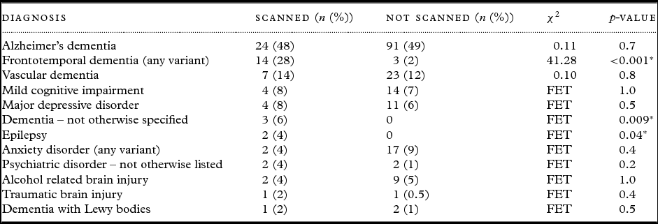
Scanning was significantly more likely if the initial diagnosis, single or multiple, included frontotemporal dementia (any variant) (28% vs. 1.6%, χ 2 = 41.28, p = <0.001). There was no difference in the proportion of patients with a diagnosis of AD between the two groups (48% vs. 49%, χ 2 = 0.11, p = 0.7). The full comparison of all initial diagnoses in the scanned population with the consensus diagnoses in the non-scanned population is included in Table 2. The percentages listed of the scanned and not scanned populations for each diagnosis exceeds 100%, in total, because mixed diagnoses are separated out for comparison.
Comparison of initial diagnoses in the scanned population with the consensus diagnoses in the not scanned population (FET indicates Fisher's Exact Test used instead of χ2)

*p < 0.05
Utility measurement
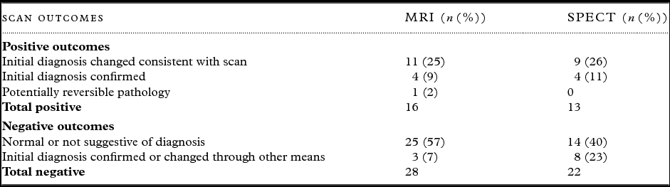
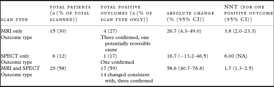
There were 16/44 (36%) positive MRI outcomes and 13/35 (37%) positive SPECT outcomes. The diagnosis changed consistent with the MRI scan findings in 11/44 (25%) and changed consistent with the SPECT scan findings in 9/35 (26%). Potentially reversible pathology was identified in a single patient, 1/50 (2%), via an MRI scan that suggested NPH.
In the scanned group, 29/50 (58%) patients completed neuropsychology testing. Of the 11 total patients who had at least one of their scan results contradict their consensus diagnosis, 8/11 (73%) underwent neuropsychology testing. In all eight patients tested, the initial diagnosis changed consistent with or was confirmed by the neuropsychology testing results. In the other three patients, the primary geriatrician was either sufficiently confident in the results of the pre-scan diagnostic process to ignore the contradictory suggestive scanned results (two patients), or the diagnosis changed consistent with the other imaging test performed (one patient). In this single patient, the SPECT was not consistent with the consensus diagnosis and suggested frontal lobe pathology, such as chronic affective disorders, alcohol-related brain injury, or frontotemporal dementia. The MRI result was consistent with the consensus diagnosis of vascular cognitive impairment, which was a change from the initial diagnosis of AD. The full list of all positive and negative outcomes is included in Table 3 below.
Scan outcomes separated by scan and type

The NNT was 3.8 (95% CI 2.0–23.3), 6.0 (95% CI NA), and 1.7 (95% CI 1.3–2.5) for MRI only, SPECT only, and MRI and SPECT together, respectively. The utility for each scan group expressed as absolute change in positive outcome and NNT is depicted in Table 4 below.
Utility for each scan group expressed as absolute change in positive outcome and NNT

Positive association testing
There were no significant associations identified between broad pre-scan variables and positive scan outcomes. The broad pre-scan variables tested included (1) initial diagnosis of AD (55% vs. 45%, χ 2 = 0.67, p = 0.4), (2) initial diagnosis of any dementia (95% vs. 5%, p = 0.06), (3) neuropsychology testing conducted (55% vs. 45%, χ 2 = 0.19, p = 0.7), (4) diagnostic certainty (mean ± SD – 1.8 ± 0.6 vs. 2.0 ± 0.7, p = 0.2).
Discussion
The significant heterogeneity in study design across similar studies (Chui and Zhang Reference Chui and Zhang1997; Geroldi et al., Reference Geroldi2008; Borghesani et al., Reference Borghesani2010; Meijs et al., Reference Meijs2015), mostly in scan types included and the definition of a positive scan outcome, limits the comparability of these results with the available international evidence.
The overall referral rate for MRI and/or SPECT in this study (26%) was significantly less than the rate in a retrospective case study conducted in the USA (Borghesani et al., Reference Borghesani2010) on MRI and SPECT scanning (49%). The same study reported that clinically meaningful information was provided in more than 80% of cases referred for scans, which is also significantly more than the positive outcome rate of 36% and 37% for MRI and SPECT, respectively, in this study. Some, but not all, of the difference is explained by the 19% of patients included in the clinically meaningful category based on an abnormal/non-diagnostic scan in the USA study. Most of these abnormal scans were included due to the presence of cerebrovascular disease, which, in the majority, was not predicted by the presence of typical cardiovascular risk factors. Abnormal/non-diagnostic scans were not felt to be sufficiently clinically relevant for inclusion as a positive outcome in this study; however, what constitutes a positive outcome is largely a clinical judgment.
The rate of initial diagnosis change consistent with the MRI scan results was 11/44 or 25% in this study. This rate is greater than reported in a Dutch study investigating the impact of additional testing, including MRI, neuropsychology testing, and cerebrospinal fluid analysis, in diagnosing dementia (Meijs et al., Reference Meijs2015). Their reported rate for MRI only diagnostic change was 24/190 or 13%. For MRI and neuropsychology, the rate increased to 37/156 or 24%; however, it remained lower than the MRI only rate and the combined MRI and SPECT rate in this study (14/29 or 48%).
The only other study (Geroldi et al., Reference Geroldi2008), conducted across multiple Italian academic centers, which has reported an NNT, measured the utility of neuropsychology testing combined with MRI or CT scanning and excluded diagnostic confirmation from the positive outcome group. Their NNT was 15.3 (95% CI 10.4–29.1) for AD, 4.1 (95% CI 3.0–6.5) for non-AD, and 8.9 (95% CI 6.9–12.9) when all initial diagnoses were considered. There were insufficient numbers of scanned patients to separate the NNT by initial diagnosis in this study. However, the NNT of 3.8 (95% CI 2.0–23.3) and 1.7 (95% CI 1.3–2.5) for MRI only and MRI and SPECT, respectively, in this study is significantly different with the difference likely partially explained by the different modalities tested and the inclusion of diagnostic confirmation as a positive outcome. Diagnostic confirmation was 25.0% of MRI positive outcomes and 30.8% of SPECT positive outcomes in this study. The remaining difference is likely related to the significantly higher proportion of patients with an initial AD diagnosis who were referred for additional testing in the Italian study (Geroldi et al., Reference Geroldi2008) (229/294 or 78%) compared to this study (23/50 or 46%).
All the comparable international studies (Chui and Zhang, Reference Chui and Zhang1997; Geroldi et al., Reference Geroldi2008; Borghesani et al., Reference Borghesani2010; Meijs et al., Reference Meijs2015) have concluded that an initial non-AD dementia diagnosis makes a positive scan outcome more likely. The Dutch study (Meijs et al., Reference Meijs2015) reported a diagnostic change rate of 54% in the initial non-AD dementia diagnosis group after their additional testing cluster. Our study was unable to significantly associate any similarly broad pre-scan variables with a positive scan outcome. This may relate to the low overall scan referral rate, resulting in lower than expected numbers of scanned patients, and a narrower definition of what constituted a positive outcome in this study. It may also relate to the lower number of patients with an initial AD diagnosis being referred for scans in this study.
The potentially reversible cause detection rate was marginally lower in this study compared with a study conducted in the USA using MRI and CT (Chui and Zhang, Reference Chui and Zhang1997) that reported a rate of 3/120 or 3%. The three cases were comprised of one meningioma requiring surgical resection and two cases of possible NPH. Potentially reversible pathology was identified in a single patient, 1/50 (2%), via an MRI scan that suggested NPH in this study. Although the associated cognitive impairment is not universally reversible post shunt insertion, there is still significant scope for improvement (Chaudhry et al., Reference Chaudhry2007), and NPH remains an important must-not-miss diagnosis. The slightly lower detection rate in this study may relate to major structural abnormalities being detected by the pre-clinic CT scan, resulting in impacted patients not proceeding to clinic assessment.
Limitations
This study was limited to a single center and local factors, such as the referral base, diagnostic criteria used, access to imaging, neuropsychology and other diagnostic resources, as well as familiarity with specific cognitive screening tests, may all affect the generalizability of these results to the broader international context. To approximate routine clinical practice, no attempt was made to standardize radiologist reporting or limit the radiology service to a single radiologist for all studies. This lack of standardization may further limit generalizability. The lower than expected referral rate reduced the number of patients scanned and the associated likelihood of identifying variables associated with positive scan outcomes. This study was also limited to identifying positive scan outcomes without determining if a positive outcome led to a clinically significant change in management.
Conclusion
The clinical utility of MRI and/or SPECT scanning in this study may be broadly superior to the available international evidence, and further research is needed to identify predictors of positive scan outcomes to further improve the utility of this limited resource. An internationally accepted consensus definition of a positive scan outcome would assist in standardizing future research. Consistent use of standardized diagnostic criteria, as well as a standardized approach to the use of cognitive screening tests would further improve the generalizability of results. Larger studies documenting whether there is significant and clinically relevant change to management as a direct result of positive outcomes from imaging would assist in quantifying the benefits of scan referral.
Conflict of interest
None.
Description of authors’ roles
J. G. designed the study, collected the majority of the data, analyzed the data, and wrote the paper. B. N. formulated the research question, collected supplementary data, and provided feedback on the paper.
Acknowledgments
Dr Sook Meng Lee, Consultant Geriatrician at Western Health (Victoria, Australia), provided valuable feedback on an alternate format pre-publication version of the paper.