INTRODUCTION
Defining monotypic genera is always a challenge because it is not clear which characters should be considered generic and which are specific. For example, Cyclophora Castracane was defined and named on the basis of the very obvious circular pseudoseptum, which in the generitype (Cyclophora tenuis Castracane) occurs on only one of the two valves. Castracane's (Reference Castracane1878) diagnosis reflected this condition, but we recently found several other species, including one in which the pseudoseptum occurs on both valves (Ashworth et al., Reference Ashworth, Ruck, Lobban, Romanovicz and Theriot2012). As new species are discovered and genera separated or joined, generic characters shift, so that each genus description is a working hypothesis subject to future change. For example, the species now known as Microtabella interrupta (Ehrenberg) F.E. Round has a complicated taxonomic history but was recently re-separated from Hyalosira Kützing, so that Microtabella F.E. Round emend Lobban & Ashworth (Reference Lobban and Ashworth2014) became a monotypic genus. In emending Round's generic description, we once again had to hypothesize which characters would be generic, and this is more difficult when there are unique and obvious characters, as in Cyclophora. These characters in Microtabella interrupta are that it forms ribbons of cells in which the plastids are confined to a lobed space in the tabulate cells, hence the specific epithet. The space is bounded on the epithecal side by the two last-formed copulae, which have strongly domed septa, and on the hypothecal side by copulae with relatively flat septa until the hypotheca approaches maturity and forms another pair of domed septa (Lobban & Ashworth, Reference Lobban and Ashworth2014). It was, therefore, interesting to discover a second species of Microtabella, with similar valve and septa structures, that supports the generic concept. The objective of this paper is to describe this new taxon as Microtabella rhombica.
MATERIALS AND METHODS
Samples studied were taken from the Guam Diatom Herbarium at the University of Guam. They had been collected by hand during scuba diving and preserved in formalin. Portions of the samples were rinsed with freshwater to dilute the preservative and then acid cleaned by adding an equal volume of concentrated nitric acid to the volume of sample and water, boiling for 10 min and then letting stand for 24 h. Acid was then removed by pipette and the residue rinsed 10× with water. Drops of the suspension of cleaned diatoms were placed on cover slips for light microscopy (LM), mounted in Naphrax® (Brunel Microscopes Ltd, Chippenham, Wiltshire, UK) or on Whatman cellulose nitrate filters for scanning electron microscopy (SEM). SEM stubs were coated with Au–Pd in a SC7620 Mini Sputter Coater (Quorum Technologies, East Grinstead, West Sussex, UK) and observed in a PhenomWorld G2 Pro desktop SEM (PhenomWorld US, Hillsboro, OR, USA) at the University of Guam and a Zeiss SUPRA 40VP SEM (Carl Zeiss Microscopy, Thornwood, NY, USA) at the University of Texas, Austin. Photomicrographs were made with a Nikon 80i microscope with differential interference contrast (DIC) illumination, a DS-Fi1 camera and an L2 Controller (Nikon Instruments, Redmond, WA, USA), as in previous studies.
Terminology follows Anonymous (Reference Simonsen1975), von Stosch (Reference von Stosch1975) and Ross et al. (Reference Ross, Cox, Karayeva, Mann, Paddock, Simonsen and Sims1979); this terminology was reviewed in Lobban & Ashworth (Reference Lobban and Ashworth2014) in relation to this and related genera.
RESULTS
SYSTEMATICS
Subclass FRAGILARIOPHYCIDAE
Order incertae sedis
Family grammatophoraceae Lobban & Ashworth
Genus Microtabella F.E. Round emend Lobban & Ashworth
Microtabella rhombica n. sp.
(Figures 1A–E, G–I & 2–4)
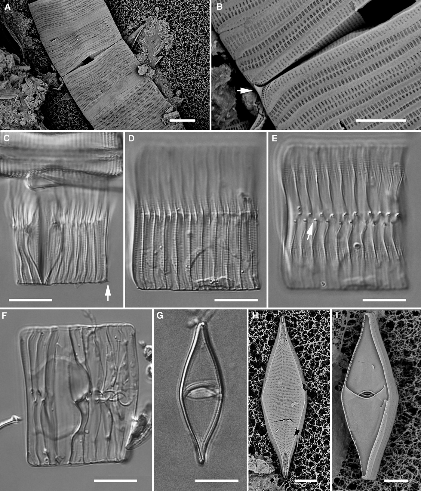
Microtabella rhombica n. sp., except (F) Microtabella interrupta. LM except (A, B, H, I) SEM: (A) joined chains, general view (GU52K-7); (B) detail of same cells, connection showing overlapping spines (arrow) (GU52K-7); (C) partial frustule (one valve on the right), showing spine (arrow) and distended copulae (GU52K-7); (D, E) copulae in girdle view, (D) focused on surface showing the striae, (E) median optical section showing the curled rim of each septum (GU52K-7). The need for two focal planes here and the lack of focus on half the frustule illustrates the transapical depth of the frustule; (F) Microtabella interrupta girdle view for comparison; (G) holotype specimen in valve view, showing the faint striae and pseudosternum (GU66G-5); (H) SEM view of valve showing striae, apical pore fields, and spines (GU66G-5); (I) several copulae in valve view showing septa (GU66G-5). Scale bars: A, C–E, 10 µm; B, F, G, 5 µm.

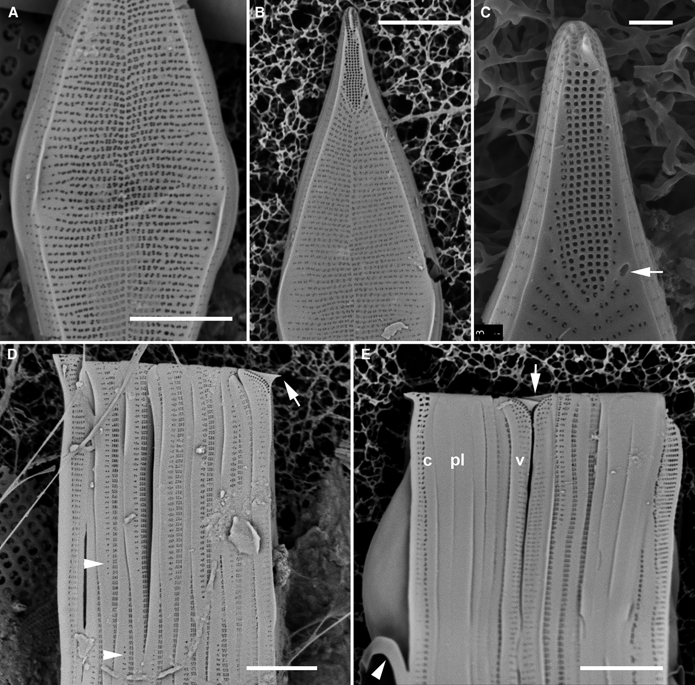
Microtabella rhombica n. sp., SEM: (A) centre portion of valve showing variation in extent to which striae extend toward the valve margin (GU66G-5); (B) apical portion of a valve showing slightly different pattern of stria extent and the apical pore field and spine (the spine seen edge-on) (GU66G-5); (C) detail of apex, showing pore field, spine and rimoportula opening (arrow) (GU52K-7); (D) portion of frustule in girdle view showing valve with apical spine (arrow) plus ~12 copulae, arrowheads indicating the alternating direction (two rows of areolae on the septate half) (GU66G-5); (E) portions of two joined frustules, showing overlapping spines (arrow); c = last copula with distended septum and ligule (arrowhead), pl = pleurae, v = valve (GU66G-5). Scale bars: A, B, D, E, 5 µm; C, 1 µm.
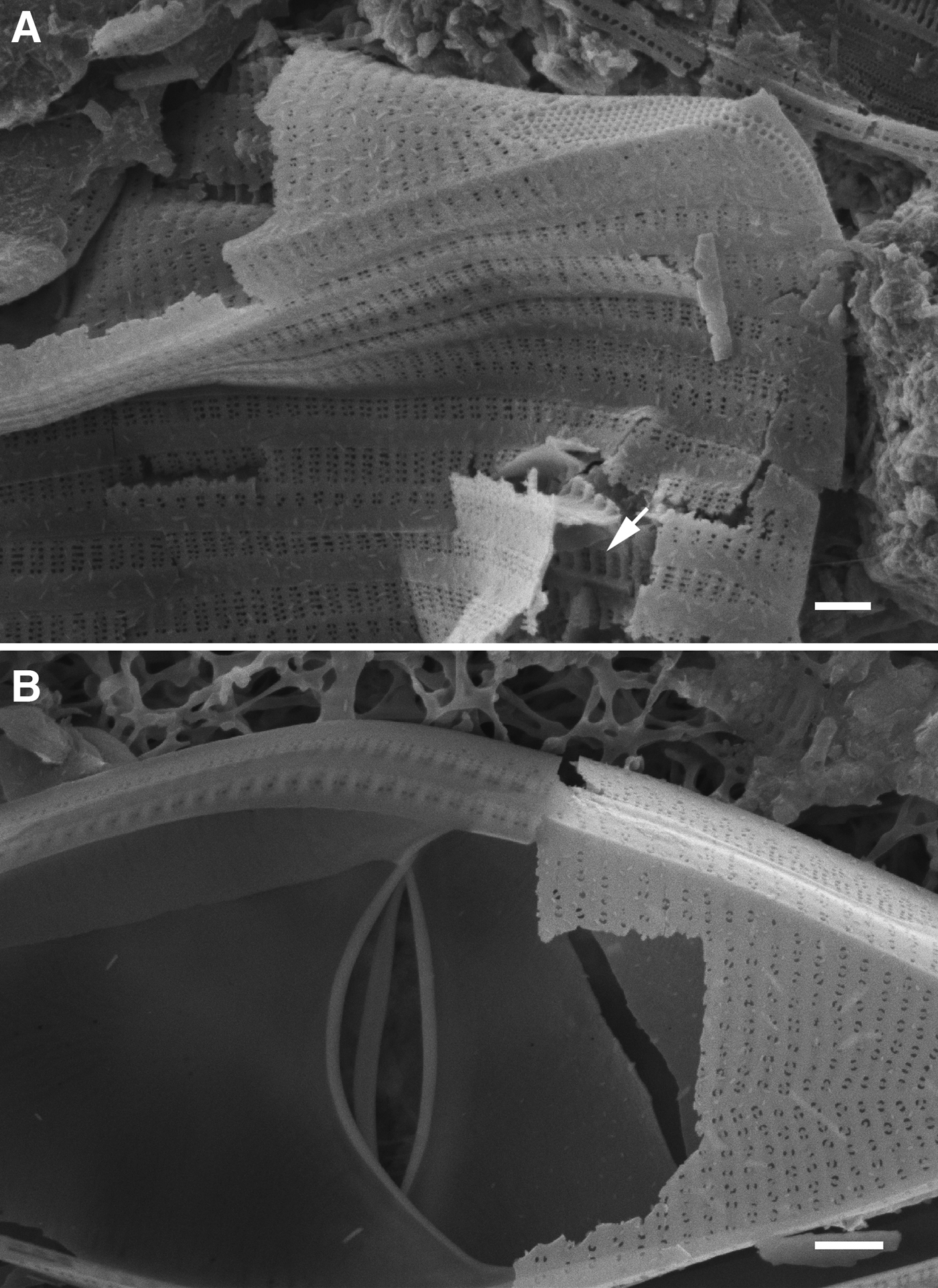
Microtabella rhombica n. sp., SEM (GU52K-7): (A) broken frustule showing biseriate striae on the copulae and thickened interstriae (arrow); (B) detail of broken frustule showing part of valve surface and the septa. Scale bars: 1 µm.
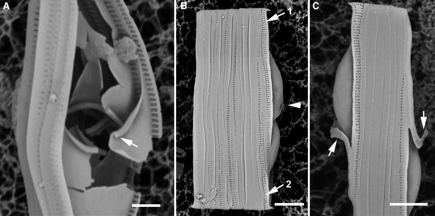
Microtabella rhombica n. sp., SEM: (A) broken copulae showing the recurved rims of the septa (arrow) (GU52K-7); (B) series of girdle bands including (from left to right) several copulae of one theca, the pleurae and the last two copulae of the other theca showing the distended septa of the last two copulae (1 = 2nd last, 2 = last) and the recurved rim on the 2nd-last copula (arrowhead) (GU66G-5); (C) series of girdle bands from a mature frustule, comprising the pleurae with the last-formed copula of each theca, showing the ligules (arrows) (GU66G-5). Scale bars: A, 2 µm; B, C, 5 µm.
DIAGNOSIS
Differing from Microtabella interrupta in the broader, rhomboidal valve, the presence of an apical spine dividing each apical pore field, the biseriate striae on the copulae and the recurved edge of the septum.
TYPE MATERIAL
Holotype: GU66G-5, slide 1694, specimen at 11.2 mm E, 11.3 mm S of reference mark (method of Sterrenburg et al., Reference Sterrenburg, Hamilton and Williams2012); deposited at California Academy of Sciences, accession # 627423, slide # 223040. Figure 1G shows the holotype specimen.
Type locality: GabGab II reef, Apra Harbor, Guam, ~15 m depth, collected 9 November 2014 by C.S. Lobban and M. Schefter. Associated with sparse algal turf in a territory of the farmer fish Plectroglyphidodon lacrymatus (Quoy & Gaimard, 1825); diatom community dominated by Biddulphiopsis membranacea (Cleve) von Stosch & Simonsen.
ETYMOLOGY
Latin: rhombic, rhomboidal, with reference to the valve shape.
ADDITIONAL MATERIALS EXAMINED
GU52K-7 (Outhouse Beach, Apra Harbor, Guam), 3 m depth, 3 May 2009; GU44Z-15 (GabGab reef, Apra Harbor, Guam), ~1 m depth, 20 June 2009.
MORPHOLOGY
Colonies ribbon-like, frustules joined by the valve faces (Figure 1A, B), not positively identified alive. Frustules tabular, with numerous septate copulae, of which the last two formed were distended (Figure 1C–E), and non-septate pleurae. Copulae with evident striae (more evident than in Microtabella interrupta; Figure 1C, D vs 1F). Valves were rhombic-lanceolate with acute apices, 24–36 µm long, 8–11 µm wide (Figures 1G–I & 2B). Striae, very faint in LM (Figure 1G), on both sides of an irregular pseudosternum, 31–32 in 10 µm,±parallel becoming strongly radiate towards the apical pore fields; central striae sometimes not reaching the valve margin (Figures 2A, B). Areolae closed by rotae with 2–4 small pores (Figures 2C & 3B). Apical pore fields extended back against the radiate striae (Figures 2B, C), pores ~60 transapical rows in 10 µm, the apical portion of the field divided by a prominent spine (Figures 1B & 2B–E), spines of adjacent cells in colonies overlapped (Figure 2E), two rows of pores extended onto the valve mantle alongside the spine. A rimoportula opening occurred at the end of the last stria, one at each apex on opposite sides of the pseudosternum (Figures 1H & 2B, C). No internal view of valve apices was found. Valve mantle sharply defined from the flat valve face by a slight ridge along the valve margin that extended as far as the apical spine (Figure 2A–C); striae along the valve mantle more or less continuous from those on the valve face.
Girdle bands: Valvocopula and several copulae all with septum starting past the middle of the valve and curving back to overlap the septa above and below with only a small central passage (Figures 1G, I & 3B); margin of the septum recurved toward the advalvar side (Figures 1D, 3B & 4A). Copulae open, the open and closed ends alternated (Figure 2D), two unequal rows of areolae, 26–27 in 10 µm, on the septate end, the advalvar row consisted of clusters of 6–8 pores (Figure 3A) and continued the full length of the copula wall; the abvalvar row, comprised clusters of 2–5 pores, diminished towards the middle of the copula. The last two copulae had distended septa (Figures 1C, 2E & 4B, C) and only a single series of striae along the whole length. Four non-porous pleurae occurred (Figures 2E & 4C).
DISCUSSION
Although I have live images of Microtabella from the sample that contained Microtabella rhombica, both species were present and I have not been able to confirm that any of those photographs are of Microtabella rhombica. Nevertheless, I infer that the plastids are confined to the space between the pair of distended septa of the epitheca and the last-formed septate copula of the hypotheca (or the hypovalve itself immediately after cell division), as in Microtabella interrupta. Microtabella rhombica is easily overlooked in LM because of its similarity to Microtabella interrupta, a widespread species which until now has been the only one with compartmentalized plastids and distended septa among a number of related ribbon-forming species (now in Hyalosira). The cell length range was 18–42 µm in Guam specimens of Microtabella interrupta versus 24–36 µm in Microtabella rhombica, and the range for Microtabella interrupta given by Navarro & Williams (Reference Navarro and Williams1991) was 15–60 µm. Thus the length ranges overlap and cannot be used to distinguish the species. The valves of Microtabella rhombica were so faintly marked that they were scarcely distinguishable from girdle bands in valve view, but they were distinctive in being twice as wide as those of Microtabella interrupta and distinctly rhombic versus lanceolate. In girdle view, copulae of Microtabella rhombica had more evident, slightly coarser areolae, 26–27 in 10 µm versus 30–35 in 10 µm in Microtabella interrupta, but the apical spines on the valves of Microtabella rhombica were not always visible. In SEM, valves of the two species were easily distinguished by the size of the apical pore field and the presence/absence of the apical spines. When only girdle bands were present, the two species were distinguished by the difference in the copular areola density and poration, and by the recurved edge of the septum in Microtabella rhombica, which in Microtabella interrupta is also thickened but flat.
In monotypic genera with strong and easily recognized characters, such as the pseudoseptum of Cyclophora tenuis (Ashworth et al., Reference Ashworth, Ruck, Lobban, Romanovicz and Theriot2012) or the necklace-like colony pattern of Bleakeleya notata (Lobban et al., Reference Lobban, Ashworth, Arai, Jordan and Theriot2011), small differences are easily overlooked and new species are found on closer inspection. In Microtabella, the distended septa are a strong feature, so that Microtabella rhombica (and perhaps other species undiscovered) may have been mistaken for Microtabella interrupta, which has been reported from many tropical areas (including Puerto Rico, Bahamas, Society Islands, Mahé and Guam; see checklist in Lobban et al., Reference Lobban, Schefter, Jordan, Arai, Sasaki, Theriot, Ashworth, Ruck and Pennesi2012).
ACKNOWLEDGEMENTS
My thanks to Matt Ashworth and Edward Theriot, University of Texas, Austin for assistance with high-resolution scanning electron microscopy there. The light and scanning electron microscopes in the University of Guam Microscopy Teaching & Research Laboratory were purchased with grants from the National Institute of General Medical Sciences, MORE Division, RISE Program. I also thank the commanding officers of Naval Base Guam for access to the study site at GabGab, and María Schefter for her continuous support and encouragement.