Introduction
Meiosis is a crucial event in oocyte development and maturation. Abnormal meiotic division in oocytes is the main cause of embryonic aneuploidy and early embryonic arrest (Webster and Schuh, Reference Webster and Schuh2017; Thomas et al., Reference Thomas, Cavazza and Schuh2021; Wu et al., Reference Wu, Li and Yang2021). In human-assisted reproduction therapy, the quality of oocytes is the key to the success or failure of in vitro fertilization. In the process of meiotic division, the spindle composed of microtubules is an extremely important structure. The correct separation of homologous chromosomes and sister chromatids under the traction of spindle microtubules is the key to ensuring normal chromosome number in daughter cells, which requires precise regulation by many factors. The microtubule-severing protein family is one of these key regulatory factors. (Vale, Reference Vale2000; Frickey and Lupas, Reference Frickey and Lupas2004).
In the mitotic division of somatic cells, spastin, one of the members of the microtubule-severing protein family, aggregates at the spindle poles and removes microtubule minus-end protective caps using the energy released by ATP hydrolysis. This exposes the free end to polymerases or depolymerases, causing dynamic changes in microtubule extension or shortening (Zhang et al., Reference Zhang, Rogers, Buster and Sharp2007; Casanova et al., Reference Casanova, Crobu, Blaineau, Bourgeois, Bastien and Pagès2009). Spastin can play a “nucleation” role by severing a long microtubule into several short segments, and then each free end exposed in each microtubule segment can produce a new long microtubule through polymerization, thereby increasing the microtubule density of the spindle (Vietri et al., Reference Vietri, Schink, Campsteijn, Wegner, Schultz, Christ, Thoresen, Brech, Raiborg and Stenmark2015).
Our previous research found that microtubule-cutting protein spastin aggregates on spindle microtubules in mouse oocytes. Oocytes lacking spastin exhibited significantly reduced spindle microtubule density. Abnormal spindle structure and function caused by lack of spastin resulted in a high proportion of chromosomal organization abnormalities and a decreased rate of first polar body extrusion (i.e. oocyte maturation defect). When the first polar body-extruded oocytes were fertilized by normal male mouse sperm and cultured in vitro, the cleavage and blastocyst rates of spastin-lacking oocytes were significantly lower (Jin et al., Reference Jin, Shou, Liu, Jiang, Shen, Cheng and Gao2022). We speculate that spastin can cut microtubules, dividing long microtubules into short segments, and under the action of microtubule polymerase, the short segments elongate into new microtubules, thereby increasing spindle microtubule density and promoting spindle assembly. In addition, the microtubule-severing activity of spastin may be regulated by collapsin response mediator protein 5 (CRMP5) (He et al., Reference He, Kooistra, Das, Oudejans, van Leen, Ziegler, Portegies, de Haan, van Regteren Altena, Stucchi, Altelaar, Wieser, Krieg, Hoogenraad and Harterink2020). The CRMP family belongs to microtubule-associated proteins and can regulate neuronal synapse growth and cell migration by promoting microtubule polymerization (Ji et al., Reference Ji, Zhang, Chen, Li, Yang, Cha, Zhang, Lin and Guo2018). As an important member of the CRMP family, CRMP5 can bind to microtubules and migrate along microtubules, promoting the function of microtubule-cutting protein spastin, while having no interaction with other members of the microtubule-severing protein family (Ji et al., Reference Ji, Zhang, Chen, Li, Yang, Cha, Zhang, Lin and Guo2018).
In neuronal cells, CRMP5 interacts with spastin on microtubules and they work together. Spastin can fragment long microtubule segments, leading to the formation of more small microtubule fragments, while CRMP5 can target specific positions on microtubules to facilitate microtubule polymerization. In mammalian oocyte meiosis, we found that CRMP5 interacts with spastin and knocking down CRMP5 results in reduced spindle microtubule density and maturation defects in oocytes (Jin et al., Reference Jin, Shou, Liu, Jiang, Shen, Cheng and Gao2022). The specific way in which CRMP5 regulates spastin function is still unknown.
Materials and methods
General chemicals, reagents, cells and animals
Unless otherwise specified, chemicals and reagents were purchased from Sigma-Aldrich; Merck KGaA. In total, 265 3-week-old female specific-pathogen-free ICR mice (weighing 18–20 g) were used in this study. The mice were obtained from Vital River Experimental Animal Technical Co., Ltd. Animals were housed at a temperature of 20–26°C and a humidity of 40–70% under a 12 h light/dark cycle. The mice were fed in feeding boxes, and the frequency of food replacement was two times a week, and the frequency of water bottle replacement was three times a week. All animal experiments were approved by the Animal Protection and Utilization Committee of Nanjing Medical University (Nanjing, China) and conducted in accordance with the institutional guidelines.
Antibodies
Mouse monoclonal anti-β-actin (cat. no. A5316–100) antibody was obtained from Sigma-Aldrich; Merck KGaA. Mouse monoclonal anti-spastin (A-4) (cat. no. sc-398264) and mouse monoclonal anti-β-tubulin (cat. no. sc-5274) antibodies were purchased from Santa Cruz Biotechnology, Inc. Human anti-centromere CREST antibody (cat. no. 15–234) was purchased from Antibodies Incorporated. Rabbit polyclonal anti-β-actin (cat. no. 30102ES40) was purchased from Yeasen Biotech Co., Ltd. Rabbit polyclonal anti-CRMP5 (cat. no. ab36203) was purchased from Abcam. Cy2-conjugated donkey anti-mouse IgG (code no. 715–225–150), rhodamine (TRITC)-conjugated donkey anti-goat IgG (code no. 705–025–147), and Alexa Fluor 647-conjugated donkey anti-human IgG (code no. 709–605–149) were purchased from Jackson ImmunoResearch Laboratories, Inc. Horseradish peroxidase (HRP)-conjugated rabbit anti-goat IgG (cat. no. 31402) and HRP-conjugated goat anti-mouse IgG (cat. no. 31430) were purchased from Invitrogen; Thermo Fisher Scientific, Inc.
Oocyte collection and culture
Immature oocytes at the germinal vesicle (GV) stage were obtained from the ovaries of 3-week-old female ICR mice. The mice were euthanized with carbon dioxide, and their ovaries were removed and placed in HEPES buffer (0.22 uM, pH = 7.4) containing 2.5 mM milrinone and 10% fetal bovine serum (Gibco, Thermo Fisher Scientific, Inc.). The follicles were punctured using injection acupuncture, and oocytes were collected from the cumulus–oocyte complexes by washing. Next, 50 separated shelled oocytes were placed in 100-μl culture medium drops, under the mineral oil in plastic containers (BD Biosciences). The medium was MEMþ [containing 0.01 mm EDTA, 0.23 mm Na-pyruvate, 0.2 mm penicillin/streptomycin, 3 mg/ml bovine serum albumin (BSA) and 20% FBS]. The oocytes were cultured at 37°C, 5% O2 and 5% CO2 in air. Before in vitro maturation (IVM), 2.5 nM milrinone was added to all media to prevent meiosis recovery.
siRNA production and microinjection
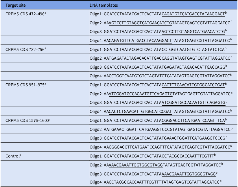
Table 1 displays the sequences of DNA templates used for generating siRNA targeting CRMP5, and a simulated sequence was used as a control template that does not bind specifically to any mRNA in the mouse genome. The templates for blocking four different coding regions (CDS) of CRMP5 mRNA were designed using BLOCK-iTTM RNAi Designer (http://rnaidesigner.invitrogen.com/rnaiexpress/) with some modifications. The specificity of the sequences was confirmed using a BLAST homology search (http://blast.ncbi). Briefly, two pairs of complementary single-stranded DNA oligonucleotides were annealed to generate two double-stranded DNA templates for each of the four CRMP5 CDS regions targeted. Two complementary single-stranded siRNAs were then synthesized based on the two templates and hybridized to produce the final double-stranded siRNA. Following standard phenol/chloroform/isopropanol precipitation, the siRNA was checked on an agarose gel for purity and calibrated before storage at −80°C. A ready-to-use siRNA mixture was generated by mixing equal molar amounts (5 μM final concentration) of siRNA targeting all four sites. Microinjection of siRNA into the cytoplasm of fully-grown immature oocytes was performed to knock down CRMP5 expression. Following injection, oocytes were arrested at the GV stage with 2.5 μM milrinone for 20 h and then cultured in milrinone-free M2 medium for maturation.
DNA oligos for siRNA production

a The numbers are the starting and ending position of the target sites in CRMP5 CDS (NM_001356948.1 in NCBI).
b Two pairs of DNA oligos are needed for each double-stranded siRNA. Oligo2 is complementary with oligo 1 except an ‘AA’ overhang at the 5′ end; oligo3 is complementary to oligo4 except an ‘AA’ overhang at the 5′ end. In each oligo, gene-specific sequences are underlined, other sequences are for recognition and binding by T7 RNA polymerase.
c Control siRNA does not target any mRNA sequence in mouse.
Immunofluorescence
Oocytes were briefly washed in PBS containing 0.05% polyvinylpyrrolidone (PVP) and subsequently permeabilized in 0.5% Triton X-100/PHEM (60 mM PIPES, 25 mM HEPES pH 6.9, 10 mM EGTA, 8 mM MgSO4) for 5 min. After three washes in PBS/PVP, the oocytes were fixed in 3.7% paraformaldehyde (PFA)/PHEM for 20 min at room temperature, followed by three washes (10 min each) in PBS/PVP. A blocking buffer containing 1% BSA/PHEM with 100 mM glycine was then added, and the oocytes were incubated overnight at 4°C with primary antibodies diluted in the blocking buffer. After three washes (10 min each) in PBS with 0.05% Tween-20 (PBST), the oocytes were incubated for 45 min in the blocking buffer with secondary antibodies (diluted 1:750 in blocking buffer) and then washed three times (10 min each) in PBST. Finally, the oocytes were stained with Hoechst 33258 (Sigma-Aldrich; Merck KGaA) at a concentration of 10 μg/ml at room temperature for 10 min. The oocytes were then mounted on slides using mounting medium (0.5% propyl gallate, 0.1 M Tris–HCl, pH 7.4, 88% glycerol), covered with a layer of glass (0.13–0.17 μm thickness) and held in place with two double-sticky bands (90-μm thick) between the slide and the cover slide to maintain their dimensions. The main antibodies were diluted as follows: anti-spastin (cat. no. sc-398264, Santa Cruz Biotechnology, Inc.), 1:200; anti-tubulin (cat. no. sc-5274, Santa Cruz Biotechnology, Inc.), 1:500; anti-human centromere CREST (cat. no. 15–234, Antibodies Incorporated), 1:500. The oocytes were examined using an Andor Revolution spinning disc confocal workstation (Oxford Instruments).
Western blotting
In total, 100 oocytes were dissolved in Laemmli sample buffer (Bio-Rad Laboratories, Inc.) containing protease inhibitors and boiled for 5 min, followed by subjecting to 10% SDS-PAGE. The separated protein was then transferred onto a polyvinylidene fluoride (PVDF) membrane, which was blocked with (Tris-buffered saline containing 0.05% Tween-20 (TBST) containing 5% nonfat dried milk for 1 h at room temperature. After blocking, the PVDF membrane was cut according to the molecular size marked by the marker, and then incubated overnight with the following primary antibodies at 4°C overnight: mouse monoclonal anti-β-actin (catalogue no. A5316–100) at a dilution of 1:1000 in blocking buffer (TBST containing 0.05% Tween-20), mouse monoclonal anti-spastin (A-4) (catalogue no. sc-398264) at a dilution of 1:500 in blocking buffer, and rabbit polyclonal anti-CRMP5 (catalogue no. ab36203) at a dilution of 1:2000 in blocking buffer. After washing in TBST, the membranes were incubated with HRP-conjugated rabbit anti-goat IgG or HRP-conjugated goat anti-mouse IgG at a dilution of 1:1000 in the blocking buffer for 1 h at room temperature, followed by treatment with ECL Plus Western Blotting Detection System (Vazyme). The data were analyzed using ImageJ software, version 1.8.0.
Image area measurement and fluorescence intensity
A focal series with 10 steps of 3 nm captured using a confocal laser microscope along the Z-axis was imported into ImageJ software, and superimposed using the Z-stacks function. The spindle outline was enclosed by a closed line chart selected using the ‘line chart’ icon in the ImageJ software. The average fluorescence intensity within the line graph was obtained by using the shortcut key ‘control+m’. The same method was used to measure the average fluorescence intensity of the background around the spindle. The average fluorescence intensity of spindle microtubules was obtained by subtracting the latter from the former. The measurements were repeated three times, and the average value was recorded.
Data analysis and statistics
All experiments were repeated at least three times. Image measurements were carried out using ImageJ software (National Institutes of Health), and results are presented as the mean ± standard error of the mean (SEM). One-way analysis of variance (ANOVA) with post hoc contrasts using the Student–Newman–Keuls test was used for multigroup comparisons. A P-value < 0.05 was considered significant.
Results
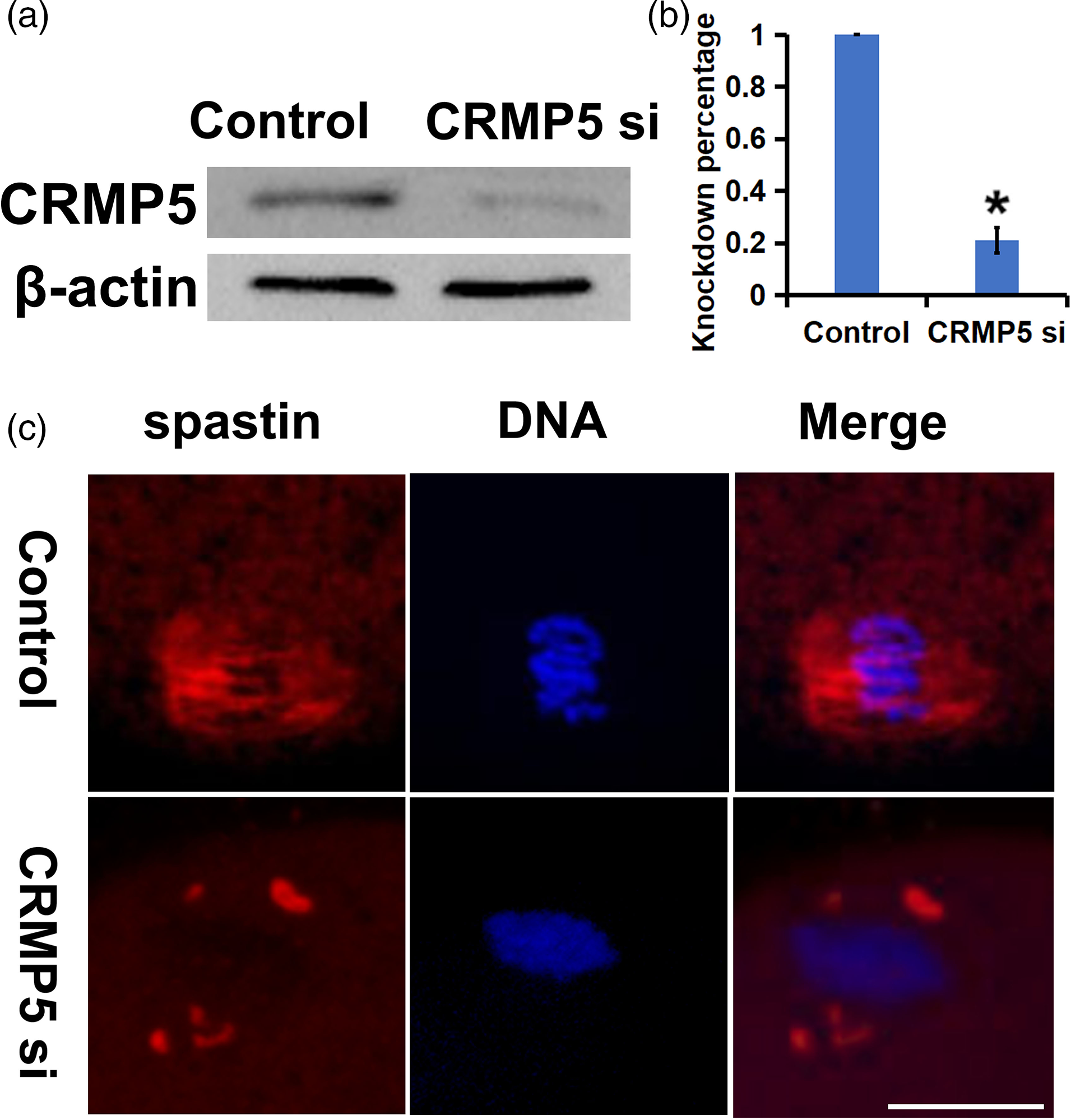
After knocking down CRMP5, the distribution of spastin migrates from spindle microtubules to spindle poles. In this study, to explore the function of CRMP5 in oocytes, we designed specific siRNAs and injected them into developmentally inhibited GV oocytes to knock down CRMP5. Western blot analysis showed that approximately 80% of the spastin was deleted (Figure 1a, b). We found that knocking down CRMP5 causes a change in spastin distribution in oocytes that developed in vitro to the MI phase, resulting in weakened spindle localization and a significant increase in accumulation at spindle poles (Figure 1c). Therefore, we propose the hypothesis that CRMP5 can bind to spastin in oocyte cells, promote its migration from spindle poles to spindle and play a role in spindle assembly by severing microtubules.
After knocking down CRMP5, spastin migrates from spindle microtubules to spindle poles. (a, b) Western blot and related statistical analysis showed that the expression of spastin decreased by ∼80% under the specific siRNA. β-Actin was used as a control; 100 oocytes were used in each group. Three independent experiments were performed for each result. (c) Immunofluorescence staining revealed that, in the control group, spastin was distributed in the meiotic spindle in oocyte cells while, in the CRMP5 knockdown group, spastin was localized to the spindle poles. Spastin is shown in red and DNA is in blue. Scale bar, 20 μm. *P < 0.05 indicates significant differences.

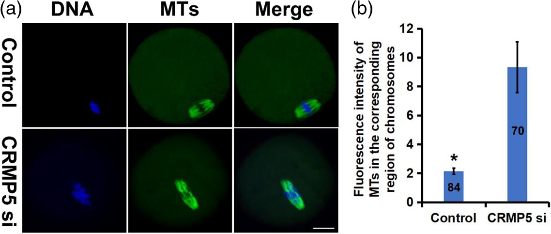
Knocking down CRMP5 results in an increase in microtubule density in the central region of the spindle in MI phase oocytes. In this study, we found that knocking down CRMP5 in oocytes and developing to the MI phase in vitro led to a significant increase in the fluorescence intensity of microtubules in the central region of the spindle (Figure 2a). This visual impression was also confirmed by statistical analysis of the data (Figure 2b). As knocking down CRMP5 causes spastin to aggregate at spindle poles and decreases its distribution near chromosomes, we speculated that the loss of spastin near chromosomes may result in abnormal activity of spindle microtubules in this region.
Knocking down CRMP5 results in an increase in microtubule density in the central region of the spindle in MI phase oocytes. (a) Immunofluorescence shows that, compared with the control group, the spindle microtubule fluorescence intensity in the corresponding region of the chromosome was significantly increased in MI phase oocytes knockdown with CRMP5. (b) Measurement and statistics of the fluorescence intensity of MTs in the corresponding region of chromosomes in the CRMP5 knockdown group and control group MI phase oocytes; 100 oocytes were used in each group. Three independent experiments were performed for each result. Tubulin is displayed in green, and DNA is in blue. Scale bar, 20 μm. *P < 0.05 indicates significant differences.

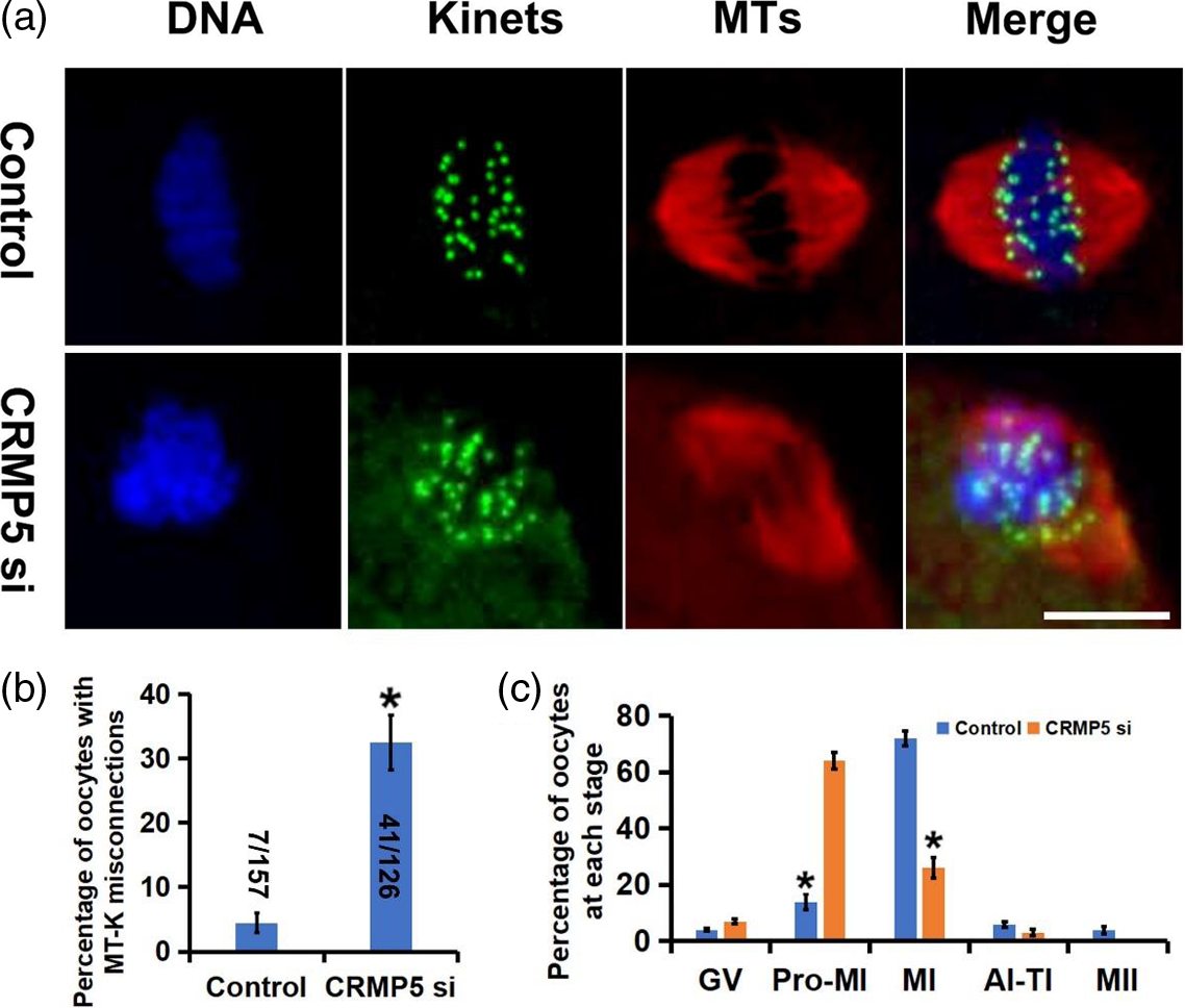
Knocking down CRMP5 can lead to microtubule–kinetochore misconnections in MI phase oocytes. Immunofluorescence staining showed that the microtubule signals of the MI-stage oocyte spindle were located on the outside of the kinetochore signals, while in the CRMP5-knockdown group, some microtubule signals were located between the kinetochore signals, suggesting incorrect microtubule–kinetochore attachments (Figure 3a). The proportion of oocytes with these erroneous attachments was significantly increased in the CRMP5-knockdown group (Figure 3b). Therefore, we speculated that spastin near chromosomes may correct microtubule–kinetochore misconnections by severing microtubules. Knocking down CRMP5 reduces the distribution of spastin near chromosomes, leading to unrepaired microtubule–kinetochore misconnections and resulting in meiotic abnormalities and chromosomal aneuploidy. In addition, we found that after 8 h of in vitro culture, the proportion of MI phase oocytes with the normal chromosomal arrangement in the CRMP5 group was significantly lower compared with that in the control group, while suspected prometaphase oocytes were significantly higher compared with that in the control group (Figure 3c). We could not rule out the abnormal distribution of chromosomes caused by spindle assembly abnormalities.
Knocking down CRMP5 can lead to microtubule–kinetochore misconnections in MI phase oocytes. (a) Immunofluorescence shows that the microtubule signals of the control group oocytes were located on the outside of the kinetochore signals while, in the CRMP5-knockdown group, some microtubule signals were located between the kinetochore signals. (b) Statistical analysis shows that the proportion of oocytes with these erroneous attachments was significantly increased in the CRMP5-knockdown group. The number of oocytes counted for each group is recorded in the bar chart. Three independent experiments were performed for each result. (c) After 8 h of in vitro development, the proportion of oocytes in the control group and CRMP5 knockdown group at each stage was determined. It can be seen that the proportion of pro-MI phase in the CRMP5 knockdown group significantly increases, while the proportion of MI phase significantly decreases; 100 oocytes were used in each group. Three independent experiments were performed for each result. Kinets refers to the kinetochore. 1pb refers to the first polar body of MII. Tubulin is displayed in red, kinetochores are shown in green, and DNA is in blue. Scale bar, 20 μm. *P < 0.05 indicates significant differences.

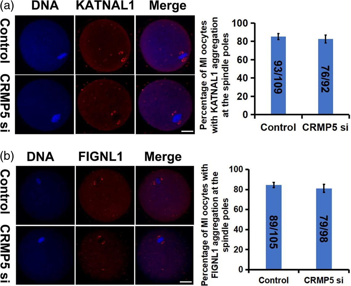
CRMP5 does not affect the distribution of KATNAL1 or FIGNL1. Notably, our prior investigations demonstrated that KATNAL1 and FIGNL1, as microtubule-severing proteins, are predominantly enriched at spindle poles where they facilitate spindle microtubule density augmentation and promote spindle assembly by inducing microtubule severing near the spindle poles (Gao et al., Reference Gao, Xu, Jin, Ying and Liu2019; Shou et al., Reference Shou, Jin, Yu, Lai, Wu and Gao2022). The immunofluorescence results showed that, when CRMP5 was specifically knocked down using siRNA, KATNAL1 (Figure 4a) and FIGNL1 (Figure 4b) still localized to the poles of the oocyte spindle with no significant change compared with the control group, indicating that CRMP5 did not alter the distribution of KATNAL1 and FIGNL1. Based on the previous results, we suggested that in the absence of CRMP5, microtubule-severing proteins tend to accumulate at the spindle poles, and spastin can be transported from the spindle poles with the assistance of CRMP5.
CRMP5 does not affect the distribution of KATNAL1 or FIGNL1. (a) Immunofluorescence and related statistical analysis showed that, when CRMP5 was knocked down, KATNAL1 localized to the poles of the oocyte spindle with no significant change compared with the control group. (b) FIGNL1 localized to the poles of the oocyte spindle with no significant change when CRMP5 was knocked down. The relevant statistical data also illustrate this point. The number of oocytes counted for each group is recorded in the bar chart. Three independent experiments were performed for each result. Scale bars, 20 μm. *P < 0.05 indicates significant differences.

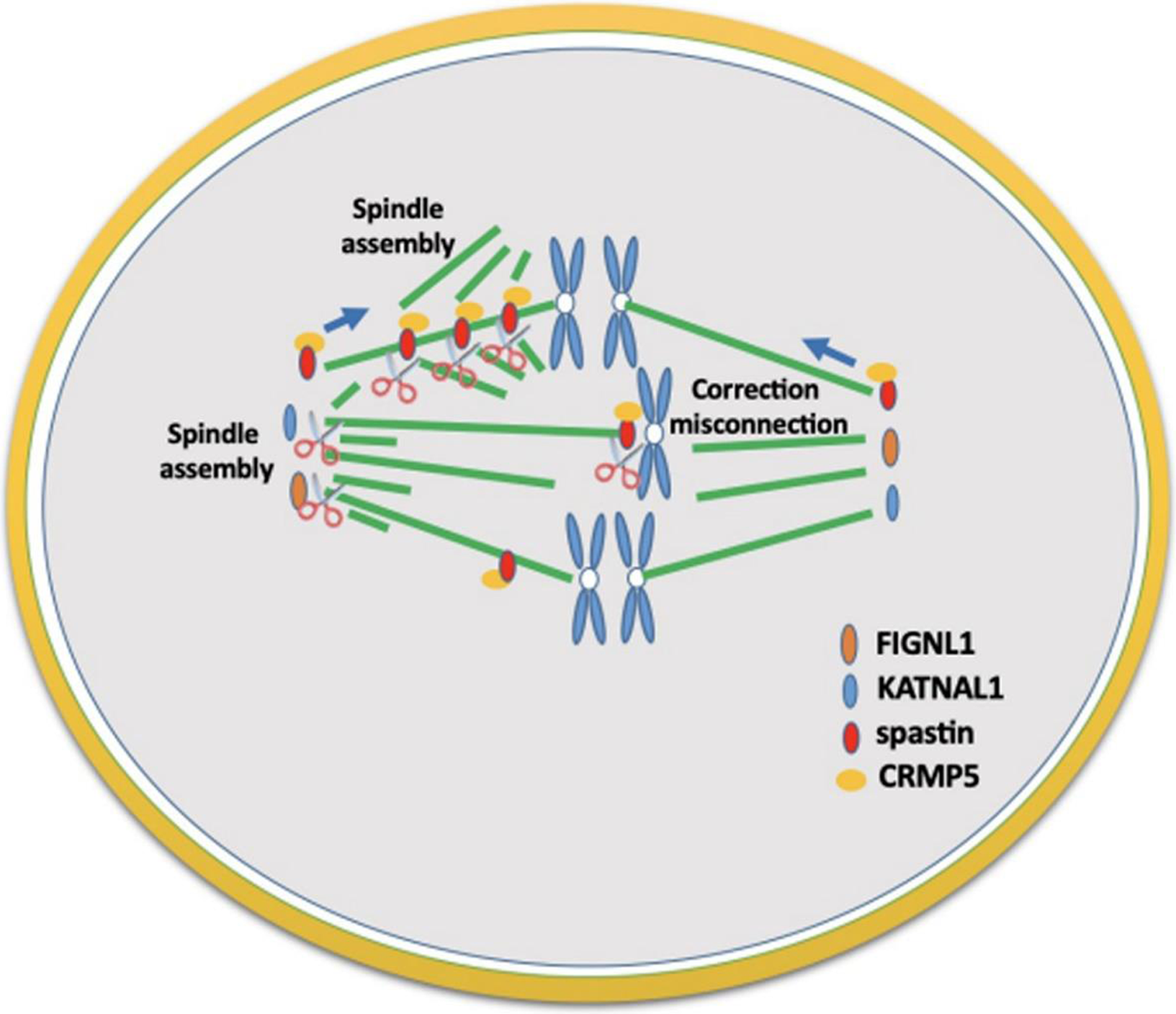
Possible model of spastin localization regulation in oocyte meiosis. In mouse oocyte cells, three microtubule-severing proteins, spastin, KATNAL1, and FIGNL1, tend to accumulate at spindle poles. CRMP5 can bind to spastin and promote its migration from spindle poles to spindle microtubules, allowing spastin to exert its microtubule-severing and nucleation activities throughout the spindle, generate more microtubule fragments, increase spindle microtubule density, and promote spindle assembly. Spastin distributed near chromosomes can correct kinetochore–microtubule misconnections, ensure the correct separation of homologous chromosomes, and avoid chromosomal aneuploidy in oocyte cells (Figure 5). The cooperation of these three microtubule-severing proteins promotes spindle assembly, corrects microtubule–kinetochore misconnections, and ensures proper oocyte meiosis and chromosome number.
Possible model of spastin localization regulation in oocyte meiosis. In mouse oocytes, three microtubule-severing proteins, spastin, KATNAL1, and FIGNL1, tend to accumulate at spindle poles. CRMP5 can bind to spastin and promote its migration from spindle poles to spindle microtubules, allowing spastin to exert its microtubule-severing and nucleation activities throughout the spindle, generate more microtubule fragments, increase spindle microtubule density, and promote spindle assembly. Spastin distributed near chromosomes can correct kinetochore–microtubule misconnections, ensure the correct separation of homologous chromosomes, and avoid chromosomal aneuploidy in oocytes.

Discussion
This study demonstrates well that the knockdown of CRMP5 led to incorrect distribution of spastin, disordered chromosome arrangement and incorrect microtubule–kinetochore connections in MI-stage oocytes. Combining the microtubule-severing effect of spastin, it was hypothesized that the knockdown of CRMP5 and aberrant distribution of spastin may contribute to erroneous chromosome segregation. The disrupted chromosome distribution in MI oocytes could be attributed to abnormal spindle assembly or incorrect chromosome separation, warranting further investigation. Chromosomal aneuploidy is the main reason for embryonic arrest (Webster and Schuh, Reference Webster and Schuh2017; Thomas et al., Reference Thomas, Cavazza and Schuh2021). Multiple studies have shown that microtubule-severing proteins play important roles in meiosis and reproductive ability. For example, katanin p80, which targets katanin 60, has been shown to be important in spindle assembly and cytokinesis during male meiosis (Pleuger et al., Reference Pleuger, Fietz, Hartmann, Weidner, Kliesch, O’Bryan, Dorresteijn and Bergmann2016). In lower organisms, the role and regulation of katanin in meiosis have been well studied (Pleuger et al., Reference Pleuger, Fietz, Hartmann, Weidner, Kliesch, O’Bryan, Dorresteijn and Bergmann2016).
As for the role of microtubule-severing proteins (MTSP) in mammalian oocyte cells, our three previous studies have all confirmed their importance. KATNAL1 and FIGNL1, two MTSP members that are dominantly expressed in mouse oocyte cells, both accumulate at spindle poles (Gao et al., Reference Gao, Xu, Jin, Ying and Liu2019; Shou et al., Reference Shou, Jin, Yu, Lai, Wu and Gao2022). The absence of these two proteins led to a decrease in spindle microtubule density and oocyte maturation disorders, suggesting that they sever microtubules and promote spindle assembly through nucleation around spindle poles (Gao et al., Reference Gao, Xu, Jin, Ying and Liu2019; Shou et al., Reference Shou, Jin, Yu, Lai, Wu and Gao2022). Spastin, another MTSP member, is distributed throughout the spindle microtubules (Jin et al., Reference Jin, Shou, Liu, Jiang, Shen, Cheng and Gao2022). Knocking down spastin led to a decrease in spindle volume and microtubule density, resulting in a lower rate of first polar body extrusion, fertilization abnormalities, and early embryonic development disorders (Jin et al., Reference Jin, Shou, Liu, Jiang, Shen, Cheng and Gao2022). Spastin may sever microtubules throughout the spindle and promote spindle assembly, a process that may be related to CRMP5 (Jin et al., Reference Jin, Shou, Liu, Jiang, Shen, Cheng and Gao2022). Studies on neurons have shown that CRMP5 can bind to the 59th amino acid residue (N-terminus of AAA domain, called the microtubule-binding domain, MTBD) of spastin, providing structural support and enhancing its microtubule-severing activity, and promoting neurite extension and neuronal branching (Ji et al., Reference Ji, Zhang, Chen, Li, Yang, Cha, Zhang, Lin and Guo2018). In mouse oocyte cells, our previous co-immunoprecipitation (Co-IP) experiment confirmed the interaction of CRMP5 and spastin, and immunofluorescence showed that they co-localize on spindle microtubules (Jin et al., Reference Jin, Shou, Liu, Jiang, Shen, Cheng and Gao2022). Knocking down CRMP5 led to a decrease in spindle microtubule density, oocyte maturation disorders, and fertilization abnormalities, similar to spastin deficiency (Jin et al., Reference Jin, Shou, Liu, Jiang, Shen, Cheng and Gao2022). Overexpression of spastin can partially rescue the phenotypic loss caused by CRMP5 knockdown (Jin et al., Reference Jin, Shou, Liu, Jiang, Shen, Cheng and Gao2022). This study shows that knocking down CRMP5 can cause a change in the localization of spastin, weaken its accumulation on the spindle, and significantly increase its accumulation at spindle poles. Based on the characteristics of spastin binding to microtubules and migrating along them with the help of CRMP5 binding, we speculated that, under the guidance of CRMP5 binding, spastin is distributed from spindle poles to spindle microtubules, promoting its microtubule-severing activity throughout the spindle, increasing microtubule density and promoting spindle assembly. In addition, spastin near chromosomes can correct microtubule–kinetochore misconnections by severing microtubules, ensuring proper separation of homologous chromosomes.
In the context of the cell cycle, microtubule-severing proteins are believed to play three distinct roles, namely exposing microtubule ends to facilitate successive depolymerization, nucleating microtubules at concentrated sites, and removing improperly attached microtubules from kinetochores. The first role has been established by numerous studies (Buster et al., Reference Buster, McNally and McNally2002; Zhang et al., Reference Zhang, Grode, Stewman, Diaz-Valencia, Liebling, Rath, Riera, Currie, Buster, Asenjo, Sosa, Ross, Ma, Rogers and Sharp2011; Sharp and Ross, Reference Sharp and Ross2012). The activity level of katanin is known to determine the difference in spindle length between X. tropicalis and X. laevis (Loughlin et al., Reference Loughlin, Wilbur, McNally, Nédélec and Heald2011). The second role is also supported by several findings (Srayko et al., Reference Srayko, O’Toole, Hyman and Müller-Reichert2006; Nakamura et al., Reference Nakamura, Ehrhardt and Hashimoto2010; Nakamura, Reference Nakamura2015). KATNAL1 localizes particularly to spindle poles in human mitotic U2OS cells; therefore knocking down KATNAL1 significantly reduces the intensity of microtubules at the poles (Sonbuchner et al., Reference Sonbuchner, Rath and Sharp2010). A similar function was observed in C. elegans meiosis, where mei-1, the homolog of katanin 60, concentrates at chromosomes and its loss causes a significant reduction in microtubules terminating at chromosomes (Srayko et al., Reference Srayko, Buster, Bazirgan, McNally and Mains2000). The third role, conversely, lacks direct evidence, but it has been suggested that MCAK, a kinesin-13 microtubule depolymerase, releases improperly attached microtubules and is regulated by Aurora B (Knowlton et al., Reference Knowlton, Lan and Stukenberg2006; Parra et al., Reference Parra, Gómez, Viera, Page, Calvente, Wordeman, Rufas and Suja2006). As disaggregase is believed to work with microtubule-severing proteins during mitosis and interphase, it is reasonable to suggest that spastin distributed near chromosomes in oocyte meiosis can use its severing activity to remove subset attachments and ensure bivalent attachments.
In addition, due to the significantly lower proportion of MI phase oocytes with the normal chromosome arrangement and a higher proportion of suspected prometaphase oocytes after CRMP5 knockout compared with the control group, based on current experimental results alone, we could not completely rule out the abnormal distribution of chromosomes caused by spindle assembly abnormalities. Further experiments are needed to confirm that CRMP5 avoids incorrect chromosome separation by correcting microtubule–kinetochore connections.
In summary, in mouse oocyte cells, the three microtubule-severing proteins, spastin, KATNAL1, and FIGNL1, tend to accumulate at spindle poles, exerting their microtubule-severing activity at the minus end of microtubules and promoting spindle assembly through nucleation around spindle poles, thereby ensuring successful meiosis and oocyte maturation. Spastin may be distributed from spindle poles to spindle microtubules, with the help of CRMP5 binding it promotes microtubule-severing throughout the spindle, increases microtubule density, and corrects microtubule–kinetochore misconnections, ensuring proper separation of homologous chromosomes and avoiding chromosomal aneuploidy. This discovery provides new insight into the cooperation and regulation mechanisms of microtubule-severing proteins in oocyte cells and provides important supplements to the mechanism of proper chromosome segregation in oocyte cells.
Acknowledgements
We thank Dr Zhang (Hangzhou Medical College) for providing plasmids.
Author contributions
ZJ and LLG conceived and designed the experiments. LLG, ZJ, ZCZ, CYX, QRL and MQL performed the experiments. LLG analyzed the data. ZJ, ZCZ, CYX, QRL, MQL and LLG contributed the reagents/materials/ analysis tools. ZJ and LLG wrote the paper. All authors read and approved the manuscript and agree to be accountable for all aspects of the research in ensuring that the accuracy or integrity of any part of the work is appropriately investigated and resolved.
Availability of data and material
The datasets used and/or analyzed during the current study are available from the corresponding author on reasonable request.
Ethics approval and consent to participate
All animal experiments were approved by the Animal Care and Use Committee of Nanjing Medical University (Nanjing, China) and performed in accordance with institutional guidelines.
Consent for publication
Not applicable.
Competing interests
The authors declare that they have no competing interests.
Funding
This present study was supported by the National Natural Science Foundation of China (82001539) and the Zhejiang Province Medical and Health Research Project Innovation Talent Support Plan (2021RC001). Zhejiang Provincial Foundation Committee provided financial support for this research and the preparation of the article. The funding bodies played no role in the design of the study and collection, analysis, and interpretation of data, and in writing the manuscript.