Introduction
There is a limited understanding about the consequences of viral infection during pregnancy for the mother and the fetus. The recent appearance and fast spreading of the new severe acute respiratory syndrome-coronavirus 2 (SARS-CoV-2) highlight the importance to understand the effects of viral infections during pregnancy. Pregnant women are considered a special group which may have higher risks to be affected by infectious agentsReference Mor and Cardenas1. Viral infections effects during pregnancy are diverse, they range from pregnancy loss by spontaneous abortion to congenital viral syndromesReference Silasi, Cardenas, Racicot, Kwon, Aldo and Mor2. There is an urgent and very relevant need to consolidate the knowledge that we have regarding the consequences of maternal viral infection over the offspring and maternal outcome given that humans are highly exposed to viral infection and there are limited tools to protect the mother and fetus during pandemics. Here, we review the effects of viral infections, with emphasis in SARS-CoV-2, during pregnancy over the mother and the offspring.
SARS-Cov-2 infection during pregnancy: what do we know so far
SARS-CoV-2 causes a respiratory syndrome known as COVID-19Reference Liu, Wang, Zhao, Kwak-Kim, Mor and Liao3–Reference Canedo-Marroquín, Saavedra, Andrade, Berrios, Rodríguez-Guilarte, Opazo, Riedel and Kalergis5, to date, present in almost in all countries around the world with the Americas presenting the higher number of cases compared to Europe and China6. This virus has a high contagiousness ratio, 2–4 new cases can appear directly from a single caseReference Bar-On, Flamholz, Phillips and Milo7. SARS-CoV-2 can be transmitted by person-to-person through saliva dropsReference Bar-On, Flamholz, Phillips and Milo7. Its infection induces mild upper respiratory tract infection, fever, cough, and lower respiratory tract infection causing pneumonia with acute respiratory distress syndrome that can be life threateningReference Singhal8. In general, COVID-19 can affect all age groups including newbornsReference Yu and Chen9. There are two groups with severe risks: older adults10 and people with previous medical conditions11. Other group that should be considered under risk is pregnant women. During pregnancy, women present an alteration of the immune system with a high tolerance and are susceptibility to infectionReference Mor, Aldo and Alvero12. In relation to SARS-CoV-2, one of four positive diagnosed pregnant women between 15 and 49 years old were hospitalizedReference Delahoy, Whitaker and O’Halloran13. A few studies realized so far showed that they do not develop a more severe illness than not pregnant womenReference Delahoy, Whitaker and O’Halloran13–Reference Segars, Katler and Mcqueen15, most of them were at the third trimester (87%) compared to the 13% were at first and second trimester of pregnancyReference Delahoy, Whitaker and O’Halloran13,Reference Li, Han and Peng14 , this high difference could be explained due to delivery date and not by COVID-19 and more further analyses needs to be done. The principal symptomatology of pregnant women with COVID-19 was cough and feverReference Li, Han and Peng14. The study by Delahoy et al. reported 598 hospitalized pregnant women, of which 54.5% of them were asymptomaticReference Delahoy, Whitaker and O’Halloran13. It has been observed that birth can trigger the development of a severe COVID-19 even if is premature birthReference Segars, Katler and Mcqueen15. In Wuhan, it has been reported that, after delivery, 6 of 118 patients develop a more severe COVID-19, this is suggested by postpartum changes as increased of volume circulationReference Chen, Li and Zheng16.
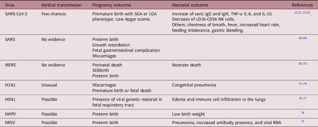
Yang et al., studied 27 COVID-19 diagnosed and confirmed pregnant women in Wuhan, China. In total, 3 of the 18 polymerase chain reaction (PCR)-confirmed patients present premature rupture of fetal membranes with one of them presenting preterm premature rupture of fetal membranes at gestational week 30. The offspring present IgG and IgM even though they have a negative PCR for SARS-CoV-2 infectionReference Yang, Hu, Zhan, Yang and Xiong17. This suggests that the newborns were not infected but they are developing an inflammatory response. Immunoglobulins are an important parameter used to evaluate the immune statusReference Özdemir, Emsen, Türk Daği and Artaç18. The origin of the high levels of immunoglobulins is not clear. It is possible that IgG can be transferred from the mother to the fetus. However, IgM does not cross the placental barrierReference Palmeira, Quinello, Ucia Silveira-Lessa, Zago and Carneiro-Sampaio19. Therefore, the increase in IgM is an interesting fact that needs to be clarified. In this sense, there is one work that intends to analyze this issue; given that, there are high rates of false positivity for the IgM determination this analysis requires more clinical evidenceReference Lamouroux, Attie-Bitach, Martinovic, Leruez-Ville and Ville20. This is suggestive of an in-utero infectionReference Fenizia, Biasin and Cetin21. Evidence related to a pro-inflammatory state was also shown by Dong et al., where a newborn girl which presented increased levels of IgM and IgG together with increased levels IL-6 and IL-10 with no pulmonary signs and negative results for SARS-CoV-2 PCR test. This elevated IgM phenotype was sustained over time being observed until 12 days after birthReference Dong, Tian and He22. A study performed in six neonates from SARS-CoV-2-infected mothers showed that all were born by C-section and had high levels of IgG, IgM, and cytokines particularly IL-6. Consistently with previous observations, all the newborns were negative in the PCR testReference Zeng, Xu and Fan23. Another study described neonatal effects observed in the offspring from SARS-CoV-2-infected mothers. Here, neonates with a negative PCR result present premature born accompanied with normal, small for gestational age (SGA), or large for gestational age (LGA) phenotypes and in some cases lower Apgar scoresReference Zeng, Xu and Fan23–Reference Trippella, Ciarcià and Ferrari26. Symptoms as shortness of breath, fever, increased heart rate, feeding intolerance, gastric bleedingReference Zhu, Wang and Fang24, rashes, and facial ulcerations have also been describedReference Chen, Peng and Wang27 showing that SARS-CoV-2 infection during pregnancy may generate several adverse effects in the offspring. There is not complete agreement about the kind of effects of SARS-CoV-2 infection over the offspring during pregnancy. For example the analysis of Yu et al., (2020) showed that after strict control measures no clinical symptoms were observed in four of seven newborns and only one of the rest presented a positive PCR test 36 h after birth and presented only mild symptoms and quickly recoveredReference Yu, Li and Kang28. A deeper analysis on the SARS-CoV-2 effects on the offspring’s immune system was performed by Liu et al., (2020). This study showed a decrease of CD16-CD56 natural killer cells (NK) together with increased levels of IgG, IgM, IL-6, IL-10, and TNF-α in neonates born from mothers infected with SARS-CoV-2 but when the neonates were tested by PCR for SARS-CoV-2 they were negativeReference Liu, Zheng and Yang29. There are few information regarding how neonates could be or not be infected by the virus or if this can be transmitted vertically by the mother. Lesser or no evidence showing that the fetus can clear the virus. The most recent evidences suggest in-utero infection and there is evidence of the presence of viral genetic material at the umbilical cord and also in the newborn nasopharyngeal tract and this issue seems to be dependent of the disease severity because this phenotype is only observed in pregnant patients that develop a severe form of COVID-19Reference Fenizia, Biasin and Cetin21.
An important increase of IL-6 was observed in one of the analyzed neonates who develop a severe necrotizing enterocolitis which is consistent with other author that observed gastric bleedingReference Zhu, Wang and Fang24,Reference Li, Han and Peng25,Reference Yu, Li and Kang28 . Increased levels of IL-6 have been proposed as an earlier marker for neonatal sepsis in newbornsReference Delanghe and Speeckaert30. A similar observation was made in neonates from human immunodeficiency virus (HIV) and hepatitis B-infected mothers which also present an increased blood levels of pro-inflammatory cytokines such as IL-6, among others, altering the immune response of the fetusReference Lohman-Payne, Gabriel and Park31,Reference Yockey and Iwasaki32 . CD16-CD56 NK cells are the first line of defense by lysing viral-infected or tumor cells which are present in higher levels in the neonateReference Lee and Lin33. A similar phenotype is observed in non-infected infants born from HIV infected mothers, here samples taken at birth showed a decreased proportion of NK cellsReference Smith, Jalbert and de Almeida34. NK cells are able to modulate the adaptative immune response, playing a key role in controlling viral infections so alterations in NK cells could alter the anti-viral defense in the neonateReference Ackermann, Verleden and Kuehnel35.
Regarding the effects of SARS-CoV-2 infection, it has been observed in autopsies from patients died from COVID-19, the presence of SARS-CoV-2 inside the endothelial cellsReference Ackermann, Verleden and Kuehnel35,Reference Varga, Flammer and Steiger36 , which can be a predictable result considering the presence of ACE2 in endothelium cellsReference Hoffmann, Kleine-Weber and Schroeder37, but it has also been observed that ACE2 expression on infected patient’s endothelial cells is higher than control healthy patientsReference Ackermann, Verleden and Kuehnel35. This phenotype is associated with vasculitis in tissues like skin, liver, kidney, among othersReference Varga, Flammer and Steiger36,Reference Colmenero, Santonja and Alonso-Riaño38 . Moreover, pulmonary tissue presents an increased angiogenesisReference Ackermann, Verleden and Kuehnel35 that can be explained by two mechanisms: one of them is growing by vessels sprouting where capillaries start to extend and the second growing by intussusceptive angiogenesis which is when one vessel is divided to form two new vessels, this phenotype has been observed in patients with chronic lung diseaseReference Voelkel, Douglas and Nicolls39.
The immune system at the maternal–fetal interface and SARS-CoV-2
Emerging viral diseases represent a significant risk for pregnant women and their infants. To avoid detrimental effects, the maternal–fetal interface which is a highly specialized tissue, contains several cell types that contribute to the development of the fetus and protect against potentially detrimental microorganismsReference Racicot and Mor40. The maternal–fetal interface is composed by maternal derived decidua and fetal derived placenta. The maternal side contains a variety of immune cells like NK cells, macrophages, neutrophils, dendritic cells (DC), and T cells and T cell subsets such as regulatory cells (Tregs) meanwhile the fetal side contains fetal blood vessels surrounded by fibroblasts and fetal macrophages, cytotrophoblasts, and syncytiotrophoblasts which are in contact with maternal bloodReference Shivhare, Bulmer and Lash41. NK cells contribute to endometrium preparation for pregnancy and implantation and are able to produce growth factors and cytokines which contribute to increase maternal blood availabilityReference Ander, Diamond and Coyne42. There is evidence in the literature that support that macrophages contribute positively to angiogenesis and tissue remodeling as have been observed by in vitro analysesReference Hazan, Smith and Jones43. Moreover, it has been thought that macrophages have a protective roleReference Vishnyakova, Elchaninov, Fatkhudinov and Sukhikh44 for their ability to respond to micro-environmental changes. DCs migrate after an inflammatory stimulus to the lymph nodes where they can present antigens to naïve T cells modulating T cell accumulation and functionReference Macri, Pang, Patton and O’Keeffe45. The density of DCs in the maternal–fetal interface is lowReference Tagliani and Erlebacher46 and there are thought to work as a surveillance system for pathogensReference Erlebacher47. Regarding T cells, Tregs proportion is maintained through almost all pregnancyReference Sasaki, Darmochwal-Kolarz and Suzuki48 and the majority of T cells found at the maternal–fetal interface are CD4+ and CD8+ T cellsReference Erlebacher47. It has been observed that a diminished number or alteration on Treg function, altering the balance Treg/Th17, can be involved in events such as abortion and infertility giving them a protective role during pregnancyReference Ander, Diamond and Coyne42,Reference Erlebacher47 . Neutrophils are activated in a pro-inflammatory state ready if there is a local infectionReference Solano49. There is another group of immune cells present called unconventional T cells that are involved in pathogen defenseReference Solano49. All these together conform a well-controlled system that gives the fetus the optimal conditions for development and regulation of the immune response. Pregnancy is a unique immunological state, which is in constant adaptation to growth and development giving to the pregnant woman a characteristic immune response which can be a potential risk for viral infection, particularly for SARS-CoV-2Reference Liu, Wang, Zhao, Kwak-Kim, Mor and Liao3. There is an increasing list of viruses that can reach maternal–fetal interface. Therefore, it is required a bigger effort for the scientific community to focus on strategies to protect and to heal the side effects of viral infection over maternal–fetal interface. Some examples of virus that are harmful to maternal–fetal interface are arboviruses like dengue, chikungunya, Zika, and respiratory viruses such as human respiratory syncytial virus (hVRS), and coronavirusReference León-Juárez, Martínez-Castillo and González-García50. The above-named viruses are able to reach the maternal–fetal interface through different mechanism. Some of these mechanisms are the direct fusion of the virus with the cell plasma membrane of the vascular endothelium or trough and internalization process using endosomes with further release in the cytoplasm affecting the function of key components of the maternal–fetal interfaceReference León-Juárez, Martínez-Castillo and González-García50. Viral entrance process is not completely understood yet, but it can have diverse sequelae for the fetus like fetal abnormalities, intrauterine death, or persistent postnatal infectionReference Koi, Zhang and Parry51. Regarding the recently described SARS-CoV-2, there is scarce evidence showing that the virus does not cross the placental barrierReference Karimi-Zarchi, Neamatzadeh and Dastgheib52,Reference Schwartz53 . By taking into consideration the available data, it has been proposed that there is no vertical transmission from the mother to the fetus. Recently, this issue was discussed in a work that argue that almost all studied women were infected during the third trimester and it is possible, as was observed for the cytomegalovirus infectionReference Goff, Griffith and Booss54, to develop a delay in fetal virus infection. Considering this point, it is possible that the time interval already studied is not enough to establish if there is a risk of vertical transmission during the first or second trimesterReference Wang, Zhou, Yang and Poon55. The ACE2, a surface receptor that allows the entry of SARS-CoV-2 to cellsReference Hoffmann, Kleine-Weber and Schroeder37, was described to be present at the human placentaReference Valdés, Neves and Anton56. In fact, a recent single-cell transcriptomic analyses reported an increased expression of ACE2 in the maternal–fetal interfaceReference Li, Chen, Zhang, Xiong and Li57 Therefore, more investigation is needed before assuming that there is not vertical transmission of SARS-CoV-2.
SARS, MERS, H1N1, and related viruses during pregnancy
Viral infections are able to cause detrimental effects over the offspring. The knowledge obtained from the most recent pandemics like SARS and MERS will allow us to understand the possible effects of SARS-CoV-2 during pregnancy. According the World Health Organization (WHO), vertical transmission of SARS-CoV-2 is still a controversial topicReference Li, Zhao and Zheng58,Reference Guan, Chen and Zhong59 . However, vertical transmission in respiratory viruses is common, in months with high frequencies of respiratory viruses. This notion changed the “sterile womb dogma” which described that unborn organs like the lungs are protected from any external agent during gestationReference Piedimonte, Walton and Samsell60,Reference Perez-Muñoz, Arrieta, Ramer-Tait and Walter61 . In the next paragraph, we analyzed the effects of other respiratory viruses in pregnancy and newborns.
In the case of SARS, there is no evidence of vertical transmission between mother and fetus, SARS-infection is produced after birth. Nevertheless, SARS-illness in pregnancy is associated to preterm births, growth retardation in the unborn, gastrointestinal complications in the fetus, and miscarriagesReference Li and Ng62–Reference Wong, Chow and Leung64. Consistently, no evidence of vertical transmission has been shown for the middle east respiratory syndrome (MERS). However, the consequences of MERS infection in gestating women are severe, even more than SARS. It has been described cases of perinatal death of women, stillbirth, neonate deaths short time after birth, preterm birth, and also there are cases of pregnant women with successful deliveries of healthy newbornsReference Payne, Iblan and Alqasrawi65–Reference Schwartz and Graham70.
In the case of influenza virus, the transmission is unusual. Specifically, avian influenza A subtype H1N1 the mother-to-fetus transmission was determinate in some cases reports, but it was infrequentReference Liu, Sha and Yu71. Similar to other respiratory viruses, H1N1 influenza produces miscarriages, prematurity birth, and even deathReference Liu, Sha and Yu71,Reference Liu72 . In Perú, was detected a case with congenital pneumonia and positive infection of H1N1 in a child that birth at 36 weeks of gestationReference Vásquez, Chávez and Gamio73. In addition in Japan, the 2% of a cohort of neonates were positive for H1N1 and the symptoms were alleviated by oseltamivirReference Takahashi, Kitajima, Kusuda, Morioka and Itabashi74. In the case of another avian influenza A (H5N1) vertical transmission has been studied, viral genetic material was determinate in the placenta and the respiratory tract of the unbornReference Gu, Xie and Gao75,Reference Korteweg and Gu76 . In another case, an analysis of the placenta of a women death due to an influenza-infection showed the presence of necrosis and analysis of the 4 months fetus showed the presence of edema and immune cells infiltration in the lungReference Shu, Yu and Li77.
Studies in pneumoviruses such as human metapneumovirus (hMPV) have shown that women infected during pregnancy present pre-term birth or children with low birth weightReference Lenahan, Englund and Katz78. In the case of hRSV, pregnant women infected with hRSV can present febrile illness, pneumonia, or both. Women who were positive cases for hRSV had preterm birth and 57% of their progeny were positive for the virusReference Regan, Klein and Langley79,Reference Chu, Katz and Tielsch80 . Also, it has been demonstrated through a preclinical model that vertical transmission of the virus can be detected after the birthReference Manti, Cuppari and Lanzafame81. hRSV-infected neonates can show pneumonia, a symptom related to acute low respiratory tract infection (ALRTI), and blood and serum samples analysis showed the presence of antibodies and viral RNAReference Manti, Cuppari and Lanzafame81. In this line, it has been determinate hRSV particles in the cord bloodReference Fonceca, Chopra and Levy82 reinforcing the concept of transmission from the mother to the fetus. Several consequences can be associated to this transmission like hyperreactivity associated to develop asthma, reinfections, and respiratory dysfunctions during infancyReference Piedimonte and Harford83. A comprehensive resume about the effects of the different respiratory viruses is shown in Table 1.
Effects of viral infections during pregnancy

Concluding remarks
Immune response during pregnancy is a very well-controlled process which is in constant adaptation due to environmental changes. So far, little is known about the effects of viral infections over the offspring, a fact that is today even more relevant because of the fast spreading of SARS-CoV-2. Even though SARS-CoV-2 and other viruses can or not be vertically transmitted, the above-described information highlights the importance of tracing pregnant women and their newborns given that the illness induced by the infection could have detrimental effects over the fetus or even worst produce death of the mother and the unborn, which is why it is needed more focus in pregnant women. For this reason, it is important to learn those mechanisms that are underling viral infections in order to develop strategies that can prevent mother–fetal damage.
Financial support
This work was supported by FONDECYT 11180739 (MCO), 1191300 (CR), Millennium Institute on Immunology and Immuno- 302 therapy (P09/016-F and ICN09_016) (MCO, CR and AMK), Nucleus project DI-03-19/N (CR and MCO).
Conflict of interest
None.
Ethical standards
The authors assert that all procedures contributing to this work comply with the ethical standards.