Introduction
Schistosomiasis is a neglected tropical parasitic disease that is endemic in 76 countries in Africa, Asia and South America. At least 258 million people were estimated to be affected by the disease in 2014 (WHO, 2016). Praziquantel (PZQ) is the main drug currently used against human schistosomiasis, and it thus has a central role in control of the disease. However, it does not have activity against juveniles of schistosomes, and there is also concern about the emergence of PZQ-resistant parasites (Cioli et al., Reference Cioli2014). PZQ causes changes to the adult tegument and muscular paralysis, leading to worm death (Cioli et al., Reference Cioli2014). Moreover, PZQ needs an effective immune response to efficiently kill the adult parasite (Doenhoff et al., Reference Doenhoff1987).
There is still a lack of agreement on the type of immune response that needs to be induced by a vaccine candidate to achieve a satisfactorily high degree of protection against schistosome infection (Dias et al., Reference Dias2014). The cellular Th1 profile characterized by an increase in IFN-γ has been shown to be more effective in preventing experimental schistosomiasis, although a strong Th2 or a mixed Th1/Th2 response was associated with reducing the worm burden (Rofatto et al., Reference Rofatto2013).
The parasite tegument is an important potential target for development of effective vaccines against schistosomes (Loukas et al., Reference Loukas, Tran and Pearson2007). Many molecules are present in the tegument, possibly concealed from the host immune system (Wilson and Coulson, Reference Wilson and Coulson2009). Anti-tegument IgE and IgG antibodies have been associated with human resistance to schistosome infection (Capron et al., Reference Capron, Capron and Rivaeau2002).
The tegument is a dynamic interface between the parasite and host blood; it extends as a syncytium around the entire parasite body (Hockley and McLaren, Reference Hockley and McLaren1973). The surface of the tegument is composed of two close bilayers: an inner apical plasma membrane and an outer membranocalyx. It is separated from the underlying musculature by the basement membrane (Abath and Werkhauser, Reference Abath and Werkhauser1996; Skelly and Wilson, Reference Skelly and Wilson2006). The tegument protects against the action of the host's immune system by incorporating immune evasion mechanisms such as antigenic mimicry and by continued membrane turnover (Abath and Werkhauser, Reference Abath and Werkhauser1996). Developing an effective anti-Schistosoma vaccine would be an important component of an integrated control strategy. However, until now no commercial vaccine is available against any human Schistosoma species (Siddiqui and Siddiqui, Reference Siddiqui and Siddiqui2017). Partial reduction in Schistosoma worm burdens could be sufficient provided it resulted in decreased morbidity, tissue pathology and restricted parasite transmission (Bergquist et al., Reference Bergquist2002).
It was previously shown that on administration of a sub-curative dose of PZQ to infected mice, some tegumental hidden antigens were exposed on the surface of the adult worm and became the target for specific antibodies that act synergistically with PZQ (Doenhoff et al., Reference Doenhoff, Modha and Lambertucci1988; Fallon et al., Reference Fallon1994). It was found that the efficacy of sub-curative dose of PZQ was significantly enhanced in mice immunized with adult worm membrane surface preparation (Fallon and Doenhoff, Reference Fallon and Doenhooff1995). Furthermore, immunization with recombinant tegument nucleotidases in association with sub-curative PZQ significantly reduced the worm burden (Rofatto et al., Reference Rofatto2013). Here, we hypothesized that the tegument from adult Schistosoma mansoni from mice treated with sub-curative PZQ might contain more immunogenic molecules derived from its interior than an untreated tegument. Therefore, in this study the protective effects of adult S. mansoni tegument preparations treated with sub-curative PZQ, either in vivo (in vivo TT) or in vitro (in vitro TT), were evaluated in experimental mice.
Materials and methods
Mice and parasites
Female Swiss albino mice, 22 ± 2 g in weight, were obtained from the Schistosome Biological Materials Supply Program, Theodore Bilharz Research Institute (SBSP/TBRI), Egypt. They were housed under standard laboratory conditions of temperature and humidity at SBSP/TBRI, where they had free access to feed on a standard commercial pellet diet and water. The mice were infected with S. mansoni cercariae of the Egyptian strain, purchased from SBSP/TBRI. This work was approved by the Ethical Review Committee of Theodore Bilharz Research Institute. The mice were not housed in a complete Specific Pathogen Free facility, so they could be representative of wild mice, thus reflecting the natural situation in humans.
Schistosoma mansoni adult tegument antigens
Tegument antigen (Teg)
The tegument of adult S. mansoni worms was isolated using the freeze–thaw method described by Roberts et al. (Reference Roberts1983), with some modifications. Briefly, the worms were washed in Hanks Balanced Salt Solution (HBSS) (Sigma, UK) before being dropped into liquid nitrogen. After thawing on ice the worms were washed with cold Tris buffered saline (TBS) supplemented with 1 mm phenylmethylsulfonyl fluoride, a protease inhibitor (Thermo Fisher Scientific, UK). The worms were subjected to 10 vortex pulses of 1 second, after which the supernatant, containing the tegumental membranes, was filtered. The filtrate was then centrifuged at 5000 g for 30 minutes (min) at 4°C and the pellet resuspended in TBS. Protein content was measured using the Coomassie Plus™ (Bradford) Assay Kit (Thermo Fisher Scientific, UK), and the preparation was then frozen at −20°C until use.
In vivo treated tegument (in vivo TT) antigen
At first the mice were infected by percutaneous exposure to 70 ± 5 S. mansoni cercariae, and 42 days after infection they were given an oral dose of 200 mg/kg of PZQ (Distocide®, EIPICO, Egypt) dissolved in 1% dimethyl sulfoxide (DMSO) (Sigma-Aldrich, UK). Twenty four hours (h) after treatment, the adult worms were perfused from the hepatic portal system and mesenteric blood vessels of the infected mice, their teguments were removed as described above and the antigen was designated in vivo treated tegument (in vivo TT).
In vitro treated tegument (in vitro TT)
The perfused adult S. mansoni worms from mice infected 42 days previously were washed with phosphate buffer saline (PBS), pH 7.4, transferred into Roswell Park Memorial Institute (RPMI) 1640 culture medium (Thermo Fisher Scientific, UK) supplemented with 100 U/ml of penicillin, 0.3 g/l of L-glutamine, 10 mg/ml of streptomycin and 10% fetal bovine serum (Sigma-Aldrich, UK), and kept at 37°C in an atmosphere of 5% CO2 for 24 h. The worms were then transferred into RPMI 1640 culture medium containing 1 μg/ml of PZQ dissolved in 1% DMSO, and incubated at 37°C and 5% CO2 for 24 h. The tegument was then removed and the antigen was prepared as described above and designated in vitro treated tegument (in vitro TT).
Scanning electron microscope examination of worms treated with sub-curative PZQ
Scanning electron microscopy (SEM) was used to examine tegumental changes in the treated worms. Eight adult S. mansoni worms treated either in vivo or in vitro were fixed in 2.5% glutaraldehyde (0.1 mol/l, pH 7.4) (Sigma-Aldrich, UK) at 4°C for 24 h. They were then fixed in 1% osmium tetroxide buffer (Sigma-Aldrich, UK) for another 1 h, dehydrated by a graded series of ethanol concentrations (50, 70, 80, 90, 95 and 100%, for 30 min each) and dried with liquid carbon dioxide. After drying, the worms were coated with gold in an ion-sputtering apparatus. They were examined and photographed using a Joel Electron Microscope (Japan) at the Theodore Bilharz Research Institute.
Analysis of the antigens by sodium dodecyl sulphate–polyacrylamide gel electrophoresis (SDS–PAGE)
The prepared antigens were analysed by SDS–PAGE (Laemmli, Reference Laemmli1970) using Mini-PROTEAN Tetra Cell (Bio-Rad, UK) apparatus. From each protein sample, 15 μl was mixed with 5 μl loading buffer (5.8 ml 1m Tris pH 6.8, 0.83 g SDS, 2.5 ml glycerol, 1 mg bromophenol blue and completed to 10 ml with dH2O) containing 5% mercaptoethanol and boiled at 95°C for 5 min. In addition to the protein samples, 10 μl of protein marker solution (Thermo Fisher Scientific, UK) was loaded in the first well and the electrophoresis was run at a constant 120 V until the dye front reached the lower end. The gel was immersed immediately in the Coomassie Blue (Sigma-Aldrich, UK) staining solution for 1 h on a rocking platform and washed in de-staining solution overnight to visualize the protein bands.
Mouse immunization and challenge infection
Two immunization experiments were performed independently for each antigen. In each experiment, 50 mice were divided into the following groups (10 mice per group): (1) uninfected control mice (UC), (2) infected mice given PBS + adjuvant (PBS + adjuvant), (3) infected vaccinated with non-treated tegument (Teg), (4) infected vaccinated with in vivo TT, and (5) infected vaccinated with in vitro TT.
For each antigen, the mice were immunized subcutaneously with 25 μg diluted in 100 μl PBS three times on days 0, 15 and 30. It was emulsified with an equal volume of complete Freund's adjuvant (CFA) (Sigma-Aldrich, UK) for the first immunization dose and incomplete Freund's adjuvant (IFA) for the boosters. PBS + adjuvant mice were injected with PBS with Freund's adjuvant following the same immunization schedule. Fifteen days after the last booster dose, the mice were infected through percutaneous exposure of abdominal skin for 1 h in water containing 80 ± 5 cercariae for each mouse (Smithers and Terry, Reference Smithers and Terry1965). Eight weeks later, all mice were sacrificed.
Use of CFA is licensed in Egypt. To minimize its inflammation-inducing potential, it was given only once and followed by IFA, the adjuvants were given via subcutaneous route. The health of the adjuvant-treated mice was closely monitored in accordance with the local ethical review process.
Antibody responses following mice immunization
Blood was collected from the tails of six mice from each group 15, 30 and 45 days after the first immunization. Sera were separated, aliquoted and stored at −20 °C until use. The level of specific IgG, IgG1 and IgG2a antibodies to Teg antigen was measured by indirect ELISA. Maxsorp 96-well microtitre plates (Nunc, Denmark) were coated with 5 μg/ml Teg antigen in carbonate–bicarbonate buffer, pH 9.4 overnight at 4°C. The plates were then blocked with 250 μl/well of 2% bovine serum albumin diluted in PBS, pH 7.2, with 0.05% Tween-20 (PBS/BSA/T-20) for 2 h at room temperature. Serum samples were diluted 1 : 100 in PBS/T-20 and loaded into the plates (100 μl/well), which were incubated at room temperature for 1 h. After washing, 100 μl/well of alkaline phosphatase (AP)-conjugated anti-mouse IgG, IgG1 or IgG2a (Southern Biotech, USA, 1 mg/ml each) diluted in PBS/T-20 (1 : 5000 for IgG and 1 : 2000 for IgG1 and IgG2a) were added for 90 min. The reaction was developed by addition of 100 μl/well of p-nitrophenyl phosphate substrate (Sigma-Aldrich, UK) and incubated until the appearance of yellow colour. Absorbance was read at 405 nm using an ELISA reader (Bio-Rad, UK). The ratio of IgG1 and IgG2a isotypes was calculated by dividing mean optical density (OD) values of IgG1 by the mean OD values of IgG2a.
Cytokines assay
Serum IFN-γ, IL-4 and IL-10 levels were measured at day 45 after the first immunization using a sandwich ELISA, which depends on capturing the cytokines, according to the manufacturer's protocol (Phar Mingen, San Diego, USA). Recombinant cytokines were used as standards. Briefly, microtitre plates were coated with the capture antibodies, then 100 μl of serum of mice or recombinant cytokine were loaded. The biotinylated detection antibody was then added, followed by streptavidin-conjugate, and the reaction was developed with nitrophenyl phosphate (Sigma-Aldrich, UK). Absorbance at 450 nm was read with an ELISA reader (Bio-Rad, UK). Assays were performed in duplicate. The cytokine concentration was calculated from a regression curve prepared using the software.
Parasitological parameters
Worm burden
After sacrificing the mice, adult worms were perfused from liver and portomesenteric vessels (Duvall and DeWitt, Reference Duvall and DeWitt1967). The numbers and sex of the adult worms (males, females and couples) were counted under a dissecting microscope.
Tissue egg load
After weighing, a piece of liver or small intestine was digested with 15 ml 5% KOH and incubated overnight at 37°C. The mean number of eggs per gram of liver and intestine was calculated (Cheever, Reference Cheever1968).
SEM of the perfused worms from vaccinated mice
The recovered worms from vaccinated mice were prepared for SEM to study the changes caused by the immunizations, as mentioned previously.
Hepatic granuloma studies
Approximately half of the livers were immediately fixed in 10% formalin and embedded in paraffin. Five paraffin liver sections (5 μm, thickness) were prepared from each mouse and stained with haematoxylin and eosin (H&E), according to the standard method. Only granulomas containing a single central egg were counted (×10) and their diameters were calculated by measuring the largest two diameters perpendicular to each other using a light microscope (Olympus, Japan). The mean number of granulomas examined and the granuloma diameter recorded for each mouse were determined, and the means for each mouse group were recorded (von Lichtenberg, Reference von Lichtenberg1962).
Statistical analysis
Statistical analysis was performed using a t-paired two-tailed Student's Test in SPSS version 20 (SPSS Inc., Illinois, USA). The results were expressed as mean ± standard deviation (SD). The percentage of reduction (PR) in worm numbers, egg counts or granuloma parameters after treatment was calculated according to Tendler et al. (Reference Tendler1986) as follows: PR = (C− T/C) × 100, where C is the mean of the parameter from the PBS + adjuvant mice group and T is the mean from the treated mice. P values of < 0.05, 0.01 or 0.001 were used to indicate statistical significance.
Results
SEM of worms treated in vivo or in vitro with sub-curative PZQ
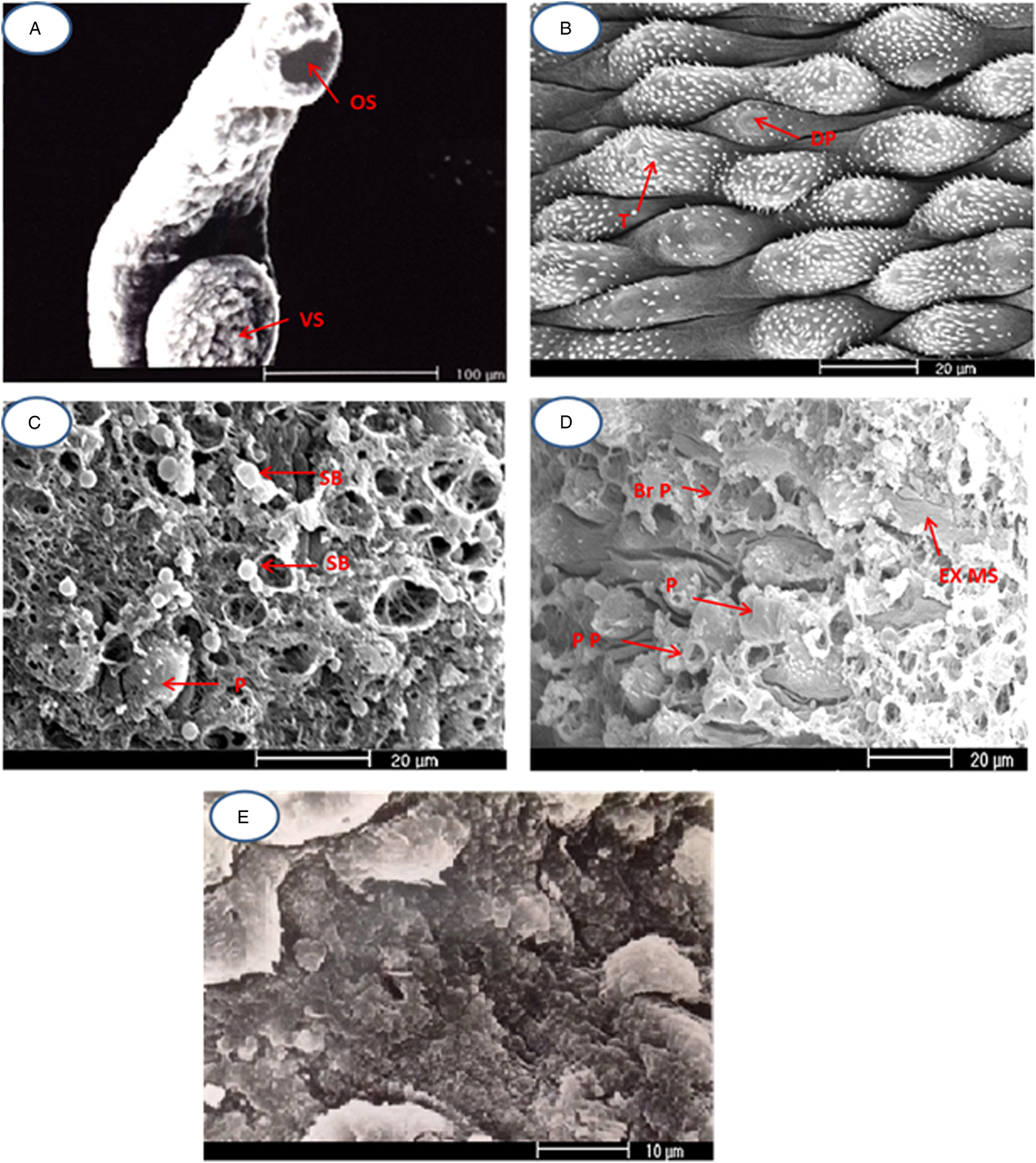
The oral and ventral suckers of the normal adult male worms were rounded to oval and had many well-developed spines (fig. 1A). The tegumental tubercles were uniform in size and distributed regularly in transverse rows, their apices being supported with many spines, in addition to the presence of dome-shaped papillae among them (fig. 1B). On exposure to sub-curative PZQ, whether in vivo or in vitro, many male adult S. mansoni showed drastic changes and damage, which were more pronounced in the in vitro treated worms. In some specimens, many blebs of variable size were found on or between the tubercles (fig. 1C). There were some protuberances, which in some instances were spherical with intact boundaries, whereas in others they were punctured, thus exposing the internal structures (fig. 1D). Some tubercles were swollen or damaged, with complete loss of spines (fig. 1D). In some regions, sloughing of the tegument was noticed and certain areas were totally disorganized and lacked recognizable structure, especially in worms exposed to PZQ in vitro (fig. 1E).
Scanning electron micrographs showing the tegument of adult Schistosoma mansoni worms. (A) Normal oral sucker (OS) and normal ventral sucker (VS); (B) tubercles (T) with spines and dome-like sensory papillae (DP); (C) and (D) the changes due to sub-curative PZQ (SB: surface blebs; P: protrusions; Br P: broken protrusion; PP: punctured protrusions; EX MS: exposed muscle layer); (E) total disorganization and lack of recognizable structure of the tegument, mainly with in vitro exposure to PZQ.

Analysis of the tegumental antigens by SDS
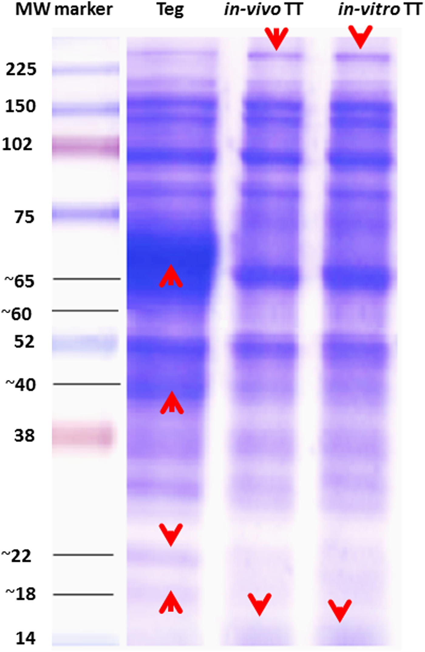
Protein fractions of Teg, in vivo TT and in vitro TT antigens were examined by SDS–PAGE analysis. Both in vitro TT and in vivo TT showed similar protein banding patterns: they both had 11 protein bands between 14 and > 225 kDa, compared with Teg, which had 13 bands. There was a protein band of 14 kDa that appeared in both treated tegument antigens but was not found in the Teg antigen preparation. Another > 225 kDa was of higher intensity in the treated than in the untreated Teg. The high intensity broad protein band spanning the molecular weight area between 75 and ~60 kDa in Teg was replaced by a well-defined band of ~65 kDa in both the treated antigens. The protein bands of ~40, ~22 and ~18 kDa in Teg were of decreased intensity and were only very faint in both the treated antigens (fig. 2).
SDS–PAGE (12%) analysis of the three tegumental antigens loaded under reducing conditions. The red arrows indicate the protein bands that differ between untreated antigen (Teg) and treated antigens (in vivo TT and in vitro TT antigens).

Antibody profile following mice immunization
All vaccinated mice groups showed higher significant (P < 0.001) specific IgG antibodies against Teg antigen when compared to PBS + adjuvant starting from day 15 after the first immunization and this response reached its peak after the third immunization. In vitro TT vaccine induced the highest IgG antibody level, followed by in vivo TT and Teg antigens (fig. 3). Regarding the IgG isotype profile, IgG1 and IgG2a increased significantly at 15, 30 and 45 days after the first immunization, compared to PBS + adjuvant mice. On calculating the IgG1/IgG2a ratio, it was found that this ratio reduced after each immunization, indicating a mixed Th1/Th2 immune response with Th1 predominance in all vaccinated mice groups (fig. 4).
Kinetics of IgG antibodies, assayed by ELISA, which were induced in mice immunized with the antigens at days 15, 30 and 45 after first immunization. Results are presented as the mean + SD of OD readings for each group of mice. ***P < 0.001 indicated statistical significance difference compared to PBS + adjuvant group. Results are representative of two independent experiments.

Kinetics of specific IgG1 and IgG2a antibodies in sera of vaccinated mice. Sera were collected from six immunized mice from each group at days 15, 30 and 45 post first immunization dose. The antibodies were assayed by ELISA. Results are presented as the mean + SD of OD readings for each group of mice. *** P < 0.001 and ** P < 0.01 indicated statistically significant difference compared to PBS + adjuvant mice. These results are representative of two independent vaccination trials.

Cytokine profile after vaccination
Following vaccination, the level of IFN-γ, IL-4 and IL-10 in the sera of mice (six selected from each group) at 45 days after the first immunization were measured (fig. 5). All vaccinated mouse groups showed significantly higher levels of IFN-γ when compared to PBS + adjuvant mice (216.4 ± 22.2 pg/ml, P < 0.001), and in vitro TT induced the highest level of IFN-γ (854 ± 53.5 pg/ml, P < 0.001), followed by in vivo TT (623 ± 21.5 pg/ml, P < 0.001), whereas Teg achieved 358.5 ± 32.6 pg/ml, P < 0.01. More IL-4 was produced in all vaccinated mice groups when compared to PBS + adjuvant (9.56 ± 2.52 pg/ml), but the difference was not significantly different to the 12.7 ± 4.1, 11.61 ± 3.21 and 13.4 ± 1.2 pg/ml found in the Teg, in vivo TT and in vitro TT vaccinated mice groups, respectively. There was also no significant difference between the vaccinated mouse groups when compared to one another. In vitro TT immunization produced the highest IL-10 cytokine serum levels at 78.85 ± 3.6 pg/ml (P < 0.001), followed by in vivo TT, which induced 64.8 ± 2.2 pg/ml (P < 0.001), whereas Teg vaccine produced 40.5 ± 4 pg/ml (P < 0.001), compared to PBS + adjuvant mice (19.77 ± 1.87 pg/ml).
These results indicate that immunization with the three tegumental preparations in conjugation with Freund's adjuvant induced a predominantly Th1 immune response, by day 45, characterized by high levels of the Th1 cytokine IFN-γ and the relative absence of the Th2 cytokine, IL-4, with an increase in immunomodulatory IL-10.
Cytokine profiles of immunized mice. Serum cytokines were detected 45 days post first immunization. Cytokines were assayed by sandwich ELISA. Results are presented as the mean ± SD of OD readings for each group of mice. ***P < 0.001, **P < 0.01 and *P < 0.05 indicated statistically significant difference. These results are representative of two independent vaccination trials.

Parasitological parameters
Total worm burden
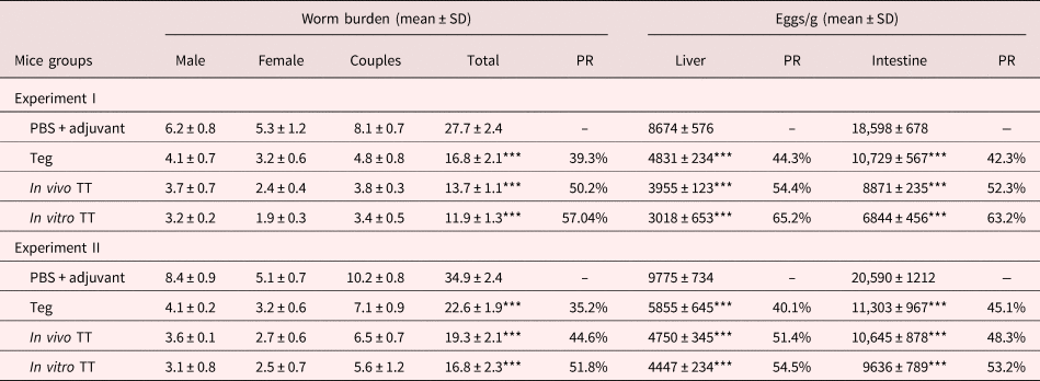
The results of the two independent vaccine trials in mice revealed a highly statistically significant reduction in worms in mice vaccinated by the tegumental antigens compared with the adjuvant control group. PBS + adjuvant mice recorded mean worm burdens of 27.7 ± 2.4 and 34.9 ± 2.4. In vitro TT vaccinated mice recorded the highest significant reduction in worm burdens (51.8 and 57.04%, P < 0.001), followed by immunization with in vivo TT (44.6 and 50.2%, P < 0.001), whereas Teg showed 35.2 and 39.3% (P < 0.001) reductions (table 1).
Effects of immunization with Teg, in vivo TT and in vitro TT antigens on worm burden and egg count per gram of tissue in Schistosoma mansoni infected mice.

*** P < 0.001 indicates statistically significant difference compared to PBS + adjuvant mice group. Teg: tegument antigen; TT: treated tegument; PR: percent of reduction; SD: standard deviation.
Tissue egg load
In vitro TT immunization induced the highest significant reduction (P < 0.001) in both mean number of liver eggs (54.5 and 65.2%) and mean number of intestinal eggs (53.2 and 63.2%), followed by in vivo TT, which achieved 51.4 and 54.4% decrease in mean total egg count in liver and 48.3 and 52.3% reduction in mean total intestinal eggs, whereas Teg showed the lowest protection values, with 40.1 and 44.3% decrease in the mean of total liver eggs and 42.3 and 45.1% decrease in mean number of intestinal eggs (table 1).
Hepatic granuloma parameters
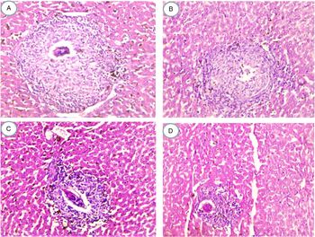
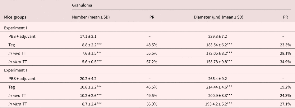
Consistent with the low egg burden in the liver, there were significant (P < 0.001) reductions in the mean number and the mean diameter of liver granulomas in all vaccinated mouse groups compared to the PBS + adjuvant group. In vitro TT vaccinated mice showed the greatest reduction value in both the mean number (56.9 and 67.2%) and the mean diameter of liver granulomas (27.1 and 34.9%), followed by in vivo TT immunized mice, which showed a 49.5 and 55.5% reduction in mean number and 24.3 and 28.1% decrease in mean diameter of hepatic granuloma, and finally Teg immunized mice, which revealed 46.5 and 48.5% reduction in the mean number and 19.2 and 23.3% decrease in the mean diameter of liver granulomas (table 2 and fig. 6).
Photomicrographs of granulomas in the livers of mice 8 weeks after S. mansoni infection (×200). Liver sections were stained with H&E. (A) PBS + adjuvant mice; (B) Teg vaccinated mice; (C) in vivo TT vaccinated mice; and (D) in vitro vaccinated TT. The images are representative of granulomas in two independent experiments.

Effect of immunization with Teg, in vivo TT and in vitro TT antigens on number and diameter of liver granuloma in Schistosoma mansoni infected mice.

***P < 0.001 indicates statistically significant difference compared to PBS + adjuvant mice group. Teg: tegument antigen; TT: treated tegument; PR: percent of reduction; SD: standard deviation.
SEM of worms perfused from the vaccinated mice groups
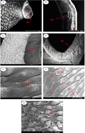
All the antigens caused various malformations in the tegument of the adult worms, but the worst deformities were shown in worms retrieved from mice receiving in vitro TT. Some adult male worms had closed oral suckers (fig. 7A), whereas others showed atrophy or contraction of the ventral sucker, and sometimes its spines were lost (fig. 7B). The widening of the gynaecophoric canal was observed in some specimens (fig. 7C), whereas others had edematous and closed canals (fig. 7D). Some tegumental segments showed swelling and oedema (fig. 7E). The tubercles showed varied changes, such as swelling, loss of ridges between them, and irregular positioning, and flattening and loss of spines were detected on the surface of some tubercles (fig. 7F). Some areas of tegument showed focal sloughing (fig. 7G).
Scanning electron micrographs showing the tegument of adult S. mansoni worms retrieved from different vaccinated mice groups. The changes were more frequent and more severe in in vitro TT immunized mice. (A) Closed oral sucker (COS); (B) retracted ventral sucker (RVS); (C) widening gynaecophoric canal (WGC); (D) closed gynaecophoric canal (CGC); (E) tubercle flattening (TF) and spine loss (SL); (F) tubercle peeling (TP) and tubercle swelling (TS); and (G) sloughing of some areas of the tegument.

Discussion
To provide effective control of Schistosoma, a vaccine would be useful in addition to mass chemotherapy and good sanitation (Siddiqui and Siddiqui, Reference Siddiqui and Siddiqui2017). The parasite tegument is an attractive source of potential vaccine molecules, and some immunogenic antigens are also found in the basement membrane secluded from the host's immune responses (Loukas et al., Reference Loukas, Tran and Pearson2007; Wilson and Coulson, Reference Wilson and Coulson2009). Schistosoma mansoni adult worms treated with sub-curative doses of PZQ show tegumental damage and exposure of many immunogenic antigens (Doenhoff et al., Reference Doenhoff, Modha and Lambertucci1988; Brindley et al., Reference Brindley1989; Fallon et al., Reference Fallon1994).
We confirmed the tegumental changes in worms treated with a sub-curative dose of PZQ either in vivo or in vitro by SEM. The changes were more severe in in vitro treated worms than in those receiving in vivo treatment. There were surface blebs, protuberance, and partial sloughing exposing the underlying muscle layer. These changes were consistent with observations in other studies that tested the effects of sub-curative PZQ on schistosomes (Modha et al., Reference Modha1990; El-Shabasy et al., Reference El-Shabasy2015). On analysing the prepared tegumental antigens on SDS–PAGE gel, the broad protein band spacing of 75–55 kDa of Teg was replaced by a well-defined protein band of ~65 kDa in both in vitro TT and in vivo TT antigens. Furthermore, some protein bands in Teg were barely apparent in both samples from treated worms, and this may be due to the shedding of some proteins of this molecular size as a result of the treatment. On the other hand, both treated antigens showed a protein band > 225 kDa, which was higher in intensity than that of Teg, and a 14 kDa protein appeared in both treated preparations. All of these changes may be attributable to drug-induced disruption of the tegument.
Teg antigen induced a 35.2–39.9% reduction in adult worms when compared to the PBS + adjuvant and this level of protection was similar to that obtained by Roberts et al. (Reference Roberts, Boot and Wilson1988), who reported 39.8% protection using purified adult tegumental membranes plus Freund's adjuvant, and with Smithers et al. (Reference Smithers1989), who showed that adult worm surface membranes in combination with saponin resulted in 18–37% (average 26%) fewer adult worms than in control mice. Also, immunization with S. mansoni schistosomula tegument yielded a 43–48% reduction in worm burden compared to the control group when used with Freund's adjuvant (Teixeira de Melo et al., Reference Teixeira de Melo2010; Araujo et al., Reference Araujo2012) and 43.1% when combined with alum/synthetic unmethylated CpG oligodeoxynucleotides (Teixeira de Melo et al., Reference Teixeira de Melo2013).
Treatment of adult schistosome worm tegument with sub-curative PZQ enhanced its protective effects: in vitro TT induced the highest protection value by 51.8–57%, and in vivo TT caused 44.6–50.2% reductions in the adult worm burden when compared to the PBS + adjuvant. This level of protection was comparable to tegumental preparations so far used in murine schistosomiasis vaccine, such as tegumental glycosyl phosphatidyl inisotol (GPI)-anchored proteins that induced 42% reduction of the adult worm burden (Martins et al., Reference Martins2012), use of an n-butanol extract of adult S. mansoni worm membrane fraction that resulted in 40.7–62.4% fewer adult worms than in control mice (Sulbaran et al., Reference Sulbaran2013), and fused Sm14/29 formulated with polyinosinic-polycytidylic acid adjuvant (polyI:C) that induced 48.4% protection (Mossallam et al., Reference Mossallam2015). Furthermore, in vitro TT induced the highest reduction in tissue egg numbers, followed by in vivo TT. Reduction in the tissue egg numbers is correlated with diminished numbers of worms, and lowering of egg numbers leads to alleviation of schistosome-induced pathology and lower disease transmission (Ranasinghe et al., Reference Ranasinghe2018).
In the current study, all the antigens were immunogenic and showed similar immune profiles: mixed Th1/Th2 with predominant Th1. The predominant Th1 immune response may be partly attributed to use of FCA, as previously reported by Heegaard et al. (Reference Heegaard2011). In natural infections, adult schistosome tegument antigens promote production of specific antibodies (Skelly and Wilson, Reference Skelly and Wilson2006), the isotypes of which are under the control of certain cytokines that selectively affect T helper cells (Spellberg and Edwards, Reference Spellberg and Edwards2001). The constant high level of specific IgG1 observed in this study could be attributed to IL-2 production, as previously demonstrated by DeKruyff et al. (Reference DeKruyff, Mosmann and Umetsu1990), while IFN-γ mediates IgG2a production by B cells (Finkelman et al., Reference Finkelman1988). The generated antibodies may recognize certain schistosomulum surface proteins, leading to activation of complement and antibody-dependent cell-mediated cytotoxicity mechanisms, causing the parasite's death (David et al., Reference David, Butterworth and Vadas1980; Joseph et al., Reference Joseph1983). Moreover, these antibodies may interfere with functions of some important surface molecules, such as alkaline phosphatase, inhibition of which will interfere with tegument repair (Fallon et al., Reference Fallon1994). The immunological importance of IgE in controlling schistosome infection and in protecting humans from parasitic re-infection was demonstrated in many studies (Dunne et al., Reference Dunne1992; Pinot de Moira et al., Reference Pinot de Moira2010). Eosinophils have the ability to kill S. mansoni larvae in the presence of IgE through the antibody-dependent cytotoxicity response (Capron and Capron, Reference Capron and Capron1994). The specific IgE antibodies against in vitro TT and in vivo TT antigens could be examined in a future work.
The induced Th1 response described here is in accordance with the protective effect of schistosomulum tegument (Teixeira de Melo et al., Reference Teixeira de Melo2010, Reference Teixeira de Melo2013). The role of IFN-γ in protection against schistosomes has been well documented. Treatment with monoclonal anti-IFN-γ antibody (Smythies et al., Reference Smythies, Coulson and Wilson1992) or use of IFN-γ knockout mice (Wilson et al., Reference Wilson1996) abolished the immunoprotective effects of irradiated cercariae. It was suggested that IFN-γ initiates leucocyte adhesion-forming foci in the infected mouse lungs, which delay schistosomulum migration (Wilson et al., Reference Wilson1996). It may also initiate recruitment and activation of pulmonary macrophages (Jankovic et al., Reference Jankovic1999). Furthermore, lymphocytes from humans with natural resistance to schistosome infection produce high levels of IFN-γ after exposure to the parasite's antigens (Oliveira et al., Reference Oliveira2012).
In vitro TT engendered the highest significant reduction in mean liver egg granuloma diameter, followed by in vivo TT, then Teg, and this down-regulation of granuloma formation could be attributed to an increase in the anti-inflammatory IL-10, which limits exacerbated schistosome granulomatous reactivity (Sundstedt et al., Reference Sundstedt1997). Schistosome-infected mice treated with recombinant IL-10/Fc fusion protein showed smaller granulomas (Flores-Villanueva et al., Reference Flores-Villanueva1996), whereas IL-10-deficient mice showed larger granulomas with less discrete boundaries, a lower leukocyte count and more diffuse collagen than in granulomas in wild-type mice (Sadler et al., Reference Sadler2003).
The alterations in the morphological structure of adult worms shown by SEM in this study suggest that the antigens were identified by the host immune system initiating immune responses against challenging Schistosoma infection. The worst deformities were shown in worms retrieved from mice receiving in vitro TT and these changes may partly explain the reduction in worm numbers, as found by El Azzouni et al. (Reference El Azzouni2017). Atrophy of the ventral sucker could interfere with male and female copulation, preventing coupling because of widening or closing of the gynaecophoric canal, which may in turn affect survival of females, as the clasping action of males around females enhances the ability of females to take their blood meals from the host (Gupta and Basch, Reference Gupta and Basch1987). Closure of the oral sucker is incompatible with life. Tegumental damage along the body will affect its protective functions, leaving worms more susceptible to the elicited antibodies and immune cells (Xiao et al., Reference Xiao2000; Mossallam et al., Reference Mossallam2015).
The higher protective efficacy of in vitro TT and in vivo TT when compared with Teg may be due to the appearance of more immunogenic antigens, these being normally cryptic inside the surface membranes, a suggestion that agrees with those of other studies reporting that in vivo sub-curative PZQ exposes some hidden highly immunogenic antigens, such as a 27 kDa worm membrane antigen (Doenhoff et al., Reference Doenhoff, Modha and Lambertucci1988), a 200 kDa glycoprotein (Brindley et al., Reference Brindley1989) and alkaline phosphatase (Fallon et al., Reference Fallon1994) that became the target for antibodies that act synergistically with the drug. Also, we suggest that PZQ may enhance immunogenicity of some tegument antigens. Moreover, treated tegumental preparations may contain more soluble antigens, enzymes and GPI-anchored proteins, which are more immunogenic.
Finally, immunization with in vitro TT achieved higher levels of worm reduction and bigger immune responses than in vivo TT and this may be attributed to some of the released antigens being preserved in the mixture during in vitro TT preparation, whereas in vivo TT preparation may result in antigens being shed in the serum of the host. Also, changes to the treated adult tegument because of in vitro exposure to sub-curative PZQ may be accompanied by the release of more cryptic antigens with high immunogenicity. In conclusion, treatment of adult S. mansoni with sub-curative PZQ enhanced its protective efficacy against experimental schistosome-infection. In vitro TT gave the best immunological response of Th1 type, causing the most prominent damage to the adult worm tegument, the highest reduction in worm burden and tissue egg count, and immunopathology associated with schistosomiasis. Therefore, in vitro TT, which is cheap to prepare, may be a potential anti-S. mansoni vaccine.
Author ORCIDs
B.E.W. El-Aswad, 0000-0001-8463-5609.
Acknowledgements
The authors sincerely appreciate Professor Mike Doenhoff, University of Nottingham, for his valuable comments and his help in reviewing this manuscript.
Financial support
This study did not receive any financial support from any private or public organization, and all costs were paid by the authors.
Conflict of interest
None.
Ethical standards
The authors assert that all procedures contributing to this work comply with the ethical standards of the relevant national and institutional guides on the care and use of laboratory animals.