Introduction
Palatine tonsil squamous cell carcinoma (SCC) can be histologically confirmed with either tonsillectomy or tonsil biopsy. Biopsies have the advantage of being quicker to perform, less invasive, and provide potentially quicker results.Reference Campbell, McLaren, Sheldon, Rock, Bracey and Malik1 Tonsillectomy has the advantage of minimising the risk of missed pathology, but with greater risk of morbidity.Reference Siddiq, Stephen, Lin, Fox, Robinson and Paleri2 The 2024 UK Head and Neck Cancer Guidelines do not recommend one approach over the other.Reference Homer, Winter, Abbey, Aga, Agrawal and Dafydd3 The guidelines recognise that assessment under a general anaesthetic has advantages in terms of assessing transoral access and feasibility of resection, but otherwise make no recommendation.Reference Homer, Winter, Abbey, Aga, Agrawal and Dafydd3 We compared whether tonsillectomy or biopsy affected surgical margin status at subsequent curative-intent surgery.
To the authors’ knowledge no previous study has compared surgical margin status in these two groups. Curative-intent treatment of palatine tonsil SCC involves either lateral oropharyngectomy and neck dissection with or without post-operative radiotherapy (RT), or primary RT (with or without chemotherapy).
The multidisciplinary team’s (MDT) recommendation for post-operative RT will be based on a range of factors including the T-stage or tumour–node–metastasis (TNM 8th edition) of the primary tumour, the margin status at curative-intent surgery, the number of affected lymph nodes and presence of extracapsular spread, and other histological features.
The surgical margin is the only factor over which a surgical team can have some measure of control. There is currently a degree of heterogeneity in the literature regarding definitions of clear, close or involved surgical margins. For the purposes of our study we defined clear as > 5 mm, close as 1–5 mm and involved as < 1 mm, as supported by the Royal College of Pathologists.Reference Helliwell and Woolgar4 Those with clear margins and other favourable prognostic factors can be considered for surgical treatment alone without the need for other modalities of treatment and hence increased morbidity.Reference Siddiq, Stephen, Lin, Fox, Robinson and Paleri2, Reference Holcomb, Herberg, Strohl, Ochoa, Feng and Abt5
We aimed to answer the question: does tonsillectomy versus biopsy influence surgical margin status at subsequent curative-intent surgery?
Methods
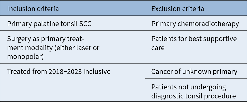
Our Business Intelligence Team provided a list of all patients with T1–T2 primary tonsil SCC, who underwent curative-intent surgery as their primary treatment modality in the last five years (2018–2023) at University Hospitals Bristol and Weston, following initial tonsil biopsy or diagnostic tonsillectomy. We undertook a retrospective case-notes review of these patients (see Table 1 for inclusion and exclusion criteria). Patients were excluded from analysis if they had not undergone a primary tonsil procedure, if no primary tumour was evident on clinical assessment or imaging, or if primary treatment was non-surgical (either best RT or best supportive care). Patients undergoing tonsillectomy were compared to patients undergoing tonsil biopsy. Ranges of demographic, surgical and tumour data were collected. Primary outcomes included margin status at curative-intent surgery and subsequent recommendation for post-operative RT.
Patient inclusion and exclusion criteria for the study

Chi-square test was used to compare the diagnostic tonsillectomy group with the tonsil biopsy group in terms of margin status, tumour staging and need for post-operative RT.
For the purposes of our study, if no tumour was detectable in curative-intent resection specimen (i.e. it had been entirely excised at initial diagnostic tonsillectomy), the thickness of the curative-intent resection specimen was added to the closest margin (if any) at diagnostic procedure to determine a final surrogate margin status. Where this was > 5 mm, this was deemed “clear.”
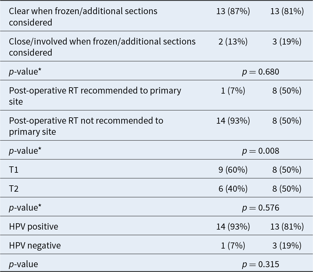
For patients that had frozen or additional margins taken in addition to their main curative-intent resection specimen, margin status was determined by adding the closest margin on the primary resection specimen to the thickness of the adjacent and orientated (frozen) additional marginal specimen. This is labelled “Clear/close/involved on frozen/additional sections” in Table 2. Where this was > 5 mm, this was deemed “clear.”
Margin status, indication for post-operative radiotherapy (RT), T stage and HPV status on main resection specimen (at curative-intent surgery); HPV= human papilloma virus

Results
Of the 31 patients that were included in the study, 16 had biopsies and 15 had tonsillectomies. The tonsillectomy group had a reduced risk of close or involved margins (< 5 mm) at curative-intent surgery (p < 0.001), and a reduced recommendation for post-operative RT to the primary site (p = 0.003). There was no significant difference in T stage between the two groups (p = 0.576). Although margins were more likely to be involved in the main resection specimen in the biopsy group, when considering subsequent frozen and additional margins, there was no statistically significant difference in final margin status between the two groups (Table 2: 13/15 [87 per cent] vs 13/16 [81 per cent]) (p = 0.68).
There was no significant difference in human papillomavirus (HPV) status in the two study groups. HPV status was positive in 13/16 (81 per cent) of biopsy patients and 14/15 (93 per cent) of tonsillectomy patients (p = 0.315). At curative-intent surgery, a tumour was detected in the main resection specimens of 2/15 (13 per cent) tonsillectomy patients, and in 15/16 (95 per cent) of biopsy patients (p < 0.001).
Discussion
To the authors’ knowledge, this study is a first to compare margins at curative-intent surgery for tonsil SCC, between initial diagnostic tonsillectomy versus biopsy. Our data demonstrate that tonsillectomy patients had significantly fewer close or involved margins at definitive surgery (p < 0.001). This was not significant when considering subsequent frozen or additional marginal specimens (p = 0.680).
Case series looking at main resection margins in patients with T1 and T2 oropharyngeal cancer report rates of involved margins of 16–38 per cent.Reference Shenouda, Rubin, Garcia, Badoual, Bonfils and Laccourreye6, Reference Zebolsky, George, Gulati, Wai, Carpenter and Van7 Our margin status rates compare favourably.
Accurate determination of margin status with frozen or additional marginal specimens requires close multidisciplinary working. Specimens must be orientated appropriately and clinical details from the surgeon regarding additional margins are crucial. This collaborative working introduces potential for more uncertainty with decision making by the MDT. Of the 15 diagnostic tonsillectomy patients, 13 (87 per cent) had no residual cancer detected in their subsequent curative-intent oropharyngeal resection. This makes determination of margin status easier for the MDT.
There was no significant difference in adjuvant RT being recommended between the two groups. However, the indication for RT in tonsillectomy patients was less likely to be the primary site (p = 0.008). As discussed previously, there are many factors that influence the MDT’s recommendation for adjuvant RT, and patients can also choose not to have it. In the authors’ experiences, uncertainty regarding final margin status may lead to the MDT recommending post-operative RT. Following the initial diagnostic tonsillectomy or biopsy, when there is no residual tumour in the subsequent curative-intent resection specimen, margin status is certain, and disease control with surgery alone can be considered more likely. Choosing tonsillectomy over biopsy may make decision making for MDTs more straightforward and help avoid post-operative RT. This has potential for reducing treatment-associated morbidity.Reference Lacas, Bourhis, Overgaard, Zhang, Grégoire and Nankivell8
We recognise that large tumours may not be suitable for tonsillectomy at initial operation and are more likely to have involved margins and require adjuvant treatment. Our data did not demonstrate a significant difference in T stage between biopsy and tonsillectomy groups.
The authors recognise the limitations of our study. We performed a retrospective analysis of patients that were not matched, although no statistically significant difference was found in T stage or HPV status. The decision whether to perform biopsy or tonsillectomy was made by the operating surgeon, before knowledge of study’s findings, and may have depended on factors not controlled for or recognised by our methods. We have also intentionally neither reported upon locoregional control nor survival outcomes, as this is beyond the scope of our study’s aims.
• Primary site palatine tonsil squamous cell carcinoma can be diagnosed with either tonsillectomy or tonsil biopsies
• Biopsies have the advantage of being quicker to perform, less invasive, and provide potentially quicker results, whereas tonsillectomies have the advantage of minimising the risk of missed pathology, but with greater associated morbidity
• At present, no clear evidence recommends superiority of one method over the other
• Margin status is a key indication for the multidisciplinary teams’ decision to recommend adjuvant treatments
• To the authors’ knowledge, no previous work has assessed if the method of initial diagnostic surgery has any influence on subsequent resection margins at curative-intent surgery
Conclusions
The authors conclude that tonsillectomy over tonsil biopsy has potential benefits in the diagnosis of palatine tonsil SCC. We would recommend further research studies be undertaken which should be prospective, multi-centred, and stratify for both diagnostic and curative-intent surgical techniques.
Acknowledgements
The Authors acknowledge the University Hospitals Bristol and Weston NHS Foundation Trust Library services for their assistance with literature review.