I. INTRODUCTION
In situ high-energy synchrotron X-ray diffraction (XRD) is a powerful tool to study the fundamentals of phase transformations. The intense beam of high-energy X-rays makes it possible to monitor change in phase fraction and unit-cell parameters of the constituent phases in the bulk of a material. Using this unique capability it is possible to analyze the transformation behavior of the austenite fraction (both at a fraction level and at the level of individual austenite grains) in low alloyed multiphase steels developed for applications requiring a combination of high strength and high ductility. This transformation of a relatively soft metastable austenite phase into a hard martensite phase upon activation by mechanical and/or thermal stimuli is called the TRIP effect. This phenomenon increases the work-hardening rate during plastic deformation and hence delays necking (Dan et al., Reference Dan, Zhang, Li and Lin2007; Jacques et al., Reference Jacques, Furnémont, Lani, Pardoen and Delannay2007). At room temperature, a typical low-alloyed TRIP steel microstructure contains three phases: ferrite, bainite, and metastable austenite (Timokhina et al., Reference Timokhina, Hodgson and Pereloma2004; Zaefferer et al., Reference Zaefferer, Olhert and Bleck2004). In our previous synchrotron X-ray diffraction studies, we have probed austenite transformation behavior in bulk TRIP microstructure during deformation at room temperature (Jimenez-Melero et al., Reference Jimenez-Melero, van Dijk, Zhao, Sietsma, Wright and van der Zwaag2011) and during cooling down to 100 K (van Dijk et al., Reference van Dijk, Butt, Zhao, Sietsma, Offerman, Wright and van der Zwaag2005; Jimenez-Melero et al., Reference Jimenez-Melero, van Dijk, Zhao, Sietsma, Offerman, Wright and van der Zwaag2007a, Reference Jimenez-Melero, van Dijk, Zhao, Sietsma, Offerman, Wright and van der Zwaag2007b, Reference Jimenez-Melero, van Dijk, Zhao, Sietsma, Offerman, Wright and van der Zwaag2009). We found that austenite stability depends on local carbon content, grain size, and orientation. In this work, the thermal and mechanical stability of retained austenite has been studied during temperature-dependent tensile tests using a near-field powder diffraction approach, i.e. analyzing the average behavior of the austenite fraction. Such analysis yields clear information on the transformation behavior, yet does not yield information on the state of deformation of individual austenite grains prior to transformation to martensite. In the literature there is considerable discussion on whether plastic deformation (i.e., the creation of dislocation or dislocation networks) precedes martensite transformation or that the martensite forms from plastically undeformed austenite grains, yet no experimental data have been presented to resolve the debate.
To monitor the material at smaller length scales, a micro-beam combined with a high-resolution far-field detector was used to try and resolve the issue. For the first time, the sub-grain structure of austenite prior to transformation has been observed. The data from near-field and far-field detectors are combined to characterize austenite stability (Jakobsen et al., Reference Jakobsen, Poulsen, Lienert and Pantleon2006, Reference Jakobsen, Poulsen, Lienert and Pantleon2007).
II. EXPERIMENTAL
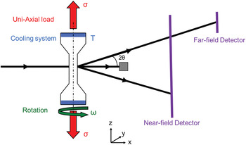
The in situ XRD experiments were performed at the 3D X-ray diffraction microscope at the ID11 beam line of the European Synchrotron Radiation Facility (Grenoble, France). Figure 1 shows the experimental setup used for the experiments. For the powder experiments, a monochromatic X-ray beam of 88 keV (wavelength of 0.141 Å) and a beam size of 100 × 100 µm2 illuminated the cylindrical sample. The diffracted beam was recorded on a two-dimensional CCD detector (FRELON) (Labiche et al., Reference Labiche, Mathon, Pascarelli, Newton, Ferre, Curfs, Vaughan, Homs and Carreiras2007) placed behind the sample. The sample was continuously rotated during exposure around the cylindrical axis perpendicular to the X-ray beam in steps of 0.25° covering an angular range of 84°. The instrument parameters of the 3D X-ray diffraction microscope were determined using a LaB6 calibrant.
Schematic representation of the experimental setup used for the high-energy micro-beam X-ray diffraction experiments on low-alloyed TRIP steels. The sample is under tensile stress (σ) and cooled from both ends. During exposure the sample was continuously rotated around the vertical axis. The far-field detector was placed in the horizontal scattering plane.

For the high-resolution experiments, a prototype HIZPAD 1-mm CdTe pixel detector (Ruat and Ponchut, Reference Ruat and Ponchut2012) was placed 3.2 m behind the sample in the horizontal scattering plane. The energy has been optimized to 69.5 keV for the far-field detector, and the beam size reduced to 40 × 40 µm2 to observe the reflections of individual grains. The {111} reflection of austenite and {110} of ferrite have been monitored simultaneously. The sample was continuously rotated around the cylindrical axis perpendicular to the X-ray beam in steps of 0.2° covering an angular range of 90°. Changes in the sample-to-detector distance during straining were determined using a CeO2 calibrant placed on the sample.
To study the mechanical stability of retained austenite, the sample was mounted on a 2-kN micro tensile tester placed on a Huber table that provided a rotation along the cylindrical axis of the sample (the loading direction was along the sample axis). For each deformation step, the strain was held constant during a complete set of measurements for the full rotation range. The mechanical stability of the austenite phase was studied at selected temperatures of 293, 213, and 153 K. The high-resolution experiments were performed at room temperature.
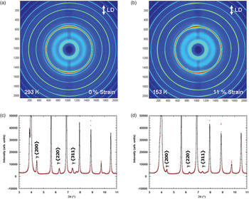
The experiment produced a series of 2D diffraction patterns as a function of temperature, sample elongation, and ω-angle. These 2D diffraction patterns were summed for the complete ω range and integrated over the azimuthal angle using the FIT2D software package (Hammersley et al., Reference Hammersley, Svensson, Hanfland, Fitch and Hausermann1996) to obtain the corresponding one-dimensional diffraction patterns as a function of elongation. Rietveld refinement of the resulting one-dimensional X-ray diffraction patterns was performed using the Fullprof package (Rodríguez-Carvajal, Reference Rodríguez-Carvajal1993) to determine the phase fraction and lattice parameter of the constituent phases as a function of macroscopic strain (Figure 2).
Two-dimensional X-ray diffraction patterns at (a) no strain and (b) the ultimate tensile strength (UTS). These patterns have been obtained by summing all the measured patterns for the whole ω-range used in the experiment. The loading direction is indicated in the figure (LD). The 2D patterns have been subsequently integrated over the azimuth angles to obtain the one-dimensional X-ray patterns as a function of scattering angle at (c) no strain, and (d) the ultimate tensile strength (red points). The corresponding Rietveld refinement of the one-dimensional X-ray patterns is also shown (black curve).

III. RESULTS AND DISCUSSION
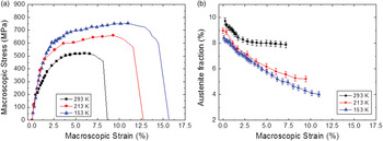
Figure 3(a) shows the macroscopic stress–strain curve of the low-alloyed TRIP steel measured step-wise at temperatures of 293, 213, and 153 K. Each data point on the curves represents a condition for which X-ray diffraction measurement of over the ω-rotation range were performed while the macroscopic strain was held constant. We recorded the diffraction patterns for a series of elongation steps up to the ultimate tensile strength. Both the yield stress and the ultimate tensile strength show an increase of more than 200 MPa when cooling the sample from 293 to 153 K. The uniform elongation also shows a significant increase at lower temperatures, taking a value of about 6.6% at 293 K and 11% at 153 K. This effect is in contrast to normal temperature-dependent mechanical behavior, where plastic deformation takes place primarily by dislocation glide and dislocation interactions resulting in an increase in strength and a reduction in maximum elongation at lower temperatures (Curtze et al., Reference Curtze, Kuokkala, Hokka and Peura2009). The reverse phenomenon observed in these samples with respect to the variation of uniform elongation with temperature may be ascribed to the presence of the TRIP effect.
(a) Macroscopic (engineering) stress–strain curves of TRIP steel measured step-wise during the in situ X-ray diffraction experiments at temperatures of 293, 213, and 153 K. (b) Austenite fraction as a function of macroscopic strain for different temperatures. Variations in the initial austenite fraction are because of transformation during cooling.

Figure 3(b) shows the austenite fraction as a function of macroscopic strain at different temperatures. The difference in the initial austenite fraction as a function of temperature is caused by austenite-to-martensite transformation during cooling. The austenite fraction decreases progressively with increasing strain because of mechanically induced austenite transformation. At all studied temperatures austenite starts to transform well before the macroscopic yield stress is reached. As reported previously, the transformation can occur in the elastic regime by adding strain energy to activate the transformation for the least stable grains (Jia et al., Reference Jia, Cong, Sun, Cheng, Nie, Ren, Liaw and Wang2009; Tomota et al., Reference Tomota, Tokuda, Adachi, Wakita, Minakawa, Moriai and Morii2004). At room temperature, the austenite fraction decreases continuously for strains up to 2.5% and then remains roughly constant until fracture. At lower temperatures the transformation continues until fracture. The amount of austenite transformed by deformation is 4.3% at 213 K and 4.5% at 153 K. At fracture, a finite amount of austenite is still present and cannot be transformed mechanically under these experimental conditions. The increase in the austenite fraction transformed by deformation at lower temperatures shows a strong correlation to increase in maximum elongation. The TRIP effect clearly becomes more pronounced at lower temperatures (Jiménez et al., Reference Jiménez, Carsí, Ruano and Frommeyer2009).
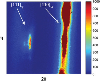
Figure 4 shows two-dimensional X-ray patterns of the unstrained sample on the far-field detector at 293 K. The small beam size allows us to monitor individual diffraction spots appearing on the austenite and ferrite diffraction rings. The far field detector allows us to clearly separate the first reflections of austenite {111}γ and ferrite {110}α. The resolution obtained, about 0.001°, is 22 times higher than the near-field detector. The diffraction spot from austenite shows intensity variations along η which reveal the presence of a rich substructure within the grain. The presence of subgrains at the initial condition indicates that the heat treatment to generate the TRIP microstructure has already induced dislocations within the retained austenite. The spread in intensity along the diffraction angle 2θ might correspond to spatial variation in carbon level within the austenite grain.
Two-dimensional X-ray patterns at no strain measured on the far-field detector, the {111}γ reflection of austenite and the {110}α reflection of ferrite are monitored simultaneously and clearly separated.

IV. CONCLUSION
The deformation induced austenite transformation is significantly enhanced at temperatures below room temperature and extends over a wider strain range. At room temperature the austenite transformation is only observed for macroscopic strains up to 2.5%, while it extends over the full deformation range at temperatures of 213 and 153 K. At lower temperatures both the ultimate tensile strength and the elongation at fracture show an increase. Higher uniform elongation at fracture is expected to be caused by the more efficient TRIP effect for lower temperatures. At all temperatures austenite transformation starts well before reaching macroscopic yield stress.
The high-resolution high-energy X-ray diffraction patterns allowed the first reflection of austenite and ferrite to be separated (they usually overlap using conventional X-ray lab source). These results show the presence of dislocation and carbon inhomogeneity in austenite grains prior to deforming the material. Their relation between the presence of dislocations in austenite grains and their transformation conditions upon further deformation of the material require further study.
Acknowledgments
We thank the European Synchrotron Radiation Facility for provision of synchrotron radiation facilities and thank the Beamline staff for assistance in using Beamline ID11 and particularly M. Ruat for assistance with the CdTe detector. This research was carried out under project number M41.5.08313 in the framework of the Research Program of the Materials Innovation Institute M2i (www.m2i.nl).