Eccentric ventricular activation in ventricular pre-excitation through right-sided accessory pathway may result in asynchronous spread of ventricular depolarisation. Abnormal electrical and mechanical conduction may mediate left ventricular dysfunction and remodelling.Reference Tomaske, Janousek and Rázek 1 – Reference Guo, Dai and Li 4 Since 2004, there have been over 60 reported cases that have suggested a possible causality between ventricular pre-excitation and dilated cardiomyopathy in the absence of sustained tachyarrhythmia.Reference Niu, Maskatia and Kim 5 – Reference Fazio, Mongiovi’ and Sutera 13 These cases were diagnosed with ventricular pre-excitation-induced dilated cardiomyopathy. However, it is only recently that ventricular pre-excitation with right-sided accessory pathways, which may cause abnormal interventricular septal motion and even dilated cardiomyopathy in patients with Wolff–Parkinson–White syndrome, has attracted attention.
We examined the clinical, electrocardiographic, echocardiographic and electrophysiological characteristics and prognosis of 25 children who presented with ventricular pre-excitation and dilated cardiomyopathy, left ventricular dilation, dysfunction, or abnormal interventricular septal motion; 23 patients were treated with ablations supplemented with medications and two patients were treated with only drug medications. The evidence and our interpretation are presented.
Materials and methods
Patients
Our study population included 25 patients and another 20 patients with idiopathic dilated cardiomyopathy. All patients met the following inclusion criteria: ventricular pre-excitation caused by right-sided accessory pathways combined with abnormal interventricular septum motion and/or left ventricular dysfunction and/or dilation; normal cardiac structure; and intracardiac electrophysiologic examination and radiofrequency catheter ablation or medication from March 2011 to June 2017. Patients with CHD, tachycardiomyopathy, atrial conduction to the ventricle via an accessory pathway while in atrial fibrillation, incessant supraventricular tachycardia, intermittent ventricular pre-excitation, hyperthyroidism, electrolyte disturbance, liver or kidney dysfunction, or acute infection were excluded. All the children stopped anti-arrhythmic drugs at least five half-lives before ablation. Periodic 24 hours dynamic electrocardiograms were performed in every patient to confirm the presence of acute episodes of tachyarrhythmia and the absence of persistent supraventricular tachycardia or tachycardia lasting >12 hours. Patients in the control group were diagnosed with idiopathic dilated cardiomyopathy without ventricular pre-excitation and were treated with routine anti-heart failure chemotherapy during the same period. All patients agreed to participate in the study, and written informed consent was granted by their parents. Informed parental consent and the child’s consent were obtained from each patient. The study protocol conformed to the ethical guidelines of the 1975 Declaration of Helsinki as reflected in a priori approval by the institution’s human research committee, and it was approved by the Institutional Research Ethics Board. All the patients who accepted ablation were 5 years of age or older.
Echocardiography
Echocardiography was postponed if a patient had experienced an episode of tachycardia within the previous 72 hours. Transthoracic echocardiography was performed with a Vivid E9 cardiovascular ultrasound system with a 3.5 MHz probe (GE Medical Systems, United States of America). All patients underwent echocardiographic examination by the same physician before ablation and 1, 3, 6, 9, and 12 months after ablation. Each echocardiographic parameter was calculated as the mean value from three cardiac cycles. The physician reviewer of the echocardiograms was blinded to the groups to avoid subjective bias. Left ventricular ejection fraction was calculated using the biplane Simpson’s method. Left ventricular end-diastolic diameter was measured via the parasternal long-axis view. Dilated cardiomyopathy was defined as left ventricular end-diastolic diameter >97th percentile of heart size, corrected for weight or body surface area, with an ejection fraction <45%.Reference Mathew, Williams and Navaratnam 14 We calculated the left ventricular end-diastolic diameter Z-value according to the method adopted by the American Boston Children’s Hospital.Reference Sluysmans and Colan 15 , Reference Colan 16 The Z-value can objectively evaluate the echocardiographic-measured value and has been widely used in recent years. The normal Z-value fluctuates between ±2 regardless of age and body surface area.
Electrophysiological studies and radiofrequency ablation
All procedures in children <14 years of age were performed under anaesthesia with propofol and analgesia administered intravenously. For older children, procedures were undertaken with local infiltration anaesthesia. Standard electrophysiologic methods were applied. A 7-Fr deflectable electrode catheter (Bard Electrophysiology, United States of America) with a 4-mm distal electrode was used for ablation with a radiofrequency current setting of 30 W and a temperature setting of 60°C. The ablation endpoints were the loss of the antegrade and retrograde conduction of the overt accessory pathways. Some ablations were accomplished using Swartz sheaths to stabilise the catheters. After ablation, the conducting system was re-evaluated to determine whether supraventricular tachycardia was still inducible and whether there was residual anterograde or retrograde conduction. The long-term indicators of successful ablation were the absence of recurrence of tachycardia and lack of evidence of a ventricular pre-excitation wave on the electrocardiogram.
Drug therapy
If patients had decreased left ventricular ejection fraction, they were treated with routine anti-heart failure chemotherapy, such as digoxin, furosemide, aldactone, and captopril, until they achieved recovery of their left ventricular function. Digoxin was only prescribed when successful ablation was finished in patients with left ventricular dysfunction. Captopril was discontinued when patients achieved normal left ventricular diameter.
Statistical analysis
Data were analysed using SPSS software for Windows (version 17.0; SPSS Inc., Chicago, IL, United States of America). Continuous variables are expressed as the mean±SD. Differences between two groups were compared using Student’s t-test. A p-value <0.05 was considered statistically significant. Pearson correlation analysis was applied to evaluate the correlation between two continuous variables.
Results
Clinical and echocardiographic manifestations
The age at diagnosis of left ventricular dysfunction, dilation, or abnormal interventricular septum movement was between 5 months and 16 years, and the mean age was 3.9±3.3 years. There were 15 females and 10 males. There was no difference in course of disease between patients with pre-excitation-induced dilated cardiomyopathy and idiopathic dilated cardiomyopathy (3.35±2.61 versus 2.87±2.44, p<0.05).
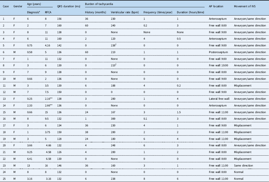
A total of 10 patients never experienced paroxysmal supraventricular tachycardia. Three cases experienced one episode of supraventricular tachycardia. The other 12 cases suffered from supraventricular tachycardia 1–6 times each year, which was sustained for approximately half an hour to several hours each episode (Table 1). None of them experienced incessant supraventricular tachycardia. Patients felt palpitations, fatigue, sicchasia, and vomiting when supraventricular tachycardia occurred. Clinical manifestations in young children included crying that made them difficult to comfort. Electrocardiogram monitoring was performed if the patient could not clearly express symptoms. Syncope did not occur in any of the cases except case 12. The daily symptoms of case 23 were chest discomfort and impaired exercise tolerance during the intervals of supraventricular tachycardia.
SVT onset, AP location, and echocardiographic characteristics of 25 cases.

AP=accessory pathway; IVS=interventricular septum; RFCA=radiofrequency catheter ablation; SVT=supraventricular tachycardia
Aneurysm: The basal segment of the interventricular segment moved similarly to an aneurysm
Same direction: The M-mode tracing from the parasternal long-axis view showed the ventricular septum and posterior LV wall moved in the same direction
Misplacement: The interventricular septum and posterior LV wall neither moved in neither the same direction nor the opposite direction
* Age at diagnosis of abnormal echocardiogram diagnosis
** Age at last follow-up (the patient did not receive ablation)
† SVT can be induced in electrophysiological studies
Cases 1–14 met the diagnostic criteria for dilated cardiomyopathy. Tachycardiomyopathy and other causes were excluded according to the chief complaints, frequency of supraventricular tachycardia, and periodic 24-hours dynamic electrocardiograms. Patients presented with the clinical signs and symptoms of chronic congestive heart failure, such as fatigue, exertional dyspnoea, and impaired exercise tolerance, and they were at the low end of physical growth compared with children in the same age group; infant patients presented with hidrosis and not enough physical strength to suckle. Case 12 had no symptoms or impaired exercise tolerance before disease onset. He suffered from a sudden cardiac origin of syncope presenting with no heartbeat. Ventricular tachycardia and fibrillation occurred when immediate chest compressions were performed. Echocardiographic examination showed signs of dilated cardiomyopathy and left ventricular dyssynchrony after successful cardiopulmonary resuscitation. Ion channel disease-related genetic testing was negative, and the possibility of inherited arrhythmogenic diseases was small. The patient received successful ablation when he was in a relatively stable condition.
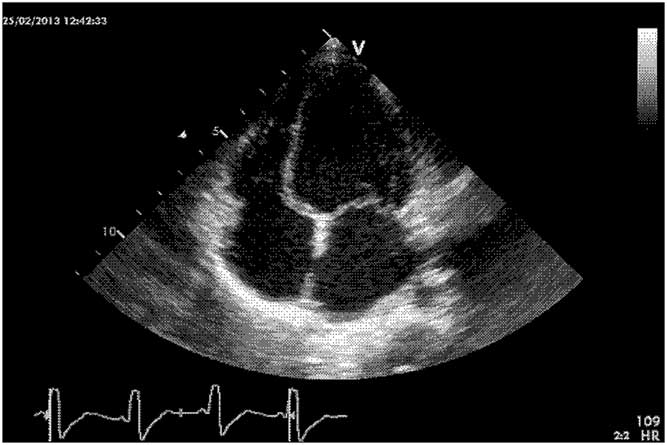
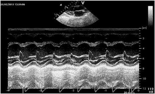
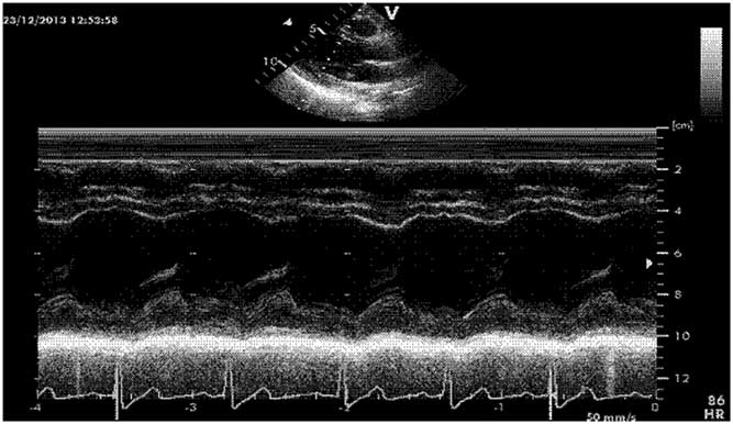
The left ventricular long-axis four-chamber view showed that the basal segment of the interventricular segment moved similarly to an aneurysm (Fig 1), and the M-mode tracing from the parasternal long-axis view showed that the interventricular septum and left ventricular posterior wall moved in the same direction (Fig 2) in the 13 cases with dilated cardiomyopathy except case 11. The echocardiogram showed misplacement of the interventricular septum and left ventricular posterior wall in case 11, which meant that the interventricular septum and left ventricular posterior wall moved in neither the same direction nor the opposite direction (Fig 3). Cases 4 and 11 combined with moderate-to-severe and moderate mitral regurgitation, respectively. Misplacement of the interventricular septum and left ventricular posterior wall was observed in 11 patients of the 20 patients with idiopathic dilated cardiomyopathy.
The basal segment of the interventricular segment moved similar to an aneurysm.

The interventricular septum and posterior wall of the left ventricle moved in the same direction.

Misplacement of the interventricular septum and posterior wall of the left ventricle.

Routine echocardiographic examination before ablation showed that cases 15–25 were characterised by abnormal interventricular septum movement (case 23) or left ventricular dilation (cases 24 and 25) or both (cases 15–22). Left ventricular ejection fraction was 50% in case 15 and normal in the other 10 cases. Three cases presented with a basal segment of interventricular septum that moved similar to an aneurysm; in two cases it was found that the interventricular septum move in the same direction with left ventricular posterior wall without basal septal aneurysmal bulging and four cases with misplacement of the interventricular septum and left ventricular posterior wall (Table 1).
Electrophysiological studies and radiofrequency ablation
Cases 13 and 14 received drug therapy and close follow-up because of the young age. The other 23 patients underwent electrophysiological studies and successful ablations. There was no evidence of multiple accessory pathways for any case. Among the 10 cases without tachycardia, eight cases were found to have no retrograde conduction of the accessory pathways, and two cases had retrograde conduction with inducible supraventricular tachycardia (Table 1). The chief complaint of case 22 was tachyrhythmia. However, the electrophysiological study showed that the accessory pathways did not have retrograde conduction and tachycardia was not induced by atrial or ventricular programmed stimulation. Orthodromic reciprocating tachycardia was induced in the other 14 cases.
The successful ablation sites were the right-sided anteroseptum in three patients, posteroseptum in one patient, anterior free wall in five patients, lateral free wall in seven patients, and posterior free wall in seven patients (Table 1). Ventricular pre-excitation recurred 24 hours after ablations in cases 3 and 5 with a return of the delta wave. The ablations were successfully repeated 1 week and 1 year later without recurrence. The retrograde conduction of the accessory pathway in case 2 recurred 4 months after the ablation. The patient accepted the second successful ablation 8 months after the first ablation. There were no complications related with ablations.
Clinical and echocardiographic improvement after catheter ablation/medication
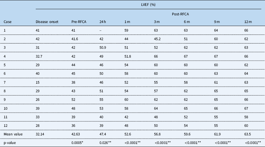
Cases 1–11 had improved physical activity, growth, and development after ablations. No life-threatening malignant arrhythmia recurred in case 12. Chest discomfort and impaired exercise tolerance during the intervals of supraventricular tachycardia in case 23 disappeared after ablation. All the abnormal movements of the interventricular septum disappeared during follow-up. Patients with decreased left ventricular ejection fraction (cases 1–12 and 15) gained normal left ventricular systolic function gradually after ablation (Table 2). A total of eight cases experienced an increase in left ventricular ejection fraction more than 5% just 24 hours after ablation. Some patients got a normal left ventricular ejection fraction of not <60% 1 month after ablation. Only three cases did not achieve normal left ventricular ejection fractions 6 months after ablation. Case 11 was severe, and he took 9 months to obtain an increased left ventricular ejection fraction of 55% (Table 2).
LVEF in patients with DCM before and after RFCA.

DCM=dilated cardiomyopathy; LVEF=left ventricular ejection fraction; RFCA=radiofrequency catheter ablation
* Compared with LVEF at disease onset
** Compared with LVEF pre-RFCA
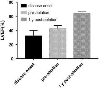
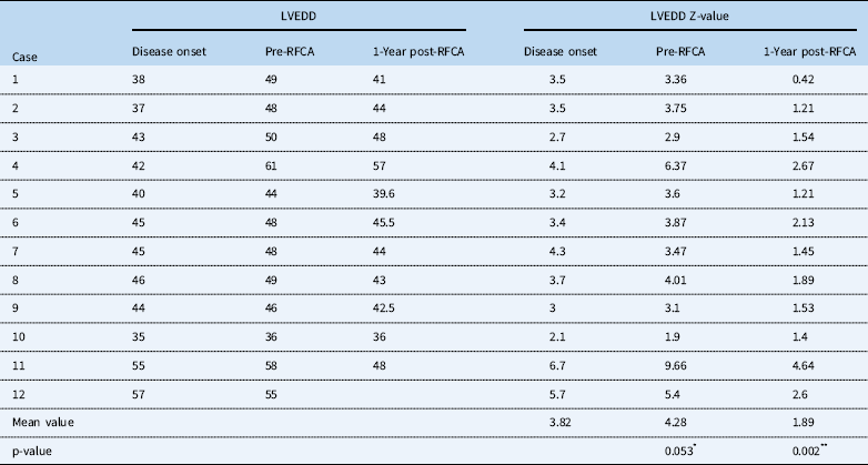
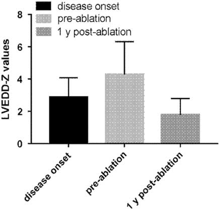
Left ventricular ejection fractions in the 12 cases with dilated cardiomyopathy were 32.14±7.71, 42.63±4.34, and 63.5±2.71% at disease onset, before ablation, and 1 year after ablation, respectively (Fig 4). Left ventricular ejection fractions increased significantly after drug therapy (t=−4.1, p<0.05) and increased to a higher level 1 year after ablation (t=−14, p<0.05). A total of 12 cases experienced a decreased left ventricular end-diastolic diameter during follow-up (Table 3). The left ventricular end-diastolic diameter Z values were 3.82±1.27, 4.28±2.04, and 1.89±1.06 at disease onset, before ablation, and 1 year after ablation, respectively (Fig 5). The left ventricular end-diastolic diameter increased after drug therapy (t=−2.05, p=0.053) and decreased 1 year after ablation compared with the diameter at disease onset (t=3.57, p=0.002). There were only four cases with left ventricular end-diastolic diameter Z-values higher than 2 during the follow-up period. A total of eight cases achieved a normal left ventricular end-diastolic diameter evaluated by Z-value. Mitral regurgitation was alleviated in cases 4 and 11.
LVEF in cases 1–12. LVEF=left ventricular ejection fraction.

LVEDD in patients with DCM before and after RFCA.

LVEDD=left ventricular end-diastolic diameter
* Compared with Z-value at disease onset
** Compared with Z-value pre-RFCA
LVEDD Z-values in cases 1–12. LVEDD=left ventricular end-diastolic diameter.

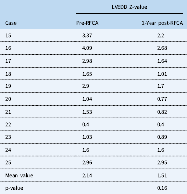
The left ventricular end-diastolic diameter did not change in patients without left ventricular dyssynchrony before and after ablation (cases 24 and 25). Cases 15–22 also experienced decreased left ventricular end-diastolic diameter after ablation during the follow-up period (Z-value: 2.14±1.16 versus 1.51±0.82, Table 4).
LVEDD Z-values in cases 15–25 before and after RFCA.

LVEDD=left ventricular end-diastolic diameter
Cases 13 and 14 were prescribed anti-heart failure drugs. Left ventricular ejection fraction increased from 50 to 54%, and the left ventricular Z-value increased from 3.26 to 3.57 through medication at 1 year and 7 months in case 13. Left ventricular ejection fraction decreased from 50 to 44%, and the left ventricular Z-value increased from 2.6 to 2.97 through medication at 7 months in case 14.
Correlation between QRS duration and left ventricular ejection fraction before and after ablation in patients with pre-excitation-induced dilated cardiomyopathy
There was no correlation between QRS duration and initial left ventricular ejection fraction, ejection fraction pre-ablation and ejection fractions 24 hours, 1 months, 3 months, 6 months, 9 months, and 1 year post-ablation (r=0.44, −0.19, −0.29, −0.12, −0.06, 0.01, 0.25, p=0.16, 0.56, 0.39, 0.7, 0.87, 0.98, 0.45, respectively).
Comparison of treatment effects between patients with pre-excitation-induced dilated cardiomyopathy and idiopathic dilated cardiomyopathy
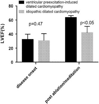
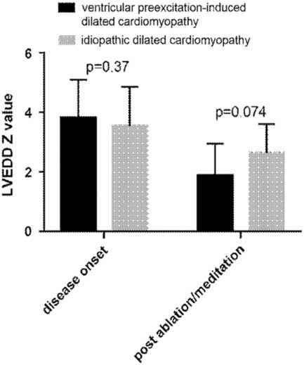
There was no difference of initial left ventricular ejection fractions and left ventricular end-diastolic diameter Z-values between patients with pre-excitation-induced dilated cardiomyopathy and idiopathic dilated cardiomyopathy. However, during follow-up treatment effects were different. Patients with pre-excitation-induced dilated cardiomyopathy got significant higher left ventricular ejection fractions and lower left ventricular end-diastolic diameter Z-values 1 year post-ablation compared with patients with idiopathic dilated cardiomyopathy just on anti-heart failure medications (Figs 6 and 7).
Comparison of LVEF between patients with pre-excitation-induced dilated cardiomyopathy and idiopathic dilated cardiomyopathy. LVEF=left ventricular ejection fraction.

Comparison of LVEDD Z-values between patients with pre-excitation-induced dilated cardiomyopathy and idiopathic dilated cardiomyopathy. LVEDD=left ventricular end-diastolic diameter.

Discussion
The first 10 cases diagnosed with ventricular pre-excitation-induced dilated cardiomyopathy were originally misdiagnosed as idiopathic dilated cardiomyopathy combined with ventricular pre-excitation or Wolff–Parkinson–White syndrome. Routine anti-heart failure chemotherapy did not produce satisfactory results in these patients. As we learned more about the diagnosis and therapy of ventricular pre-excitation-induced dilated cardiomyopathy, we performed detailed echocardiographic examinations on them and found that the interventricular septum and left ventricular posterior wall moved in the same direction, and the basal segment of the interventricular septum moved similarly to an aneurysm. These patients achieved complete recovery of left ventricular systolic functions after successful ablations. Their excellent prognosis confirmed our initial diagnosis. We summarised the characteristics of these patients as follows: the diagnostic criteria of dilated cardiomyopathy were met; incessant tachycardia and other causes of dilated cardiomyopathy could be excluded; electrocardiogram indicated ventricular pre-excitation with right-sided accessory pathways; left ventricular dyssynchrony could be detected by M-mode echocardiography or other methods; and the prognosis of ventricular pre-excitation-induced dilated cardiomyopathy after ablation was excellent. Some clinicians differed in the diagnosis. They diagnosed such cases as idiopathic dilated cardiomyopathy combined with ventricular pre-excitation. They held that ventricular pre-excitation was just an adverse factor but not the cause of dilated cardiomyopathy. We considered that left ventricular systolic function may completely recover through 1 year of treatment with regular anti-heart failure chemotherapy in mild cases of idiopathic dilated cardiomyopathy; however, recovery was less probable in moderate or severe cases. The rapid recovery of left ventricular ejection fraction after cessation of the antegrade conduction while drugs were not altered suggested a causal relationship between the overt right-sided accessory pathway and the development of dilated cardiomyopathy. In addition to recovery of disease, echocardiography can provide information and contribute to differential diagnosis. Patients with idiopathic dilated cardiomyopathy may present with dyssynchronous ventricular wall contraction; however, it is rare to observe the thinning of the basal segment and a movement pattern similar to that of an aneurysm, which is typical for ventricular pre-excitation-induced dilated cardiomyopathy.Reference Tomaske, Janousek and Rázek 1 , Reference Fazio, Mongiovi’ and Sutera 13 , Reference Ko 17 The prognosis of pre-excitation-induced dilated cardiomyopathy was better than idiopathic dilated cardiomyopathy, which manifested the value of ablation in the therapy of pre-excitation-induced dilated cardiomyopathy. In our opinion, anti-heart failure meditations were needed post-ablation. Left ventricular ejection fraction of case 12 increased by 12%, from 36 to 48%, 1 month after ablation performed in another hospital. However, after the initial increase, his recovery of left ventricular systolic function was slow without the help of anti-heart failure drugs. He came to our department 4 months after ablation, and we prescribed anti-heart failure drugs to accelerate the recovery and they produced a satisfactory effect.
Since the diameter of the left ventricular did not decrease after ablation in cases 24 and 25, we supposed that there was not a causal relationship between ventricular pre-excitation and left ventricular dilation if dyssynchronous ventricular wall contraction was not observed. However, this finding should be further confirmed by more cases and detailed examinations.
For case 4, retrograde conduction of the AP recurred 4 months after the ablation. She experienced six episodes of supraventricular tachycardia, with each episode sustained for approximately 1 hour. Nonetheless, her left ventricular systolic function continued to improve, and left ventricular dyssynchrony disappeared during this period despite frequent tachycardia. Therefore, this evidence indicated that abnormal ventricular activation alone, caused by anterograde conduction, in patients with overt right-sided accessory pathways had adverse effects on cardiac function. Since ablation can eliminate the pathogeny, the prognosis of ventricular pre-excitation-induced dilated cardiomyopathy is much better than idiopathic dilated cardiomyopathy.Reference Tomaske, Janousek and Rázek 1 , Reference Guo, Dai and Li 4 , Reference Udink ten Cate, Kruessell and Wagner 9 Ablation was the first therapeutic choice for ventricular pre-excitation-induced dilated cardiomyopathy in patients if their ages were suitable for ablation.Reference Udink Ten Cate, Wiesner, Trieschmann, Khalil and Sreeram 18
Most of the patients with ventricular pre-excitation-induced dilated cardiomyopathy could benefit from regular anti-heart failure chemotherapies, which can increase left ventricular ejection fraction to a certain extent. However, left ventricular ejection fraction would not be satisfactory until successful ablation was performed. The left ventricular end-diastolic diameter seemed to increase with chemotherapy alone. It is worthy of mention that anti-heart failure drugs were still needed for the recovery after ablation.
At present, the cause of ventricular pre-excitation-induced dilated cardiomyopathy, left ventricular dysfunction or dilation is thought to be abnormal interventricular septal motion and left ventricular dyssynchrony induced by electromechanical dyssynchrony.Reference Udink ten Cate, Kruessell and Wagner 9 The location of the accessory pathway is predictive of this association.Reference Udink ten Cate, Kruessell and Wagner 9 Of 63 reported cases with ventricular pre-excitation-induced dilated cardiomyopathy, 50 cases had right-sided septal accessory pathways and 13 cases had right-sided free wall accessory pathways.Reference Tomaske, Janousek and Rázek 1 – Reference Winter, Meyer and Martinek 12 Septal dyskinesia was reported by Kwon BS as the only significant risk factor for reduced left ventricular ejection fraction, which was identified in a multi-variate analysis that included septal dyskinesia, age at diagnosis, accessory pathway location, and QRS duration.Reference Kwon, Bee and Kim 19 Septal dyskinesia refers to basal septal aneurysmal bulging with paradoxical septal motion and septal wall thinning. It can be observed in all our cases with ventricular pre-excitation-induced dilated cardiomyopathy. Its mechanism is similar to left ventricular dysfunction and remodelling that is induced by left ventricular dyssynchrony in cases of right ventricular pacing or complete left bundle branch block.Reference Cheng, Helm and Abraham 20 , Reference Tantengco, Thomas and Karpawich 21 In our study, QRS duration also had no correlation with left ventricular ejection fraction.
The pathogenic mechanisms responsible for the development of left ventricular dysfunction and remodelling in patients with overt right-sided accessory pathways have not yet been fully elucidated. A dyssynchronous septal myocardial segment may function similar to an aneurysm, as observed in most of our cases. Some authors postulated that in cases with right free wall accessory pathways, left ventricular activation is almost synchronous over the normal conduction pathway, with a pre-contraction area being limited to the right ventricular free wall. In contrast to right free wall pathways, septal and posteroseptal pathways may induce earlier interventricular activation, and subsequently, a substantial part of septal pre-excitation and delayed activation of the left ventricular free wall.Reference Tomaske, Janousek and Rázek 1 Four of our cases had septal accessory pathways, and 19 cases had free wall accessory pathways. We considered that prominent segmental dyskinesia and dyssynchronous left ventricular motion can be induced by right free wall accessory pathways similar to septal accessory pathways. We hold that not only septal or paraseptal accessory pathways but also free wall accessory pathways in patients with ventricular pre-excitation may induce left ventricular dysfunction, even dilated cardiomyopathy, if serious dyssynchronous left ventricular contraction and segmental dyskinesia occur. We suspect that the extent of left ventricular dyssynchrony and abnormal ventricular septal motion may be critical determinants in the onset of left ventricular dysfunction and remodelling.
Cases 15–23 had abnormal ventricular septal movements, left ventricular dilation, or systolic dysfunction. It is unknown whether these patients would develop dilated cardiomyopathy during follow-up if ablations were not performed in time. Abnormal ventricular wall motion, left ventricular systolic dysfunction, or left ventricular dilation did not occur in most of the patients with overt right-sided accessory pathways. Further studies are needed to understand the detailed mechanisms.
The incidence of ventricular pre-excitation-induced dilated cardiomyopathy, left ventricular dysfunction, and dilation has most likely been underestimated. Some patients escaped diagnosis due to a lack of careful echocardiographic examinations. We speculated that abnormal ventricular wall motion manifested as misplacement of the interventricular septum and left ventricular posterior wall in the early stage of disease and then changed to moving in the same direction; basal septal paradoxical movement similar to an aneurysm was the manifestation at the serious stage.
With respect to patients with Wolff–Parkinson–White syndrome, tachyarrhythmia-induced cardiomyopathy has been well known worldwide. However, the causality between ventricular pre-excitation with right-sided accessory pathways and dilated cardiomyopathy or left ventricular dysfunction in the absence of recurrent or incessant tachycardia has just been realised and has not attracted much attention. Therefore, patients with overt right-sided accessory pathways need an echocardiographic examination in daily clinical work to observe whether they have dyssynchronous left ventricular contraction, dysfunction, or enlargement. Newer echocardiographic techniques such as tissue Doppler imaging and speckle-tracking echocardiography may demonstrate more details about ventricular dyssynchrony and function.Reference Winter, Meyer and Martinek 12 , Reference Szmit, Stec, Szymański and Opolski 22
Limitations
In our study, left ventricular ejection fraction was calculated using the biplane Simpson’s method, which was most reliable by two-dimensional echocardiography. However, the abnormal ventricular wall motion may affect the measurement of left ventricular ejection fraction. Cardiac MRI could resolve this issue. For economic reasons, cardiac MRI was not adopted. MRI will be considered in future studies.
Conclusions
We consider that ventricular pre-excitation with right-sided accessory pathways may lead to left ventricular dysfunction, dilation, or even dilated cardiomyopathy through dyssynchronous ventricular contraction. Echocardiographic examinations help to recognize left ventricular dyssynchrony. Not only septal or paraseptal accessory pathways but also free wall accessory pathways in patients with ventricular pre-excitation may have such adverse effects. Electromechanical resynchronisation can reverse left ventricular dysfunction and remodelling. Ventricular pre-excitation-induced dilated cardiomyopathy should be suspected in all patients who present with heart failure and ventricular pre-excitation with right-sided accessory pathways. Left ventricular dyssynchrony detected by echocardiographic examination supports the diagnosis. The cases in which ventricular pre-excitation was not considered to be the cause of dilated cardiomyopathy may benefit from ablation because dyssynchronous electrical and mechanical conduction may be an adverse factor for the recovery of cardiac function.
Acknowledgements
None.
Financial support
This research received no specific grant from any funding agency, or from commercial or not-for-profit sectors.
Conflicts of interest
None.
Ethical standard
The authors assert that all procedures contributing to this work comply with the Helsinki Declaration of 1975, as revised in 2008, and has been approved by the institution’s human research committee, Institutional Research Ethics Board.