Introduction
The efficiency of in vitro embryo production still needs to be improved in domestic animals. For example, in bovine species, oocytes aspirated from slaughterhouse ovaries have heterogeneous cellular and molecular features and, consequently, the rate of oocytes that reach the blastocyst stage is ∼35% (Lonergan and Fair Reference Lonergan and Fair2008). As recently reviewed by Bezerra et al. (Reference Bezerra, Dau, Van Den Hurk and Silva2021), there is a clear relationship between follicle size and the quality of the enclosed oocyte. Bovine oocytes from small antral follicles (<3.0 mm) still do not have the capacity to undergo meiotic maturation (Hyttel et al., Reference Hyttel, Greve and Callesen1989; Lequarre et al., Reference Lequarre, Vigneron, Ribaucour, Holm, Donnay, Dalbiès-Tran, Callesen and Mermillod2005). Chromatin morphology of bovine oocytes from small-sized and mid-sized antral follicles (3.0–6.0 mm) have four discrete stages of germinal vesicle (GV), that is from GV0 to GV3 (Dieci et al., Reference Dieci, Lodde, Labreque, Dufort, Tessaro, Sirard and Luciano2016). According to these authors, approximately one-third of oocytes obtained from slaughterhouse ovaries have GV2 and GV3 status, whereas two-thirds of the oocytes are at the GV0 and GV1 stages. GV0 oocytes are transcriptionally active and unable to progress through metaphase II of the meiotic division (Luciano et al., Reference Luciano, Franciosi, Modina and Lodde2011). Therefore, many oocytes from mid-sized follicles (3.0–6.0 mm) may not have completed cytoplasmic maturation, which can negatively influence the blastocyst rate after in vitro fertilization, showing the importance of studying this oocyte population.
The presence of functional transzonal projections (TZP) mediating bidirectional communication between oocyte and cumulus cells is responsible for keeping high levels of cAMP in the oocyte, as well as its chromatin uncondensed (GV0). When this communication is interrupted, the chromatin rapidly condenses and RNA synthesis suddenly ceases (Luciano et al., Reference Luciano, Franciosi, Modina and Lodde2011). Therefore, an in vitro delay in meiotic resumption during a prematuration period has been used to increase the efficiency of oocyte development, the oocyte then gains more time to accumulate important molecules (Lima et al., Reference Lima, Bezerra, Souza, Matos, van den Hurk and Silva2018). Among the transcripts that are stored are kinase c-MOS (Wu et al., Reference Wu, Ignotz, Currie and Yang1997), histone H1Foo (Fu et al., Reference Fu, Ghadam, Sirotkin, Khochbin, Skoultchi and Clarke2003; Yun et al., Reference Yun, An, Ning, Zhao, Yang and Lei2015), cyclin B1 (CCNB1; Wu et al., Reference Wu, Ignotz, Currie and Yang1997) and growth and differentiation factor 9 (GDF9) (Biase and Kimble, Reference Biase and Kimble2018). These transcripts play important roles during oocyte maturation and early embryo development (Wu et al., Reference Wu, Ignotz, Currie and Yang1997). It is also important to consider that oxidative stress during in vitro culture of oocytes can cause negative effects, mainly due to the accumulation of reactive oxygen species (ROS). To minimize the damage caused by oxidative stress, non-enzymatic antioxidants are commonly added to culture medium. For example, N-acetylcysteine (NAC) is a precursor of intracellular cysteine and glutathione that reduces oxidative stress by eliminating free oxygen radicals. Mahmoodi et al. (Reference Mahmoodi, Soleimani Mehranjani, Shariatzadeh, Eimani and Shahverdi2015) reported that NAC regulates the expression of antioxidant enzymes. Among these enzymes, superoxide dismutase (SOD) promotes a dismutation of superoxide radicals to H2O2, which is further detoxified to water and oxygen by catalase (CAT) or glutathione peroxidase. CAT is responsible for the conversion of H2O2 into water and oxygen, whereas glutathione peroxidase (GPx) catalyzes the degradation of lipid peroxides and H2O2 (Kala et al., Reference Kala, Shaikh and Nivsarkar2016). Periredoxin-6 (Prdx6) is another enzyme with the ability to bind and reduce phospholipid hydroperoxides (Fisher et al., Reference Fisher, Vasquez-Medina, Dodia, Sorokina, Tao and Feinstein2018). However, it is still not known if NAC reduces the levels of ROS during bovine oocyte maturation and if it influences TZP, mitochondrial activity, and gene expression.
The aims of this study were to evaluate the effects of NAC on oocyte maturation, mitochondrial activity and TZP distribution, as well as on the levels of ROS and mRNA for CAT, SOD, Prdx6, GPx, GDF-9, H1Foo, CCNB1 and c-Mos in bovine oocytes matured in vitro.
Material and methods
All chemicals were purchased from Sigma Chemicals Company, St. Louis, MO, USA unless otherwise stated in the text.
Ovaries and cumulus–oocyte complexes (COC) recovery
Cow ovaries (n = 200) were collected in slaughterhouses and transported to the laboratory in TCM-199 containing antibiotics (100 IU/ml penicillin and 100 mg/ml streptomycin) at 32°C, for a maximum period of 1 h. The COC of mid-sized antral follicles (3.0–6.0 mm) were recovered and, after morphological evaluation, destined for in vitro prematuration. This study was approved and carried out in accordance with the rules and guidelines of the Ethics and Animal Welfare Committee of the Federal University of Ceará (no. 016/20).
Prematuration and maturation of COC
For in vitro prematuration, COC were cultured in TCM-199 containing Earle salts and l-glutamine (Sigma) supplemented with 0.2 mM pyruvic acid, 5.0 μg/ml luteinizing hormone (LH) (Lutropin®-V, Bioniche, Belleville, ON, Canada), 0.5 µg/ml follicle-stimulating hormone (FSH) (Follitropin®-V, Bioniche, Belleville, Canada), 0.4% BSA, 100 IU/ml penicillin, 50 µg/ml streptomycin, 100 µM forskolin (FSK) and 100 nM C-type natriuretic peptide (CNP). The COC were cultured in four-well plates for 8 h at 38.5°C, with 5% CO2 in air, and then used for in vitro maturation (IVM).
For IVM, the culture medium was the same as used during prematuration, but without FSK and CNP. For the treatments, COC were cultured in control medium alone or supplemented with 0.1, 0.5 or 2.5 mM NAC. The NAC concentrations were chosen according to previous studies (Whitaker et al., Reference Whitaker, Casey and Taupier2012). COC were matured in vitro for 22 h (Bezerra et al., Reference Bezerra, Silva, Rissi, Rosa, Cesaro, Costa, Gonçalves and Silva2016), and then, chromatin configuration, TZP, mitochondrial activity, and the levels of ROS and RNAs were evaluated. This experiment was repeated eight times.
Assessment of chromatin configuration of oocytes
To assess meiotic progression after IVM, the cumulus cells were removed with the aid of a vortex and the oocytes were fixed in 4% paraformaldehyde for 15 min and transferred to 0.1% Triton X-100. The chromatin configuration was evaluated after the addition of 10 μg/ml of Hoechst 33342 under an inverted epifluorescence microscope (Nikon, TS100). Oocytes with a nuclear membrane were considered to be at the GV stage, whereas those without a nuclear membrane, that is that were at germinal vesicle break-down (GVBD), metaphase I, anaphase I, telophase I, or metaphase II stages, were considered to have resumed meiosis (Bezerra et al., Reference Bezerra, Silva, Rissi, Rosa, Cesaro, Costa, Gonçalves and Silva2016).
Assessment of mitochondrial activity in oocytes
After in vitro culture in the different treatments, the oocytes were recovered and stained for mitochondrial evaluation. Briefly, oocytes were incubated in PBS with 100 nM Mitotracker Red (Mitotracker® Red, CMXRos, Molecular Probes, Melbourne, Victoria, Australia) at 37°C for 20 min. After this period, the oocytes were washed in PBS and evaluated with the aid of an epifluorescence microscope (TS100; Nikon Corp.), as described by Lima et al. (Reference Lima, Bezerra, Souza, Matos, van den Hurk and Silva2018). Fluorescence intensity was measured using ImageJ software (version 1.46; National Institutes of Health, Bethesda, MD).
Assessment of ROS levels in oocytes
The oocytes were washed in 0.1% polyvinyl alcohol in phosphate-buffered saline (PBS-PVA) and incubated with 10 mM 6-carboxy-2,7-dichlorodihydrofluorescein diacetate (H2DCFDA, Molecular Probes®, Eugene, OR, USA), which measures singlet oxygen, superoxide, hydroxyl radical, peroxide, hydroperoxides and other types of ROS. After incubation at 38.5°C for 30 min, in the dark, the oocytes were washed with PBS-PVA and placed on glass slides in ProLong® Gold (Molecular Probes, Eugene, OR, USA), as described by Sovernigo et al. (Reference Sovernigo, Adona, Monzani, Guemra, Barros, Lopes and Leal2017). The slides were evaluated using an epifluorescence microscope (Nikon, TS100) under 460 nm wavelength. The fluorescence intensity of ROS staining was analyzed individually using ImageJ software (version 1.46; National Institutes of Health, Bethesda, MD, USA). Relative fluorescence intensity was considered to be directly proportional to ROS concentration.
Analysis of TZP in COC
After IVM, oocytes were fixed in 4% paraformaldehyde for up to 1 h at room temperature. Then, they were transferred to a solution composed of PBS plus 0.1% BSA and 0.1% Tween 20 for 8–12 h at 4°C. After this period, COC were incubated in blocking solution composed of PBS plus 0.5% BSA, 0.2% sodium azide, 1% milk powder, 10% goat serum, 1% of donkey serum, 0.1 M glycine and 0.1% Triton X-100 for 1 h, under agitation and protected from light. Then, the oocytes were incubated for 2 h at room temperature with Alexa 488 Phalloidin antibody (Invitrogen, cat. no. T6199, 1:50 dilution) also under shaking and protected from light. After this incubation period, the oocytes were subjected to three washes in blocking solution, for 5 min, as described by Lopes et al. (Reference Lopes, Rodrigues, de Brito, Rocha, Ferreira, de Sá, Silva, de Alcântara, Alves, Figueiredo, Zelinski and Rodrigues2020). Finally, oocytes were placed on 60-well slides (µ-Slide Angiogenesis IbiTreat, Ibidi GmbH, Germany) containing 10 µl of mounting medium (50% glycerol and 50% PBS) with DAPI (ABCAM-104139) and examined under a confocal laser scanning microscope (Zeiss LSM 700 META, Weimer, Germany). The TZP distribution throughout the zona pellucida of oocytes was evaluated.
Quantification of mRNA in oocytes
Quantification of mRNA was performed in oocytes cultured with 0.5 mM NAC, and had higher rates of resumption of meiosis when compared with those culture in control medium alone or supplemented with 0.1 mM NAC. Total RNA isolation and DNA synthesis were performed using TRIzol (Invitrogen, São Paulo, Brazil), according to the manufacturer’s instructions. For this, 1 ml of TRIzol was added to each sample, and the lysate was then aspirated through 20G needles before being centrifuged at 10,000 g for 3 min. Then, the lysates were diluted 1:1 in 70% ethanol and placed in a mini-column. After binding the RNA to the column, DNA digestion was performed using RNase-free DNase (340 units/ml) for 15 min. After washing the columns three times, RNA was collected in 30 µl of ultrapure water. Total RNA concentration was assessed using a spectrophotometer (Instrutherm) and 1 µg of total RNA was used for reverse transcription. Before the reverse transcription reaction, the RNA samples were incubated for 5 min at 70°C, and then cooled in ice. Reverse transcription was performed in a total volume of 20 μl, composed of 10 μl of sample RNA, 4 μl of reverse transcriptase buffer (Invitrogen, São Paulo, Brazil), 8 RNasin units, 150 units of Superscript reverse transcriptase, 0.036 U random primers, 10 mM DTT and 0.5 m of each dNTP (Invitrogen, São Paulo, Brazil). The mixture was incubated at 42°C for 1 h, then at 80°C for 5 min, and then stored at −20°C. A negative control was prepared under the same conditions, but without the addition of reverse transcriptase.
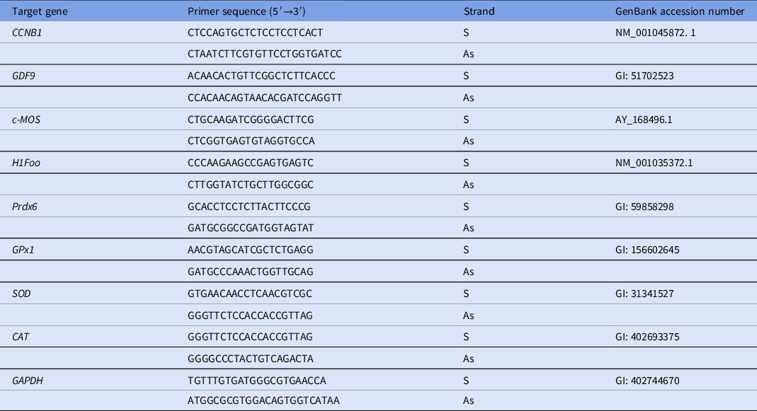
The quantification of messenger RNAs was performed using SYBR Green. Each real-time reaction (15 µl) contained 7.5 µl of SYBR Green Master Mix (PE Applied Biosystems, Foster City, CA), 5.5 µl of ultrapure water, 1 µl of cDNA and 0.5 µM of each primer. Real-time PCR was performed on a thermocycler (Master Cycler, Eppendorf, Germany) using oocytes samples from four repetitions of the experiment. The primers (Table 1) were designed to amplify specifically messenger RNAs for CAT, SOD, Prdx6, GPx, GDF9, H1Foo, CCNB1 and c-Mos. The glyceraldehyde-3-phosphate dehydrogenase (GAPDH) gene was used to normalize mRNA levels. The specificity of each primer pair was confirmed by analyzing the melting curve of PCR products. The thermal cycling profile for the first round of PCR was initially denaturation and activation of the polymerase for 10 min at 95°C, followed by 40 cycles of 15 s at 95°C, 30 s at 58°C, and 30 s at 72°C. The final extension was performed at 72°C for 10 min. All reactions were performed in a Step One Plus instrument (Applied Biosystems, Foster City, CA, USA). The 2−ΔΔCt method was used to transform the Ct values into mRNA expression relative levels (Livak and Schmittgen, Reference Livak and Schmittgen2001).
Primer pairs used for quantification of messenger RNAs

As, Anti-sense; S, sense.
Statistical analysis
Statistical analysis was performed using GraphPad Prism (5.0) software. The percentages of oocyte at the GV stage and with resumption of meiosis in the different treatments were evaluated using the chi-squared test. The data of expression of mRNAs for CAT, SOD, Prdx6, GPx, GDF-9, H1Foo, CCNB1 and c-Mos, as well as those of ROS and mitochondria in the oocytes, were analyzed using the Kruskal–Wallis test, followed by Dunn’s multiple comparison test. Differences were considered significant when the P-value was < 0.05.
Results
Assessment of meiotic progression in the oocytes
During IVM, the presence of different concentrations of NAC significantly reduced the percentages of GV oocytes when compared with control medium. Additionally, lower percentages of GV oocytes were observed after using culture medium supplemented with 0.5 and 2.5 mM NAC, when compared with those oocytes matured in the presence of 0.1 mM NAC. The presence of 0.5 and 2.5 mM NAC increased the percentages of oocytes with resumption of meiosis when compared with those cultured in control medium alone or supplemented with 0.1 mM NAC (Table 2).
Percentages of oocytes in germinal vesicle (GV) stage and that had meiotic resumption after IVM in the presence of different concentrations of NAC

A,B,CDifferences between treatments in each column, P < 0.05.
Mitochondrial activity in the oocytes
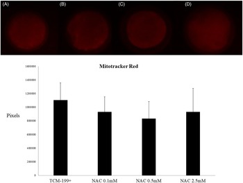
Oocytes matured in control medium alone or containing NAC had mitochondria uniformly distributed throughout the cytoplasm. No significant differences in fluorescence intensity were observed between the treatments (Figure 1).
Fluorescence staining intensity and distribution of mitochondria in oocytes after IVM in control medium alone (A) or supplemented with 0.1 mM (B), 0.5 mM (C), or 2.5 mM (D) NAC.

Intracellular levels of ROS
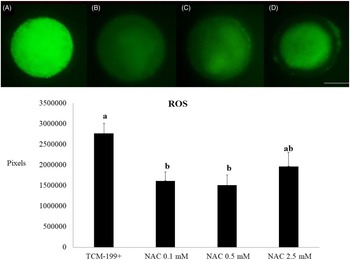
The intracellular levels of ROS in oocytes matured with 0.1 and 0.5 mM NAC were lower (P < 0.05) than those observed in oocytes matured in control medium (Figure 2). No significant differences were observed in ROS levels between oocytes matured in the presence of 2.5 mM NAC and those from the control group (P > 0.05) (Figure 2).
Fluorescence staining intensity for ROS in oocytes after IVM in control medium alone (A) or supplemented with 0.1 mM (B), 0.5 mM (C), or 2.5 mM (D) NAC. a,bSignificant differences between treatments (P < 0.05).

Analysis of TZP in the oocytes
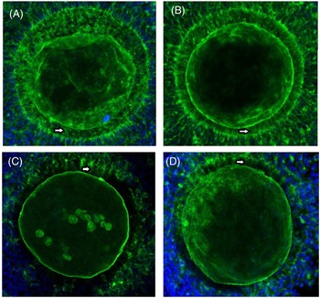
Fluorescence analysis showed that the distribution of TZP throughout the zona pellucida decreased in oocytes matured in the presence of 0.5 mM NAC when compared with those matured in control medium alone or supplemented with 0.1 or 2.5 mM NAC (Figure 3).
Confocal microscopy images showing TZP (arrows) distribution in oocytes after IVM in control medium alone (A) or supplemented with 0.1 mM (B), 0.5 mM (C), or 2.5 mM (D) NAC. Green staining: F-actin; blue DAPI staining: cellular nuclei.

Levels of mRNA for CAT, SOD, PRDX6, GPX, GDF-9, H1FOO, CCNB1 and c-MOS in the oocytes
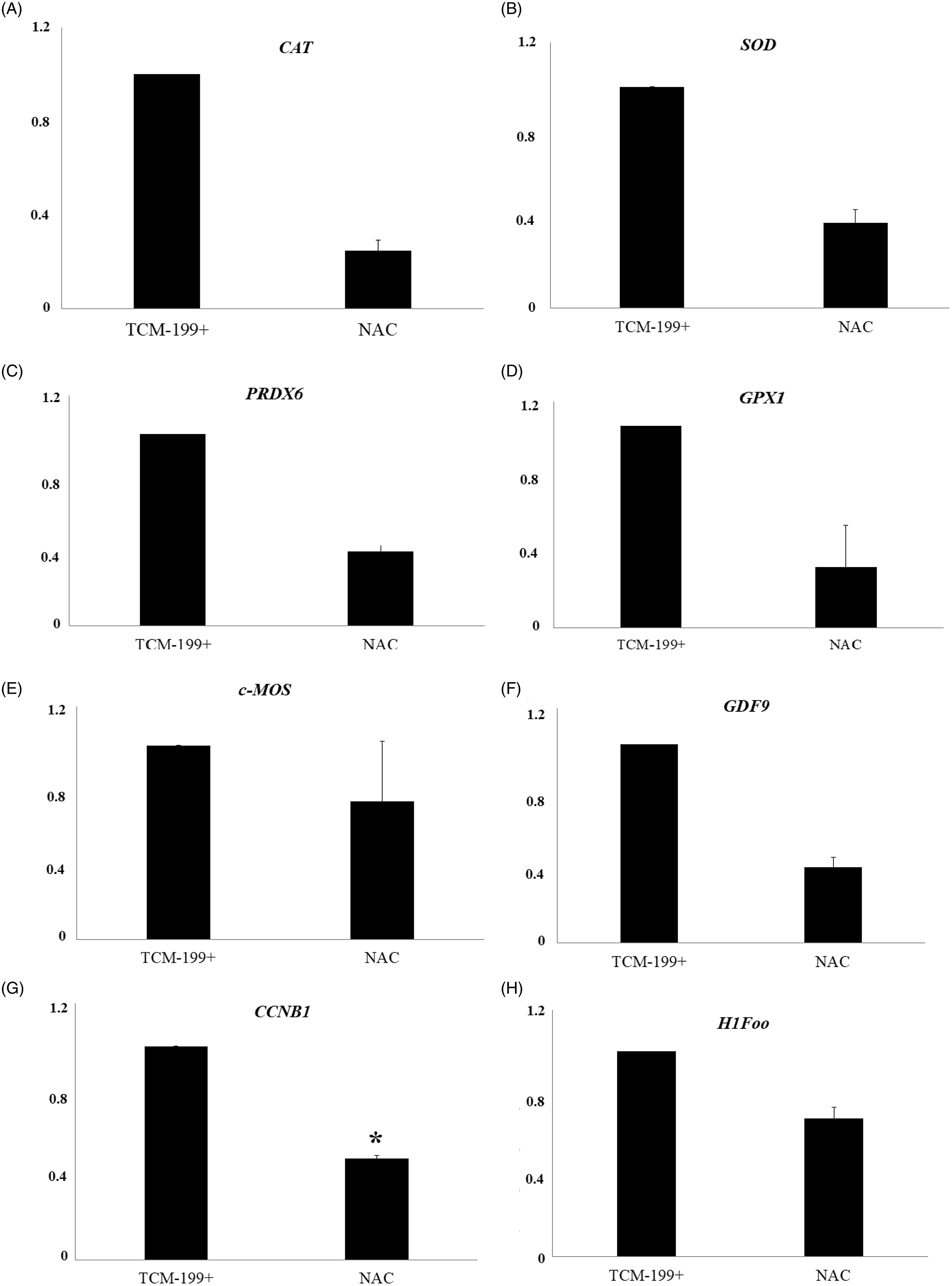
Figure 4 shows that 0.5 mM NAC significantly reduced the levels of messenger RNA for CCNβ1 after IVM. Despite an apparent reduction in the levels of mRNA for CAT, SOD, Prdx6, GPx, c-MOS, GDF9 and H1Foo in oocytes matured in the presence of NAC, the differences were not significant when compared with the control group (P > 0.05).
Levels of mRNA for CAT (A), SOD (B), Prdx6 (C), GPx1 (D), c-MOS (E), GDF9 (F), CCNB1 (G), and H1FOO (H) in bovine oocytes after maturation in TCM-199+ alone or supplemented with 0.5 mM NAC. *Statistically significant differences (P < 0.05).

Discussion
This study shows that NAC increases the meiotic progression and reduces the levels of ROS during oocyte maturation. It is known that during IVM, oocytes are exposed to high tensions of oxygen leading to increased production of ROS, which can damage DNA, RNA and proteins. Considering that oxidative stress in oocytes is often determined by measuring ROS levels (Li and Zhao, Reference Li and Zhao2019), NAC contributed to maintain the redox balance during IVM and had a positive effect on oocyte meiotic resumption. These effects may be associated with the capacity of NAC to scavenge free radicals and to chelate metals (Atkuri et al., Reference Atkuri, Mantovani, Herzenberg and Herzenberg2007). The ability of NAC to control oxidative stress by glutathione (GSH), is well known (Güntürk et al., Reference Güntürk, Yazici, Köse, Dağli, Yücel and Yay2019). Li and Zhao (Reference Li and Zhao2019) revealed that 0.1 mM NAC was able to reduce intracellular levels of bisphenol-induced ROS in in vitro matured mouse oocytes, restoring oocyte nuclear maturation and the subsequent competence of embryonic development after in vitro fertilization. Recently, Sun et al. (Reference Sun, Jang, Park, Oh, Lee, Hwang, Xu, Hwang and Lee2021) reported that treatment of oocytes with 1.0 mM NAC for 8 h during IVM attenuated intracellular ROS. Considering that antioxidant enzymes are synthesized in response to oxidative stress, a reduction in ROS in oocytes matured in the presence of NAC can explain the tendency to reduce the levels of mRNAs for enzymes that control oxidative stress such as Prdx6, SOD, CAT and GPx. The mechanism of action of NAC to control oxidative stress and to improve oocyte IVM was recently reviewed by Barrozo et al. (Reference Barrozo, Paulino, Silva, Barbalho, Nascimento, Neto and Silva2021).
The present study shows that oocytes cultured in vitro had a homogeneous distribution of mitochondria in the cytoplasm, but NAC did not influence mitochondrial activity. During oocyte maturation, mitochondria are a key component of the metabolic machinery responsible for supplying energy that is consumed during the maturation process and is also the main generator of free radicals in mammals (Ferreira et al., Reference Ferreira, Vireque, Adona, Meirelles, Ferriani and Navarro2009; Brand et al., Reference Brand, Orr, Perevoshchikova and Quinlan2013). The distribution of mitochondria to areas of high energy consumption is crucial for the oocyte and embryo during critical periods of the cell cycle. For this reason, the mitochondrial distribution pattern has been associated with the quality and developmental capacity of mammalian oocytes (Cajas et al., Reference Cajas, Cañón-Beltrán, Ladrón de Guevara, Millán de la Blanca, Ramos-Ibeas, Gutiérrez-Adán, Rizos and González2020). Mastrorocco et al. (Reference Mastrorocco, Cacopardo, Martino, Fanelli, Camillo, Ciani, Roelen, Ahluwalia and Dell’Aquila2020) demonstrated that dispersed mitochondrial distribution throughout the cytoplasm is associated with maturity and competence of oocytes. Furthermore, Takahashi et al. (Reference Takahashi, Hashimoto, Yamochi, Goto, Yamanaka, Amo, Matsumoto, Inoue, Ito, Nakaoka, Suzuki and Morimoto2016) revealed that mitochondria in human oocytes at the GV stage have a heterogeneous distribution, but after GVBD this configuration quickly changed to a homogeneous distribution. In rats, Dumollard et al. (Reference Dumollard, Duchen and Sardet2006) found that mitochondria appeared uniformly distributed in the periphery of the cytoplasm in immature oocytes. Romek et al. (Reference Romek, Gajda, Rolka and Smorąg2011) also described that there was an accumulation of mitochondria in the periphery before maturation and in the formation of mitochondria clusters located in the centre after oocyte maturation in cattle.
During development, growing oocytes engage in a prolonged phase of intensive RNA synthesis necessary to produce essential transcripts for oocyte growth and early embryonic development (Bezerra et al., Reference Bezerra, Lima, Paulino, Silva, Silva, Souza, van den Hurk and Silva2019). In the follicular microenvironment, cell communication between oocytes and cumulus cells is complex, and both sides have active regulatory functions. Macromolecules, such as RNAs (Macaulay et al., Reference Macaulay, Gilbert, Caballero, Barreto, Fournier, Tossou, Sirard, Clarke, Khandjian, Richard, Hyttel and Robert2014), can also be transported from cumulus cells to oocytes. In the present study, the distribution of TZP throughout the zona pellucida was reduced in oocytes matured with 0.5 mM NAC. Recently, Abbassi et al. (Reference Abbassi, El-Hayek, Carvalho, Wang, Yang, Granados-Aparici, Mondadori, Bordignon and Clarke2021) showed that TZP retraction is not regulated by oocyte-derived signals, but is instead controlled by epidermal growth factor receptor (EGFR) signalling within the granulosa cells. Previously, Wang et al. (Reference Wang, Hou, Yi, Wang, Ding, Chen, Long, Liu and Wu2013) reported that NAC enhances mRNA levels for EGF and amphiregulin in porcine epithelial cells. Fully grown oocytes within antral follicles are held in meiotic arrest through the action of cyclic GMP (cGMP) secreted only by the mural granulosa cells (Jaffe and Egbert, Reference Jaffe and Egbert2017). Consequently, the reduction in the distribution of TZP in oocytes cultured with 0.5 mM NAC led to a drop in cGMP transported to the oocyte, enabling the latter to initiate meiotic maturation.
Bezerra et al. (Reference Bezerra, Lima, Paulino, Silva, Silva, Souza, van den Hurk and Silva2019) showed that prematuration of oocytes from mid-sized antral follicles increased the levels of mRNA for CCNB1 and c-Mos, but did not influence the levels of mRNAs for GDF9, PARN, eIF4E and H1Foo. Conversely, these authors showed an increase in the levels of all this transcripts during in vivo and in vitro growth of preantral follicles, emphasizing that they were actively synthesized during follicular growth, but not at the late stages of oocyte maturation. Our study showed that NAC reduced the levels of CCNB1 during oocyte maturation, which can be associated with the higher rate of oocyte meiotic resumption in oocytes matured in the presence of NAC. This reduction can be associated with the translation of CCNB1 mRNA to proteins, which are required during meiosis (de Vantéry et al., Reference de Vantéry, Gavin, Vassalli and Schorderet-Slatkine1996). The apparent reduction in the levels of mRNA for c-MOS, GDF9 and H1Foo in oocytes matured in the presence of NAC can also be associated with their translation to proteins that will have an important role during oocyte fertilization and early embryo development. Regarding the role of these proteins, c-MOS is a kinase expressed exclusively in germ cells that have an important role in oocyte maturation (Wu et al., Reference Wu, Ignotz, Currie and Yang1997). The c-MOS–MAPK1/3 pathway is important for maintaining oocytes arrested at the MII stage (Phillips et al., Reference Phillips, Petrunewich, Collins, Booth, Liu and Baltz2002). GDF9 is involved in the process of oocyte maturation by regulating the function of cumulus cells from preovulatory follicles (Gui and Joyce, Reference Gui and Joyce2005). H1Foo plays a key role in the control of chromatin configuration and is essential for oocyte maturation (Furuya et al., Reference Furuya, Tanaka, Teranishi, Matsumoto, Hosoi, Saeki, Ishimoto, Minegishi, Iritani and Yoshimura2007).
In conclusion, the presence of 0.5 mM NAC in maturation medium increases the percentage of oocytes with resumption of meiosis and reduces the levels of ROS and the distribution of TZP in the oocyte, showing that supplementation of culture medium with NAC is very important to optimize oocyte IVM. NAC also reduces mRNA levels for CCNB1, but does not influence the expression of CAT, GPX, Prdx-6, SOD, GDF9, H1Foo and c-Mos. Understanding the mechanisms involved in oxidative stress control and how NAC is able to reduce the damage caused by oxidative stress can contribute to the improvement of oocyte IVM.
Financial support
This research was supported by grants from the National Council for Scientific and Technological Development (CNPq, Brazil, grant number 308737/2018-0). The authors would like to thank the Central Analítica-UFC (funded by Finep-CT-INFRA, CAPES-Pró-Equipamentos, and MCTI-CNPq-SisNano2.0) for microscopy measurements.
Statement of interest
None of the authors have any conflict of interest to declare.
Ethical standards
The authors declare that all procedures were performed according to national and institutional guidelines on the care and use of animals.