Introduction
Intracytoplasmic sperm injection (ICSI) is an assisted reproduction technique that involves the injection of a single sperm into the cytoplasm of a mature oocyte arrested in metaphase II (MII) (García-Roselló et al., Reference García-Roselló, García-Mengual, Coy, Alfonso and Silvestre2009; Salamone et al., Reference Salamone, Canel and Rodríguez2017; Fuentes et al., Reference Fuentes, Muñoz, Contreras, Arias and Felmer2022). ICSI does not present major problems in humans, rabbits and mice (Keefer, Reference Keefer1989; Van Steirteghem et al., Reference Van Steirteghem, Nagy, Joris, Liu, Staessen, Smitz, Wisanto and Devroey1993; Kimura & Yanagimachi, Reference Kimura and Yanagimachi1995), since the injection is a sufficient stimulus to activate the oocyte. However, in the bovine species, oocytes are not efficiently activated by the sperm after injection, resulting in low developmental rates in vitro. Importantly, successful activation not only relies on the intrinsic competence of the oocyte, but also on the proper sperm membrane breaking and the release of sperm-borne oocyte-activating factors, such as phospholipase C zeta (PLCζ). Inefficiencies in this process have been linked to incomplete Ca2+ oscillations and failure of pronuclear (PN) formation in bovine ICSI (Saunders et al., Reference Saunders, Larman, Parrington, Cox, Royse, Blayney, Swann and Lai2002; Kashir et al., Reference Kashir, Nomikos and Lai2018).
To exit from MII arrest, bovine oocyte activation protocols must include a chemical or physical stimulus that increases intracellular Ca2+ concentrations, generally employing ionomycin (Rho et al., Reference Rho, Wu, Kawarsky, Leibo and Betteridge1998a; Hamano et al., Reference Hamano, Li, Qian, Funauchi, Furudate and Minato1999), Ca2+ ionophore (A23187), or ethanol (Fissore & Robl, Reference Fissore and Robl1993). During fertilization, the repetitive Ca2+ oscillations induced by the sperm trigger a cascade of biochemical events that reduce the activity of the maturation-promoting factor (MPF), a key complex formed by cyclin B and p34^cdc2, the latter being specifically regulated by phosphorylation and dephosphorylation (Alberio et al., Reference Alberio, Zakhartchenko, Motlik and Wolf2001). The decrease in MPF kinase activity, mediated by Ca2+-dependent pathways, leads to the inactivation of the metaphase II–arresting molecules and allows resumption of the cell cycle (Nakada & Mizuno, Reference Nakada and Mizuno1998; Malcuit et al., Reference Malcuit, Kurokawa and Fissore2006; Abdalla et al., Reference Abdalla, Shimoda, Hirabayashi and Hochi2009). However, in bovine oocytes, the Ca2+-mediated MPF inactivation induced by these artificial stimuli is only transient, which often results in insufficient sperm head decondensation and poor embryonic development (Susko-Parrish et al., Reference Susko-Parrish, Leibfried-Rutledge, Northey, Schutzkus and First1994). To overcome this limitation, activation protocols have incorporated inhibitors that prolong MPF inactivation, such as dimethylaminopurine (DMAP), which blocks protein kinase phosphorylation, or protein synthesis inhibitors including cycloheximide (CHX) and anisomycin (ANY) (Rho et al., Reference Rho, Kawarsky, Johnson, Kochhar and Betteridge1998b; Li et al., Reference Li, Hamano, Qian, Funauchi, Furudate and Minato1999; Chung et al., Reference Chung, Keefer and Downey2000; Oikawa et al., Reference Oikawa, Takada, Kikuchi, Numabe, Takenaka and Horiuchi2005; Bevacqua et al., 2010; Vichera et al., 2010; Arias et al., Reference Arias, Sánchez and Felmer2016; Akagi et al., Reference Akagi, Tani and Matsukawa2020; Valencia et al., Reference Valencia, Pérez, Matus, Felmer and Arias2021).
Another problem that ICSI presents in cattle is the low rate of implantation and births. In fact, only one calf has been generated with DMAP (Oikawa et al., Reference Oikawa, Takada, Kikuchi, Numabe, Takenaka and Horiuchi2005), while the birth of calves generated with CHX has not been described to date. These data contrast with more than 40 calves born from ethanol-activated oocytes (Hamano et al., Reference Hamano, Li, Qian, Funauchi, Furudate and Minato1999; Horiuchi et al., Reference Horiuchi, Emuta, Yamauchi, Oikawa, Numabe and Yanagimachi2002; Oikawa et al., Reference Oikawa, Takada, Kikuchi, Numabe, Takenaka and Horiuchi2005; Ashibe et al., Reference Ashibe, Miyamoto, Kato and Nagao2019). One of the most effective ethanol-based oocyte activation protocols involved incubating oocytes for 5 min with 7% ethanol, after 4 h of sperm injection. This approach yielded blastocysts ranging from 14 – 29%, depending on the study and the generation of live offspring (Horiuchi et al., Reference Horiuchi, Emuta, Yamauchi, Oikawa, Numabe and Yanagimachi2002; Fujimani et al., 2004; Oikawa et al., Reference Oikawa, Takada, Kikuchi, Numabe, Takenaka and Horiuchi2005). In the present study, we also evaluated different combinations of oocyte activation protocols based on ethanol and proposed a new strategy that incorporates Glutathione (GSH), Cysteamine (Cys) or dithiobutylamine (DTBA) during the incubation of injected oocytes as sperm head decondensation compounds. GSH is a major intracellular redox regulator that accumulates in the ooplasm during maturation and reduces disulphide bonds in sperm protamines, thereby facilitating sperm chromatin decondensation and male pronucleus formation. Cys, as a precursor of GSH, enhances intracellular GSH synthesis and contributes to maintaining the reducing environment required for this process (Sutovsky & Schatten, Reference Sutovsky and Schatten1997; De Matos & Furnus, Reference De Matos and Furnus2000).
In cattle, the efficiency of ICSI has remained limited compared with other species, largely due to difficulties in achieving reliable oocyte activation. While mechanical penetration or sperm injection alone is insufficient to trigger PN formation (Rho et al., Reference Rho, Wu, Kawarsky, Leibo and Betteridge1998a; Hwang et al., Reference Hwang, Lee, Yoon, Yoon, Lee and Choi2000), several activation methods have been tested with variable outcomes. Calcium ionophores, ethanol treatment and inhibitors of protein synthesis can induce oocyte activation, yet their capacity to support embryo development to the blastocyst stage or to term remains inconsistent (Keskintepe et al., Reference Keskintepe, Pacholczyk, Machnicka, Norris, Curuk, Khan and Brackett1997; Chung et al., Reference Chung, Keefer and Downey2000). Given the central role of oocyte activation in determining ICSI success in cattle, optimizing these protocols is essential to advance the efficiency and applicability of this technique in reproductive biotechnology.
The aim of this study was to evaluate the developmental competence of bovine oocytes activated by different combinations of protocols, including parthenogenetic haploid and diploid activation and oocytes subjected to ICSI with or without additional activation treatments. Furthermore, we proposed a new strategy incorporating GSH, Cys or DTBA during the incubation of injected oocytes to enhance sperm head decondensation and improve PN formation and embryo development.
Materials and methods
Unless stated otherwise, all chemicals were purchased from Sigma-Aldrich (St Louis, MO, USA).
Ovary collection
The ovaries were obtained from adult multiparous Holstein-Friesian cows (3–7 years old) at a local slaughterhouse (Frigorífico Temuco, Chile). After collection, they were transported to the laboratory in saline solution (0.9% sodium chloride) supplemented with penicillin and streptomycin, maintained at 37°C. Only ovaries with a normal external appearance, free of visible pathological lesions, were included in the study. In the laboratory, the ovaries were washed and, subsequently, cumulus-oocyte complexes (COCs) were aspirated from 2–6 mm diameter follicles using a 10 mL syringe attached to an 18G needle. The COCs were selected under stereoscopic image, choosing those with more than six layers of compact cumulus cells and homogeneous cytoplasm.
In vitro maturation of oocytes
Selected oocytes were matured in a 4-well NUNC plate with TCM-199 medium supplemented with 6 µg/ml luteinizing hormone (LH) (Sioux Biochemical, Inc. Sioux City, IA, USA), 6 µg/mL follicle stimulating hormone (FSH) (Bioniche Life Science Inc. Belleville, Ontario, Canada), 1 μg/mL of oestradiol, 0.2 mM of pyruvate, 0.025 mg/ml of gentamicin and 10% FBS (HyClone Laboratories, Inc., Logan, UT, USA) for 24 h at 38.5°C, 5% CO2 in air and saturation humidity.
Semen preparation and ICSI procedure
Commercial frozen semen from a single Holstein-Friesian bull (5 years old) was used throughout all experiments to minimize variability associated with paternal factors. The semen, provided by a certified AI centre (Alta Genetics Inc., Alberta, Canada), had proven in vivo and in vitro fertilization capacity and was collected and cryopreserved under standardized conditions. Prior to use, spermatozoa were selected by Percoll gradient. This approach ensured consistency and reproducibility of ICSI. Oocytes were denuded by exposure to 0.1% hyaluronidase (Sigma-Aldrich) for up to 3 min with gentle vortexing (speed 5), followed by repeated pipetting until all cumulus cells were removed. For ICSI, individual motile spermatozoa with normal morphology (intact head, midpiece and tail) were selected under 400× magnification using Hoffman optics. Only spermatozoa free of visible structural abnormalities were used for injection.
ICSI was developed in microdroplets of HECM-Hepes (HH) medium with an inverted microscope (Nikon TS100, Nikon Instruments, New York, NY, USA) and hydraulic micromanipulators (Narishige International, New York, NY, USA). Microinjections were carried out at × 200 magnification with the stage warm set at 37.5°C with a piezo-ICSI (PMAS-CT150 piezo-drill; Prime Tech, Japan).
The spermatozoa were selected according to their normal morphology from 5% PVP drops (Fisher Scientific). Each spermatozoon was immobilized with a pulse caused by the Piezo with an intensity 10 and speed 7. Oocytes were located with their polar body in the orientation of 6 or 12 o’clock position, to perform the microinjection at the 3 o’clock. The zona pellucida was crossed using a Piezo pulse of intensity 10 and speed 7 and to rupture the oolemma an intensity of 4 and speed 3 was used. After this rupture, the spermatozoon was deposited into the oocyte.
Oocyte activation protocols
Mature oocytes were selected to generate haploid parthenotes. Selected oocytes were activated with 5 μM ionomycin (Io) for 4 min and incubated for 4 h in KSOM medium. After incubation, they were activated again with a second pulse in 1%, 3%, 7% and 10% ethanol (Et) for 5 min. To generate diploid parthenotes, mature oocytes were activated with 5 µM Io for 4 min or 7% Et for 5 min and then incubated for 4 or 5 h in KSOM medium supplemented with 2 mM DMAP or 1 µg/mL ANY with cytochalasin B (CB) 5 µg/mL, respectively.
During oocyte activation protocols of sperm-injected zygotes the activation medium was supplemented with 15 mM GSH, 1 mM Cys and 5 mM DTBA. The concentrations of the different decondensation compounds tested were based on previous literature (Zambrano et al., Reference Zambrano, Águila, Arias, Sánchez and Felmer2017, Canel et al., Reference Canel, Suvá, Bevacqua, Arias, Felmer and Salamone2018 and Suttirojpattana et al., Reference Suttirojpattana, Somfai, Matoba, Nagai, Parnpai and Geshi2016, respectively).
Embryo culture
The parthenotes and presumptive zygotes generated by ICSI were cultured for the first 72 h in 50 µL drops of KSOM/0.4% BSA-(FAF) medium (Millipore) covered with mineral oil and on day 3 were supplemented with 5% FBS. The culture was carried out with a low O2 tension atmosphere (5% O2, 5% CO2 and 90% N2) at 38.5ºC and saturation humidity. The cleavage rate was evaluated after 72 h of culture while the blastocyst rate was evaluated at day 8.
Pronuclear evaluation
Presumptive zygotes were fixed at 16–18 h post-ICSI in 4% paraformaldehyde (PFA) in PBS (15 min, RT), permeabilized with 0.1% Triton X-100 in PBS (5 min) and counterstained with Hoechst 33342 (5–10 µg/mL in PBS, 10 min, RT, protected from light). Samples were mounted in anti-fade medium and examined using an epifluorescence microscope Nikon TS100F equipped with DAPI/Hoechst filter set; 365–405 nm excitation, with a 40× or 60× objective.
Pronuclear (PN) scoring criteria. Zygotes were classified as: 0PN: no visible pronuclei; chromatin condensed or irregular. 1PN: single pronucleus present. 2PN (normal): distinct male and female pronuclei with visible nucleolar precursor bodies when present. >2PN (abnormal): polypronuclear. When no PN were observed, chromatin status was recorded as condensed, partially decondensed or fragmented.
Experimental design
Experiment 1 was designed to evaluate the effect of different concentration of ethanol (1%, 3%, 5% and 7%) in a base protocol of haploid parthenote activation (Io 4 min + 4 h incubation + 5 min). The DMAP control group consisted of oocytes activated with Io 4 min followed for 4 h in KSOM medium supplemented with 2 mM DMAP.
Experiment 2 was designed to evaluate the effect of activating diploid parthenotes. For this, different conditions were evaluated to allow the generation of diploid parthenotes. These conditions included a first pulse of Ca2+ with Io followed by the incubation with DMAP for 4 h or ANY+CB for 5 h, respectively, and a first pulse of Et followed by the incubation with DMAP for 4 h or ANY+CB for 5 h.
Experiment 3 was designed to evaluate the effect of applying an additional activation step in diploid parthenotes. For this, different conditions were evaluated to allow the generation of diploid parthenotes. These conditions included a first pulse of Ca2+ with Io followed by incubation with DMAP for 4 h or ANY+CB for 5 h, respectively, and a first pulse of Et followed by incubation with DMAP for 4 h or ANY+CB for 5 h. After these treatments, an additional pulse of 7% Et for 5 min was applied.
Experiment 4 was designed to evaluate the effect of different sperm head decondensation compounds (GSH, Cys and DTBA) added during oocyte activation on the PN formation of bovine zygotes generated by Piezo-ICSI. After sperm injection, presumptive zygotes were activated with ionomycin (5 μM, 4 min) followed by incubation in KSOM medium supplemented with ANY (1 μg/mL) and CB (5 μg/mL) for 5 h. During this incubation period, GSH (15 mM), Cys (1 mM) or DTBA (5 mM) were added as decondensation agents. At the end of the activation, oocytes were exposed to 7% ethanol for 5 min, washed and then cultured in KSOM medium under the same conditions described above until PN evaluation.
Experiment 5 was conducted under the same conditions as Experiment 4, but instead of PN assessment, embryos were cultured in KSOM medium and evaluated for cleavage at 72 h and blastocyst formation at day 8.
Statistical analysis
Quantitative data sets are presented as means and standard deviation (± S.D.) and analyzed using one-way ANOVA. Post hoc analysis to identify differences between groups was performed using Tukey test. Binomial data sets, such as PN formation, were analyzed by using Fisher test. Differences were considered significant at p < 0.05.
Results
In vitro development of haploid parthenogenetic embryos
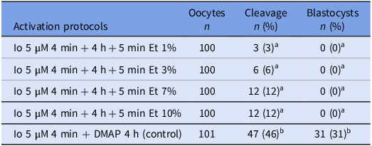
In the first experiment, 501 oocytes were parthenogenetically activated with different concentrations of ethanol. The results showed no differences in embryo cleavage (range 3 – 12%) among treatments. Differences were only observed between the Et treatments and the DMAP control (46% cleavage). Regarding the development of blastocysts on day 8 (D8), none of the treatments reached this stage of development, which contrasts with the control with 30.6% blastocysts (Table 1). Thus, these data indicate that ethanol pulse after ionomycin activation is not sufficient for the parthenogenetic activation of haploid oocytes.
In vitro development of haploid parthenogenetic embryos

Io = Ionomycin; DMAP = Dimethylaminopurine; Et = Ethanol.
Different letters above the columns indicate significant differences (P < 0.05).
In vitro development of diploid parthenogenetic embryos
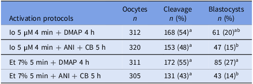
In the second experiment, the generation of diploid parthenotes embryos was evaluated. In this experiment, 1248 oocytes were used, of which 632 were distributed in 2 treatments activated with Io, while the remaining 616 were distributed in 2 treatments activated with ET 7%. The results of this experiment showed no differences in embryo cleavage between all treatments (Table 2). In this experiment, all treatments achieved the blastocyst stage (Table 2). However, the treatments (Io 5 μM for 4 min + DMAP 2 mM for 4 h) and (Et 7% for 5 min + DMAP 2 mM for 4 h) showed higher cleavage and blastocyst rates, and the treatment (Et 7% for 5 min + DMAP 2 mM for 4 h) achieving the highest blastocyst rate (Table 2). The results suggest that ethanol pulse before incubation with DMAP is effective inducing parthenogenetic development.
In vitro development of diploid parthenogenetic embryos

Cleavage and blastocyst rates were recorded on D3 and D8, respectively (12 replicates for each treatment).
Et = Ethanol; Io = Ionomycin; DMAP = Dimethylaminopurine; ANI = Anisomycin; Cito B = Cytochalasin B.
In vitro development of diploid parthenogenetic embryos activated with a second pulse of ethanol
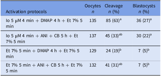
In this experiment, the generation of parthenogenetic diploid embryos was evaluated as in experiment 2, with the difference that a second pulse with 7% ethanol was performed at the end of the incubation. Differences in the cleavage were only found between the protocols (Io 5 μM 4 min + DMAP 2 mM 4 h + Et 7% 5 min) and (Et 7% 5 min + DMAP 2 mM 4 h + Et 7% 5 min) (63 and 19%, respectively) (Table 3). The blastocyst rates were higher in the treatments with two pulses of Ca2+ at the beginning and at the end of the protocols (Io 5 µM 4 min + DMAP 2 mM 4 h + Et 7% 5 min) and (Io 5 µM 4 min + ANY 1 mg/mL + CB 5 h + Et 7% 5 min), the first treatment showing the highest blastocyst rates (Table 3). Both treatments with Et at the beginning and at the end (Et 7% 5 min + DMAP 2 mM 4 h + Et 7% 5 min and Et 7% 5 min + ANY 1 µg/mL + CB 5 h + Et 7% 5 min), presented the lowest rates of cleavage and blastocyst formation (Table 3). Overall, these data indicate that two pulses of ethanol are deleterious to induce oocyte activation.
In vitro development of diploid parthenogenetic embryos with 2 pulses of Ca2+

Cleavage and blastocyst rates were recorded on D3 and D8, respectively (5 replicates for each treatment).
Et = Ethanol; Io = Ionomycin; DMAP = Dimethylaminopurine; ANI = Anisomycin; CB = Cytochalasin B.
Different letters above the columns indicate significant differences (P < 0.05).
Pronuclear formation of injected oocytes incubated with sperm head decondensation compounds (GSH, Cys and DTBA) during oocyte activation
In this experiment, 281 presumptive zygotes were used to evaluate the PN formation. The highest rates of male PN formation were observed in the control (Io 4 min ANY + CB 5 h Et 7% 5 min) with 21%, and the treatment (Io 4 min ANY + CB DTBA 5 h Et 7% 5 min) with 10% (Table 4 and Fig. 1 Panel A). It was also observed that approximately half of the presumptive zygotes had a condensed sperm head indicating that these zygotes had not been activated (Table 4 and Fig. 1 Panel B). The treatment that presented the highest rate of inactivated oocytes with condensed sperm heads was (Io 4 min ANY + CB GSH 5 h Et 7% 5 min) with 75%, which was significantly higher than the other treatments (P < 0.05), while the treatments with the lowest rate of inactivated oocytes were (Io 4 min ANY + CB 5 h Et 7% 5 min) with 44% and (Io 4 min ANY + CB + DTBA 5 h Et 7% 5 min) with 40% (Table 4). However, the treatment including DTBA exhibited more pronounced nuclear alterations (Table 4 and Fig.1 Panel C).
Pronuclear formation of injected oocytes incubated with sperm head decondensation compounds (Gsh, Cys and DTBA) during activation

Pronuclear evaluation was analyzed after 14 and 20 h post activation.
Io = Ionomycin; Any = Anisomycin; CB = Cytochalasin; Et = Ethanol; Gsh = Glutathione; Cys = Cysteamine; DTBA = Dithiobutylamine; FP = Female pronucleus; PB = Polar body; CSH = Condensed sperm head; PDSH = Partially decondensed sperm head; MP = Male pronucleus. Other nuclear configurations: multinucleated zygote (as the presence of more than one pronucleus in haploid groups and more than 2 pronuclei in diploid zygotes).
Different letters above the columns indicate significant differences (P < 0.05).
Representative images of the pronuclear evaluation 18 h postactivation. Panel A. FP, Female pronucleus; MP, Male pronucleus; PB, Polar body. Panel B. CSH, Condensed sperm head; FP, Female pronucleus; PB, Polar body (PB). Panel C. PN, Pronucleus.

In vitro development of injected oocytes incubated with sperm head decondensation compounds (GSH, Cys and DTBA) during oocyte activation
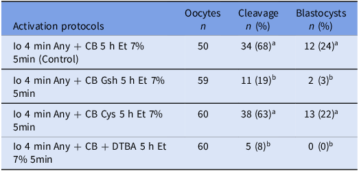
In this experiment, 229 injected oocytes were generated by Piezo-ICSI in the same conditions as the experiment above and the embryos analyzed at D3 and D8 of the embryo culture. The control without sperm head decondensation treatment (Io 4 min ANY 5 h Et 7% 5 min) and (Io 4 min ANY + Cys 5 h Et 7% 5 min) showed the highest cleavage rates (68% and 63%) and blastocyst rates (24% and 22%), respectively (Table 5), compared to GSH and DTBA treatments, which showed the lowest cleavage and blastocyst rates. In fact, the treatment in the presence of DTBA had the lowest performance among the treatments evaluated with no development to the blastocyst stage. Thus, these data suggest that the inclusion of sperm head decondensing agents does not negatively impact the early in vitro development of ICSI-generated embryos.
In vitro development of injected oocytes incubated with sperm head decondensation compounds (Gsh, Cys and DTBA) during activation

Et = Ethanol; Io = Ionomycin; DMAP = Dimethylaminopurine; ANI = Anisomycin; Cito B = Cytochalasin B; Gsh = Glutation; Cys = Cysteamine; DTBA = Dithiobutylamine (3 replicates for each treatment).
Different letters above the columns indicate significant differences (P < 0.05).
Discussion
The activation of the oocytes and the decondensation of the sperm head are two relevant factors for the low efficiency of ICSI in bovines. In addition, the efficiency of bovine ICSI also depends on the correct breakdown of the sperm plasma membrane to allow the release of oocyte-activating factors, notably PLCζ, which are essential to trigger the physiological Ca2+ oscillations required for meiotic resumption and PN formation. Therefore, both the oocyte’s responsiveness and the sperm’s ability to deliver activating signals must be considered when optimizing activation protocols (Sounders et al., 2002; Kashir et al., Reference Kashir, Nomikos and Lai2018).
To address these factors, we evaluated different oocyte activation strategies using ethanol as the basis, since ethanol-based protocols are the only ones consistently reported to generate live offspring (Hamano et al., Reference Hamano, Li, Qian, Funauchi, Furudate and Minato1999; Horiuchi et al., Reference Horiuchi, Emuta, Yamauchi, Oikawa, Numabe and Yanagimachi2002; Oikawa et al., Reference Oikawa, Takada, Kikuchi, Numabe, Takenaka and Horiuchi2005; Ashibe et al., Reference Ashibe, Miyamoto, Kato and Nagao2019) in bovine species.
In the first part of this study, different concentrations of ethanol (1, 3, 7 and 10%) were used to evaluate its effectiveness in the activation of haploid parthenogenetic oocytes after an initial Ca2+ pulse with ionomycin and 4 h of incubation. Cleavage and blastocyst rates were noticeably lower (range 2 to 12% and 0%, respectively), than the control using Io + DMAP for 4 h (46% and 30%, respectively). This experiment revealed that a pulse of ethanol after ionomycin activation is not sufficient for the parthenogenetic activation of haploid oocytes. This finding is consistent with previous studies suggesting that optimal oocyte activation requires more complex stimuli (Méo et al., Reference Méo, Leal and Garcia2004; Abdalla et al., Reference Abdalla, Shimoda, Hirabayashi and Hochi2009).
Although the cleavage and blastocyst rates observed with ethanol alone were low, it should be noted that ethanol is the only chemical activation protocol consistently associated with the birth of live calves after ICSI (Hamano et al., Reference Hamano, Li, Qian, Funauchi, Furudate and Minato1999; Horiuchi et al., Reference Horiuchi, Emuta, Yamauchi, Oikawa, Numabe and Yanagimachi2002; Oikawa et al., Reference Oikawa, Takada, Kikuchi, Numabe, Takenaka and Horiuchi2005; Ashibe et al., Reference Ashibe, Miyamoto, Kato and Nagao2019). In fact, our data (Table 1) suggests a slight increase in cleavage rates at higher ethanol concentrations (10%), raising the possibility of dose-dependent effects. Moreover, ethanol activation has shown synergistic effects when combined with inhibitors such as DMAP, supporting more efficient and sustained MPF inactivation (Fujinami et al., Reference Fujinami, Hosoi, Kato, Matsumoto, Saeki and Iritani2004; Abdalla et al., Reference Abdalla, Shimoda, Hirabayashi and Hochi2009). Therefore, while ethanol alone may be insufficient to ensure complete activation, its potential advantages in combinatorial strategies warrant further consideration.
However, a significant improvement was observed in the rates of embryonic development in diploid parthenotes, using ethanol combined with DMAP and ionomycin combined with DMAP. These treatments showed the highest rate of cleavage and blastocyst rates (55% and 27% and 54% and 20%, respectively). Suvá et al. (Reference Suvá, Canel and Salamone2019) used ionomycin as a control instead of ethanol plus 4 h of incubation in DMAP and obtained high rates of cleavage and development to blastocysts (79% and 40%, respectively). However, the same conditions evaluated by Abdalla et al. (Reference Abdalla, Shimoda, Hirabayashi and Hochi2009) showed low rates of cleavage and blastocysts (57% and 11%, respectively), suggesting that this combination (Io+DMAP) lack of reproducibility.
With the aim of improving these oocyte activation treatments, the effect of adding a second Ca2+ pulse with ethanol 7% was evaluated under the same conditions. In this experiment, the best treatment was Io - DMAP for 4 h and Et, a treatment that achieved a high cleavage rate (63%) and a blastocyst development of 27%. Importantly, in this experiment, using two ethanol oocyte activation pulses, at the beginning and at the end of the protocol, respectively, resulted in a low cleavage rate and very low blastocyst rates (5%), suggesting a deleterious effect. Excess of ethanol provided by the two pulses was apparently detrimental to the embryonic development, confirming previous findings by Gurtovenko et al. (Reference Gurtovenko and Anwar2009). However, two sequential activations with lower ethanol concentrations may help avoid toxicity while still ensuring effective MPF inactivation and sperm head decondensation. Although not evaluated in this study, this strategy represents a promising approach for future investigation. Finally, the effects of different sperm head decondensing agents were evaluated. During fertilization, the condensed sperm chromatin undergoes decondensation to generate the male pronucleus. This process is supported by reduced GSH, which accumulates in the ooplasm during oocyte maturation (Sutovsky & Schatten Reference Sutovsky and Schatten1997). In fact, in in vitro embryo production 0.1 mM cysteamine (Cys) is generally added to the in vitro maturation (IVM) medium as a primary precursor of GSH (De Matos & Furnus C. Reference De Matos and Furnus2000). For this experiment, presumptive zygotes were incubated with GSH, Cys, or DTBA after following Piezo-ICSI, rather than pre-treating the sperm with these compounds as is usually done (Sekhavati et al., Reference Sekhavati, Shadanloo, Hosseini, Tahmoorespur, Nasiri, Hajian and Nasr-Esfahani2012; Suttirojpattana et al., Reference Suttirojpattana, Somfai, Matoba, Nagai, Parnpai and Geshi2016; Zambrano et al., Reference Zambrano, Águila, Arias, Sánchez and Felmer2017). Under these conditions, between 40% and 75% of the injected oocytes in the different treatments were not activated and therefore did not initiate decondensation of the sperm head, regardless of the treatment. This result contrast with the high male PN formation reported by Zambrano et al. (Reference Zambrano, Águila, Arias, Sánchez and Felmer2016) when GSH was used as sperm pretreatment before ICSI. Regarding sperm head decondensation, the control group (without decondensation treatment) and the DTBA treatment, yielded the highest rates of female and male pronuclei formation, with 21% and 10%, while the GSH and Cys treatments resulted in the lowest rates of PN formation (3% and 5%, respectively).
Finally, embryos were generated by Piezo-ICSI with the same treatments as the previous experiment to assess the development up to the blastocyst stage. In this experiment, the Piezo-ICSI-Cys treatment achieved good cleavage and blastocyst rates (63% and 22%, respectively), like the control without sperm head decondensation treatment. These results are consistent with those reported by Abdalla et al., (Reference Abdalla, Shimoda, Hirabayashi and Hochi2009), who used dithiothreitol as a decondensing agent. However, DTBA and GSH treatments did not yield favourable cleavage or blastocyst rates. In fact, DTBA treatment failed to produce any blastocysts. This effect could be attributed to the turbidity caused by the reagent or the concentration used, which may have had some embryotoxic effects, an issue previously reported by Suttirojpattana et al. (Reference Suttirojpattana, Somfai, Matoba, Nagai, Parnpai and Geshi2016) with sperm pretreated with DTBA before ICSI. Interestingly, DTBA in this previous study increased the efficiency of blastocyst production by ICSI. However, the use of DTBA in our conditions (during the activation) was associated with a higher incidence of nuclear configurations such as the presence of more than 2 pronuclei in diploid zygotes, possibly explaining the poor outcomes of this treatment. The GSH treatment also resulted in low cleavage and blastocyst rates. In this study, a high concentration of GSH (15 mM) was used based on previous reports where this level had been applied in bovine ICSI (Zambrano et al., Reference Zambrano, Águila, Arias, Sánchez and Felmer2017). Nevertheless, no further optimization was attempted, and the poor outcomes observed here may reflect the embryotoxic effects of excessive GSH, as concentrations above 5 mM have been reported to negatively affect sperm function and embryo development (Gadea et al., Reference Gadea, Gumbao, Matas and Romar2005; Zhang et al., Reference Zhang, Liu, Wang, Yang and Hu2016). In agreement with these observations, our results showed that supplementation with 15 mM GSH during the activation period did not significantly improve pronucleus formation, suggesting that under these conditions GSH was unable to favour chromatin decondensation and male pronucleus development. This is consistent with findings in boar sperm head decondensation treatments prior to ICSI, where high concentrations of GSH excessively reduced ROS levels, leading to unfavourable outcomes (Gadea et al., Reference Gadea, Gumbao, Matas and Romar2005; Zhang et al., Reference Zhang, Liu, Wang, Yang and Hu2016). By contrast, Zambrano et al. (Reference Zambrano, Águila, Arias, Sánchez and Felmer2017) reported that the use of 15 mM GSH in combination with sperm pretreatment prior to injection improved sperm head decondensation and increased pronucleus formation, thereby enhancing preimplantation embryo development. These methodological differences suggest that the timing of GSH exposure, whether applied to sperm before injection or to oocytes during activation, may be a critical factor determining its effect on pronucleus formation and subsequent development.
Similarly, DTBA employed by Suttirojpattana et al., (Reference Suttirojpattana, Somfai, Matoba, Nagai, Parnpai and Geshi2016) proved to be more efficient than dithiothreitol, a widely used reducer of disulphide bonds in spermatozoa. In their study, DTBA enhanced both PN formation rates and development to the blastocyst stage. These data highlight the need for careful standardization of the concentration and time of incubation of these reducing agents when used during the oocyte activation period. Finally, the Cys treatment demonstrated embryonic development rates comparable to the control without sperm decondensing agents, suggesting that Cys could be a promising alternative for improving embryo generation coupled to ICSI.
The present results suggest that the efficiency of bovine ICSI remains constrained by incomplete oocyte activation and inefficient sperm head decondensation. Biologically, this may reflect the transient nature of Ca2+-mediated MPF inactivation in cattle (Malcuit et al., Reference Malcuit, Kurokawa and Fissore2006; Abdalla et al., Reference Abdalla, Shimoda, Hirabayashi and Hochi2009) and the limited availability of oocyte cytoplasmic factors to remodel sperm chromatin. Our findings with cysteamine support its role in enhancing intracellular GSH and promoting decondensation, whereas excessive concentrations of GSH or DTBA appear detrimental, in line with previous reports (Suttirojpattana et al., Reference Suttirojpattana, Somfai, Matoba, Nagai, Parnpai and Geshi2016; Zambrano et al., Reference Zambrano, Águila, Arias, Sánchez and Felmer2017). These observations emphasize the importance of fine-tuning activation protocols to balance efficacy and cytotoxicity. From an applied perspective, ethanol-based protocols remain attractive given their proven ability to support live births (Hamano et al., Reference Hamano, Li, Qian, Funauchi, Furudate and Minato1999; Horiuchi et al., Reference Horiuchi, Emuta, Yamauchi, Oikawa, Numabe and Yanagimachi2002; Oikawa et al., Reference Oikawa, Takada, Kikuchi, Numabe, Takenaka and Horiuchi2005; Ashibe et al., Reference Ashibe, Miyamoto, Kato and Nagao2019), and their potential combination with protein synthesis or kinase inhibitors may represent a promising direction. However, our study is limited by the use of only a single concentration for each decondensation agent and by the absence of long-term developmental assessments beyond the blastocyst stage. Future work should focus on optimizing dose and timing, as well as evaluating post-implantation development and offspring production. Overall, these data contribute to refining bovine ICSI activation protocols and highlight both the opportunities and challenges in translating these findings into reproductive biotechnology applications.
In conclusion, a single calcium pulse followed by ethanol is insufficient to activate haploid parthenotes, while ethanol is as effective as ionomycin in generating diploid parthenotes when combined with inhibitor of protein synthesis or phosphorylation. Applying two Ca2+ pulses does not improve diploid parthenogenetic development rates and may be detrimental when both involve ethanol, likely due to toxic overexposure.
Notably, a protocol involving two Ca2+ pulses with ionomycin, ANY and ethanol in the presence of Cys, yielded satisfactory embryonic development by ICSI comparable to the control. In contrast, treatments with GSH and DTBA yielded poor results, including low cleavage rates and the lack of blastocyst formation in the case of DTBA. These findings highlight the challenges of oocyte activation and sperm head decondensation in bovine ICSI, emphasizing the importance of careful standardization of the concentration and incubation time of reducing agents during the oocyte activation period to avoid adverse effects on embryonic development.
Acknowledgements
The authors gratefully acknowledge the support of our local Slaughterhouse (Frigorifico Temuco, Chile).
Author contributions
Conceptualization, Funding acquisition: Ricardo Felmer; Data curation, Formal analysis: Víctor Gallardo; Analysis: Víctor Gallardo; Methodology: Víctor Gallardo, Ricardo Felmer, Luis Aguila; Project administration and Resources: Ricardo Felmer, Luis Aguila; Supervision: Ricardo Felmer, Luis Aguila, Maria Elena Arias; Writing – original draft: Víctor Gallardo; Writing – review and editing: Víctor Gallardo, Ricardo Felmer, Luis Aguila, Maria Elena Arias.
Funding statement
This work was supported by the Chilean National Agency for Research and Development (ANID), through project FONDECYT 1201166 Chile, and FONDECYT 11230091 Chile.
Competing interests
None of the authors have conflict of interest to declare.
Ethical standards
With reference to this article, no animals were involved.