Patent ductus arteriosus is reported in approximately one-third of babies weighing 501–1500 g, and almost half of infants weighing <1000 g require medical treatment for symptomatic ductal patency.Reference Hamrick and Hansmann 1 , Reference Sehgal and McNamara 2 Preterm ductus remains one of the most challenging clinical conditions in neonatologyReference Hamrick and Hansmann 1 – Reference Sehgal 7 because of individual variability regarding the behaviour of the ductReference Lai and McCrindle 8 and that the benefits of treating larger ducts remain undetermined. Although spontaneous closure is achieved in most instances by proper metabolic and respiratory managementReference Koch, Hensley, Roy, Brown, Ramaciotti and Rosenfeld 9 and there is an increasing body of evidence that proposes ductal patency as a secondary phenomenon,Reference Benitz 10 – Reference Laughon, Simmons and Bose 12 there is a subgroup of cases in which patent ductus arteriosus results in significant haemodynamic consequences that increase neonatal morbidity and mortality.Reference Evans and Kluckow 13 – Reference Noori, McCoy and Friedlich 15
Considering the systemic effects of cyclooxygenase inhibitors on the developing systems of preterm infants,Reference Pezzati, Vangi, Biagiotti, Bertini, Cianciulli and Rubaltelli 16 – Reference Patel, Roberts, Azzopardi, Hamilton and Edwards 18 predicting cases that will later achieve spontaneous closure will help avoid unnecessary intervention. On the other hand, given that delayed medical therapy is associated with lower success rates,Reference Abdel-Hady, Nasef, Shabaan and Nour 19 early identification of preterm infants who subsequently fail to achieve ductal closure allows early initiation of intervention and reduced treatment failure.
The diagnosis of haemodynamically significant ductal patency requires echocardiographic assesment;Reference Evans 20 however, the definition of haemodynamically significant patent ductus arteriosus remains controversial.Reference De Waal and Zonnenberg 21 , Reference McNamara and Sehgal 22 Controversy also remains regarding the benefit of treating patent ductus arteriosus in preterm infants, with studies providing contradictory results.Reference Sinha 23
Our aim was to evaluate preterm infants on the second day of life in order to determine the sensitivity and specificity of twelve separate echocardiographic parameters in infants who subsequently developed haemodynamically significant patent ductus arteriosus compared with those who spontaneously achieved ductal closure and to identify those who require subsequent intervention. We also aimed to identify the optimal cut-off value with the lowest rate of misclassification for each echocardiographic parameter on the second day of life, and we compared the diagnostic performances of these markers as well.
Materials and methods
This prospective study was approved by the institutional ethics committee of Adnan Menderes University Hospital, and infants were enrolled to the study after obtaining written parental consent. The study was conducted at the neonatal intensive care unit of Adnan Menderes University Hospital. We included preterm infants born at ⩽32 weeks of gestational age and they were followed-up during the period between January, 2012 and 2013. We excluded patients who died in the first week of life and those who had major congenital abnormalities, grade 3 or 4 intra-ventricular haemorrhage, or congenital cardiac malformations. The outcome of interest was the sensitivity and specificity of the echocardiographic measurements to identify individuals requiring intervention when comparing infants who subsequently developed haemodynamically significant patent ductus arteriosus with those who successfully achieved ductal closure.
Blood cultures using 1 ml blood sample at birth, complete blood count, peripheral smear, and serum C-reactive protein measurements at six hours of life were performed as part of the routine premature care in our unit. Serum C-reactive protein levels were measured with a commercially available kit using the turbidimetric method (Tokyo Boeiki, Prestige 24i, Tokyo, Japan). Early onset neonatal sepsis was defined as clinical sepsis in the first 48 hours after birth, with elevated immature/total neutrophil count, C-reactive protein, and positive blood culture.Reference Polin 24 Empirical antibiotic choices for preterm infants were ampicillin and gentamicin. Nasal continuous positive airway pressure or mechanical ventilation was initiated, depending on the severity of respiratory distress, and a surfactant (Survanta®, Abbot Laboratories, Chicago, Illinois, United States of America) was administered to babies with respiratory distress syndrome within the first 2 hours of life at a dose of 4 ml/kg.
In all cases, parenteral nutrition was initiated on the first day at 100 ml/kg/day initially, and was increased by 20 ml/kg/day up to 160–180 ml/kg/day based on urinary output and density. Along with parenteral nutrition, enteral feeding was initiated on the first day of life and was increased gradually. Dopamine was used if blood pressure dropped below the fifth percentile for gestational and postnatal age.Reference Cayabyab, McLean and Seri 25
Echocardiography measurements
Echocardiographic evaluation was performed by a single paediatric cardiologist, starting on the second day of the infant’s life, and cases were subsequently followed-up every other day until ductal closure was achieved or until there were clinical signs of re-opening – that is, re-appearance of any of the clinical signs of ductus arteriosus. Measurements were performed with an ultrasonography system (HD11 XE; Philips Medical Systems, Eindhoven, the Netherlands) using an 8–13 MHz transducer. We measured the following parameters: transductal diameter; pulmonary arterial diastolic flow; presence of retrograde diastolic flow in the aorta; ductal diameter by body weight; left pulmonary artery pulsatility index; left ventricle to aortic root ratio; left atrium to aortic root ratio; left ventricular output; left ventricular flow velocity time integral; mitral early/late diastolic flow ratio; descending aorta pulsatility index; and superior caval vein diameter and flow. Infants were placed in the supine position during the echocardiographic assessment without sedation. Ductal diameters were measured using two-dimensional and colour flow at the pulmonary end, the median section, and the aortic end of the ductus on high left parasternal view. The size of the ductus was measured in mm at its narrowest part on a colour Doppler image. The ductal gradient was obtained using continuous wave Doppler. Peak systolic velocity less than 2 m/second was considered as a sign of non-restrictive pulsatile flow.Reference Kluckow and Evans 26 , Reference Evans and Iyer 27
M-mode measurements of the left atrium/aortic root and left ventricle/aortic root (left atrium/left ventricle) ratios and two-dimensional measurements of the left ventricle and ductus arteriosus diameters were assessed from parasternal views. Aortic and left pulmonary arterial blood flows, transmitral inflow, and ductal shunting data were obtained by Doppler studies.Reference El Hajjar, Vaksmann, Rakza, Kongolo and Storme 28 , Reference Suzmura, Nitta and Tanaka 29
In the apical four-chamber view, peak early filling velocity, peak atrial systolic velocity, and early/late diastolic flow ratio were recorded for the mitral valve by placing the Doppler sample volume at the tip of the mitral valve.Reference Schmitz, Stiller and Koch 30
The superior caval vein flow velocity time integral was assessed from a low sub-costal view.Reference Kluckow and Evans 31 The diameter of the superior caval vein was obtained by M-mode echocardiography from the right or left parasternal long-axis view at the junction of the superior caval vein and the right atrium. The mean of the maximum and minimum diameter was recorded. Superior caval vein flow was calculated as follows:Reference Kluckow and Evans 31
Superior caval vein flow (ml/kg/minute)=(heart rate×superior caval vein cross-sectional area×superior caval vein velocity time integral)/body weight.
The diameter of the left ventricular outflow tract was imaged from a parasternal long-axis view, using the leading edge technique. The flow velocity time integral was assessed from the apical view with the sample volume placed in the ascending aorta.Reference El Hajjar, Vaksmann, Rakza, Kongolo and Storme 28 , Reference Sehgal and McNamara 32 , Reference Groves, Kuschel, Knight and Skinner 33 The heart rate was quantified from peak-to-peak intervals of the Doppler velocity time signals. Left ventricular output was calculated using the following formula:Reference El Hajjar, Vaksmann, Rakza, Kongolo and Storme 28
Left ventricular output (ml/kg/minute)=(heart rate×aortic cross-sectional area×aortic velocity time integral)/body weight.
Descending aorta flow velocity time integral was assessed twice, both from a low sub-costal and a high parasternal sagittal view. Any reversal of diastolic flow in the descending aorta was taken into consideration, and was subtracted from the antegrade flow to reveal an integrated velocity time integral. Systolic and diastolic descending aorta diameters were assessed by M-mode echocardiography using the trailing edge-leading edge procedure.Reference Groves, Kuschel, Knight and Skinner 33
Left pre-ejection period was described as the time from the start of the QRS complex on electrocardiography to the opening of the aortic valve on a long-axis M-mode view. Left ventricular ejection time was determined by measuring the time between aortic valve opening and closure from a pulse-wave aortic Doppler trace. The left pre-ejection period/left ventricular ejection time ratio accounted for the left ventricular systolic time interval.Reference Sehgal and McNamara 32 The pulsatility index of the left pulmonary artery and that of the descending aorta were calculated as follows:Reference Freeman-Ladd, Cohen and Carver 34
Pulsatility index=(peak systolic velocity−peak diastolic velocity)/peak systolic velocity.
All the echocardiographic data were digitally recorded and reviewed subsequently by the same investigator. Measurements for all 12 parameters were repeated twice in a random subset of 15 patients with the second measurement performed after a 30-minute interval. The intra-observer variability had an overall mean percentage difference of 2.9±1.6%.
Diagnosis of haemodynamically significant patent ductus arteriosus
Although the definition of a haemodynamically significant duct varies in randomised controlled trials, we chose the four most commonly used parameters.Reference De Waal and Zonnenberg 21 – Reference McNamara and Sehgal 22 Infants who fulfilled all of the four following criteria on day 5 were diagnosed as having haemodynamically significant patent ductus arteriosus:Reference McNamara and Sehgal 22 ductal diameter >1.5 mm with left-to-right shunt; left atrium/aortic root ratio >1.5; diastolic flow in the pulmonary artery >0.20 m/second; and the presence of retrograde diastolic flow in the aorta. In the event where a haemodynamically significant patent ductus arteriosus was detected, treatment decisions were made according to the infant’s clinical status. Increased respiratory distress, need for increased mechanical ventilation parameters, apnoea, resting tachycardia, metabolic acidosis, hyperdynamic precordium, bounding pulses, systolic or continuous murmur at the upper left sternal border, hypotension, signs of pulmonary oedema on chest X-ray, or pink secretions from the endotracheal tube were the clinical signs considered for treatment decisions. Oral ibuprofen (Pedifen®, Atafarm, Istanbul, Turkey) was administered for medical treatment (initial dose of 10 mg/kg, followed by 5 mg/kg at 24 and 48 hours).
Statistical analysis
Statistical analysis was performed using MedCalc statistical software (MedCalc Software, Belgium). Comparison was carried out using Fisher’s exact test for nominal variables and the Mann–Whitney independent samples U-test for continuous variables. The Bland–Altman test was used to calculate intra-observer variability. Sensitivity, specificity, positive and negative likelihood ratios, and best cut-off values were calculated by obtaining the receiver operating characteristic curves for each echocardiographic parameter. The area under the receiver operating characteristic curve ranged from 0.50 to 1.00, depending on the efficiency of the test. The larger the area under the curve, the better the differentiation power of the test.Reference Moons, de Groot, Linnet, Reitsma and Bossuyt 35 We computed the sample size for cases with and without patent ductus arteriosus as 10/40 with type I error at 0.05 and type II error rate ⩽0.10 (power⩾0.90) if the area under the curve was >0.80.Reference Obuchowski, Lieber and Wians 36
Results
There were 427 infants hospitalised during the study period, and 58 infants were ⩽32 weeks of gestational age during the study period. Of these, three were excluded because of congenital cardiac malformation and two cases were excluded because of death on the second day of life and presence of grade 4 intra-ventricular haemorrhage. A total of 53 infants were finally eligible for evaluation.
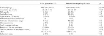
The infants’ demographic data are shown in Table 1. Of the 53 infants enrolled in our study, 12 (22%) were diagnosed with haemodynamically significant patent ductus arteriosus based on the echocardiographic assessment on days 5–7; three infants required dopamine treatment for hypotension, and all three were in the patent ductus arteriosus group. Ductal closure was achieved in eight cases with a single course of oral ibuprofen, four cases required a second course, and none of the infants required surgical intervention.
Demographic characteristics in the two groups.

CRP=C-reactive protein; PDA=patent ductus arteriosus
Quantitative data are presented as median (IQR) and qualitative data as counts (percentages)
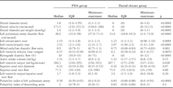
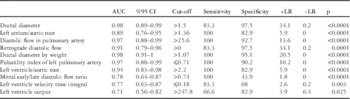
The demographic findings are summarised in Table 1. Echocardiographic parameters on the second day are shown in Table 2, with the parameters compared using a receiver operating characteristic curve analysis (Table 3). Transductal diameter; pulmonary arterial diastolic flow; retrograde aortic diastolic flow; ductal diameter by body weight; left pulmonary artery pulsatility index; and left ventricle to aortic root ratio had high sensitivity and specificity (p<0.0001, area under the curve >0.9). Parameters that displayed moderate sensitivity and specificity on the second day were left atrial to aortic root ratio; left ventricular output; left ventricular flow velocity time integral; and mitral early/late diastolic flow ratio (p<0.05; area under the curve, 0.7–0.88). Parameters that displayed low sensitivity and specificity on the second day were descending aortic pulsatility index and superior caval vein diameter and flow (p>0.05).
Echocardiographic parameter measurements in the two groups on the second day of life.

IQR=interquartile range; PDA=patent ductus arteriosus
Comparison of the echocardiographic markers by receiver operating characteristic curves.

AUC=area under curve; CI=confidence inverval; LR=Likelihood ratio
Discussion
We compared 12 echocardiographic parameters on the second day after birth using a receiver operating characteristic curve analysis. Our results showed that ductal diameter by body weight, left pulmonary artery pulsatility index, and left ventricle/aortic root parameters on the second day had excellent sensitivity and specificity for identifying those infants requiring subsequent intervention.
Johnson et alReference Johnson, Breart and Gewitz 37 evaluated 3559 premature infants shortly after birth and compared the echocardiographic parameters of 415 infants who subsequently developed clinically significant patent ductus arteriosus with those of 1496 infants without signs of haemodynamically significant patent ductus arteriosus. The authors stated that the best discrimination between the two groups was provided by a left atrium/aortic root ratio of 1.40, a left ventricle/aortic root ratio of 2.10, or a left ventricular systolic time interval of 0.27. Multivariate analysis demonstrated that better separation between the two groups occurred when the left atrium/aortic root ratio and left ventricular systolic time interval were considered jointly. The authors also suggested that echocardiographic variables should be used together to improve the power of this diagnostic tool, as there was a large degree of overlap between the normal group and the group with patent ductus arteriosus. We found similar cut-off points for left atrial and left ventricle to aortic root ratios; however, left ventricle to aortic root ratio appears to have better diagnostic relevance than the latter (area under the curve values were 0.94 and 0.88, respectively).
Sehgal and McNamaraReference Sehgal and McNamara 32 found that left ventricular systolic time interval closely correlated with myocardial function, and they proposed this parameter as an alternative method of assessing left ventricular performance. As ductal patency results in reduced left pre-ejection period and increased left ventricular ejection time, an overall decline in left ventricular systolic time interval is expected.Reference Sehgal and McNamara 32 Left ventricular systolic time interval had an area under the curve of 0.77 in our study. Similarly, the area under the curve for left ventricular output/superior caval vein ratio was also low in our study. Kluckow and Evans previously found that left ventricular output/superior caval vein flow had a high correlation with diastolic flow rate in the duct, left atrium/aortic root, ductal diameter, and average flow velocity in the left pulmonary artery before the second day of life,Reference Kluckow and Evans 31 and proposed that, as it is directly proportional to ductal flow, left ventricular output/superior caval vein flow should be considered an additional precise indicator for the magnitude of the ductal shunt. El Hajjar et al,Reference El Hajjar, Vaksmann, Rakza, Kongolo and Storme 28 assessing the relationship between left ventricular output/superior caval vein ratio and left atrium/aortic root ratio, diameter of the ductus, and the mean end-diastolic flow velocity of the left pulmonary artery on the second day of life, also suggested left ventricular output/superior caval vein ratio as an ideal criterion for evaluating patent ductus arteriosus. The authors found an area under the curve for left ventricular output/superior caval vein ratio of 0.94, and recommended a cut-off point of ⩾4. In our study, the median value for this parameter was 3.7 in the ductus arteriosus group. Left ventricular output also had a lower sensitivity and specificity to predict haemodynamically significant ductus in our study (area under the curve for left ventricular output, 0.79).
Pulsatility index refers to a variable reflecting the downstream resistance to blood flow in the vascular bed; therefore, the pulsatility index of the left pulmonary artery provides insight into right ventricular contractility, pre-load, and afterload.Reference Johnson, Breart and Gewitz 37 We found that when the left pulmonary artery pulsatility index was <0.72 on the second day of life, it was highly probable that future intervention would be required, with a sensitivity of 100% and a specificity of 90.2%. Our findings are consistent with those of Freeman et al,Reference Freeman-Ladd, Cohen and Carver 34 who proposed dividing the pulsatility index of the left pulmonary artery by that of the descending aorta as a precise indicator of ductal shunting. The authors discussed that minimal shunting is possible with a large duct and emphasised the importance of the amount of shunting for accurate assessment; however, they evaluated their study group based on suspected haemodynamically significant patent ductus arteriosus at different times, ranging from 0 to 29 days of life. The haemodynamic situation of the duct can be influenced by time and by alterations in the pulmonary dynamics after birth. In contrast, we evaluated the pulsatility indices of the left pulmonary artery and descending aorta on one specific day – the second day of life – and before the clinical signs of haemodynamically significant patent ductus arteriosus developed. We found pulsatility index of the left pulmonary artery to be a highly valuable indicator, with an area under the curve of 0.97, whereas the pulsatility index of the descending aorta was not relevant. The sensitivity of the index to changes in pulmonary arterial resistance and compliance should be investigated further.
A possible explanation for the discrepancy between our results and previous reports may be variation in the parameters with respiration and the impact of mechanical ventilation on measurement quality. Infants with patent ductus arteriosus, when compared with infants who successfully achieve ductal closure, usually have lower gestational age, more frequent respiratory distress syndrome, and a need for mechanical ventilation. The presence of respiratory distress syndrome and the use of mechanical ventilation may alter pulmonary vascular resistance and have a remarkable influence on left-to-right shunting through the open ductus.Reference Kluckow and Evans 26 Intra-observer and inter-observer variability are also limitations in ductal studies, making it difficult to draw final conclusions from a particular study or to make comparisons between studies.
A second reason for the difference in our results compared with previous studies is that we included patients with gestational age ranging from 26 to 32 weeks; however, the more immature the baby, the more time it takes for it to be stabilised haemodynamically. Relatively, mature infants usually need less inotropic and pulmonary support and they face less metabolic and biochemical derangements, resulting in different consequences, interfering with ongoing ductal closure at the cellular level. Immature babies not only have different lung mechanics but also require different fluid protocols. The behavioural pattern of the duct may also vary in babies at different stages of maturation. Sonographic examination of the ductus on the second day of life may reveal a totally different dynamic picture between, for instance, a baby at 26 weeks of gestation and one at 32 weeks of gestation. A comparison of measurements carried out at less than 28 weeks with those carried out at greater than 28 weeks would be helpful; however, given that the most important predictor of ductal patency is gestational age, a more comprehensive approach would be to classify cases according to their degree of maturation, instead of including all premature cases below a particular gestational week in a single group. Perhaps, every echocardiographic parameter should be evaluated relative to the specific gestational age, which would facilitate comparisons of the data gathered from studies with different designs.
Another possible improvement to our study would be having a second cardiologist perform the final echocardiographic assessment, blinded to the previous sonographic findings. This approach could have avoided errors arising from bias, but it was not feasible for our study group because there was only one paediatric cardiologist in our centre during the study period.
Early intervention for haemodynamic consequences of left-to-right shunting across the ductus, including pulmonary over-circulation and systemic (particularly cerebral) hypoperfusion, decreases the risk of respiratory and neurologic morbidities in preterm infants. On the other hand, if early prophylaxis is given to all preterm infants, more than half will be exposed to the side-effects of cyclooxygenase inhibitors unnecessarily because spontaneous closure is achieved in most instances.Reference Sinha 23 Regarding ductal patency, both the ideal timing and the necessity for treatment remain to be determined.Reference Sinha 23 Earlier and accurate identification of cases requiring subsequent medical intervention from those that can be managed with watchful waiting will greatly help the practicing clinician to develop individualised therapeutic strategies.
Conclusion
We identified the optimal cut-off points for echocardiographic markers on the second day of life to predict subsequent ductal patency. We suggest that left pulmonary arterial pulsatility index, left ventricle/aortic root ratio, and left pulmonary artery diastolic flow are useful adjuncts to conventional variables to predict haemodynamically significant patent ductus arteriosus. These indicators can offer a broader outlook when used with conventional parameters.
Acknowledgements
None.
Financial Support
This research received no specific grant from any funding agency, commercial, or not-for-profit sectors.
Conflicts of Interest
None.
Ethical Standards
The authors assert that all procedures contributing to this work comply with the ethical standards of the relevant national guidelines on human experimentation (Turkey) and with the Helsinki Declaration of 1975, as revised in 2008, and has been approved by the institutional committee (Adnan Menderes University Hospital).