In Brazil, sickle cell anaemia is the most prevalent hereditary disease,Reference Naoum 1 – Reference Zago and Costa 3 affecting from 0.1% to 0.3% African descents.Reference Di Nuzzo and Fonseca 4 , Reference Watanabe 5 The Brazilian Ministry of Health currently estimates about 25,000 persons with sickle cell anaemia in the country. According to the National Neonatal Screening Programme, one in every 1000 newborn babies has sickle cell anaemia, and one in every 200 has the sickle cell trait. Annually, 3500 children are born with sickle cell anaemia, 6 and in the State of Acre, Eastern Amazon, one child is born with sickle cell anaemia for every 3480 live births.
In sickle cell anaemia, it is the echocardiogram that mainly shows the cardiac cavities’ enlargement due to increased cardiac output, primarily resulting from the systolic volume increase.Reference Ahmed, Siddiqui, Sadiq, Shahid, Patel and Russo 7 – Reference Lester, Sodt, Hutcheon and Arcilla 10 This increase becomes more evident in the second year of life.Reference Lester, Sodt, Hutcheon and Arcilla 10 Ventricular hypertrophy appears to be a compensatory mechanism for the volume overload, in an attempt to reduce stress on the myocardial wall.Reference Grossman, Braunwald, Mann, McLaurin and Green 11 The dilatation levels of the left ventricle and of the hypertrophy are associated with the severity of the anaemia and the age of the patient,Reference Covitz, Esperland, Gallagher, Hellenbrand, Leff and Talner 8 , Reference Lester, Sodt, Hutcheon and Arcilla 10 , Reference Batra, Acherman and Wong 12 , Reference Hutz 13 and are observed in virtually all patients,Reference Martins, Mesquita, Cunha, Pinheiro, Romêo and Pareto 14 even in paediatric populations.Reference Estrade, Poitrineau, Bernasconi, Garnier and Donatien 15 – Reference Sachdev, Machado and Shizukuda 17
One of the largest studies on cardiac function in adults with sickle cell anaemia revealed that left ventricular diastolic dysfunction and pulmonary hypertension are independent risk factors. They contribute to mortality, and these findings would be enough for the recommendation of echocardiographic screening of adults with sickle cell anaemia in order to identify high-risk patients.Reference Sachdev, Machado and Shizukuda 17
The study of heart abnormalities in sickle cell anaemia has become essential, because these cardiac alterations are occurring in sickle cell anaemia patients so frequently and at an early stage. This has been observed despite them becoming more evident in adulthood, and the current context of sickle cell anaemia care, in which improvements to health conditions have decreased mortality. There has been a reduction in mortality rates, especially in the first five years of life, thus leading to a delayed mortality.Reference Hassell 18 These children have been increasingly able to reach adulthood, hence suffering the consequences of chronic cardiac damage. Overall, the search for better understanding of this issue has increasingly become more relevant.
This study aims to describe the distribution of echocardiographic, radiological and electrocardiographic alterations for a sample of children and adolescents in the presence or absence of sickle cell anaemia.
Materials and methods
We conducted a descriptive epidemiological study to determine the prevalence of echocardiographic alterations in two groups of children and adolescents <20 years of age who had an echocardiogram performed between January, 2011 and January, 2012. They were differentiated according to the presence (n=45) or absence of sickle cell anaemia (n=109).
The study included all the children and adolescents diagnosed with sickle cell anaemia, which was confirmed by the heel prick test or by haemoglobin electrophoresis. These patients were recruited during routine treatment at the only two paediatric haematology clinics that offer free and universal care in the city of Rio Branco, Acre, Brazil. Children were excluded if they had other haemoglobinopathies, congenital heart diseases, arrhythmias or other acquired heart diseases, or if they had been hospitalised with sickle cell crisis or had received a blood transfusion within the previous 15 days.
The comparison group comprised 108 children and adolescents who had echocardiograms performed to screen for heart disease and whose cardiac assessment result revealed the absence of heart disease. Children were excluded if they had a history of anaemia requiring treatment, transfusions, or if they had not undergone the heel prick test.
The examinations for cardiac assessment adopted for use in this study were the echocardiogram, the electrocardiogram, and chest radiography. The ratio of patients without sickle cell anaemia who required echocardiographic examinations to each patient diagnosed positive for sickle cell anaemia was 2.4:1. An interview between the physician and the relative responsible for the patient was also conducted, with questions pertaining to physical characteristics, personal and family history of disease, and medications used. In addition, results of recent blood tests were gathered from medical records.
The transthoracic echocardiogram was performad by the same examiner, using a Philips HD11XE device (Amsterdam, Noord-Holland, Netherlands), without sedation. The cardiac dimensions were obtained in accordance with the guidelines of the American Society of Echocardiography,Reference Lang, Bierig and Devereux 19 and the assessment of systolic function, ventricular mass and heart valve function was performed. The estimation of pulmonary pressure was carried out towards the tricuspid regurgitant jet whenever detected. Left and right ventricle diastolic diameters, septum thickness and posterior wall measurements were taken in three subsequent opportunities, and their arithmetic average obtained.
For data analysis, height and weight measurements were converted to body surface. The patients’ cardiac dimensions were compared with data distribution among non-sickle cell anaemia patients with similar body surfaceReference Daubeney, Blacksone, Weintraub, Slavik, Scanlon and Webber 20 towards the use of z-scores, which express the standard deviation from the predicted normal value. Measurements with a z-score>2 were considered to be altered.
To assess systolic function, we obtained the ejection fraction, left ventricular shortening, and the myocardial performance index of the left and right ventricles, using the Tei method.Reference Tei, Nishimura, Seward and Tajik 21
The left ventricular mass and left ventricular mass index were calculated according to the formula described by Devereux et al.Reference Devereux, Alonso and Lutas 22 Left ventricular hypertrophy was defined as a left ventricular mass index above the 95th percentile, which corresponds to a value higher than 95 g/m2.Reference Gladwin, Sachdev and Jison 23 The relative wall thickness was defined as: (Posterior wall×2)/Left ventricular diastolic diameter.Reference Ganau, Devereux and Roman 24 Patients with left ventricular hypertrophy were considered to have concentric hypertrophy when the relative wall thickness was >0.41 and to have eccentric left ventricular hypertrophy when the relative wall thickness was normal, that is, ≤0.41.
The pulmonary pressure value was considered normal for systolic right ventricular pressure <31 mmHg or a tricuspid regurgitation jet velocity <2.5 m/second; velocities of 2.5 to 2.9 m/second indicated mild pulmonary hypertension; and velocities ≥3 m/second reflected moderate to severe pulmonary hypertension.Reference Gladwin, Sachdev and Jison 23
The cardiac evaluation by chest radiography was performed by measuring the cardiothoracic ratio. A cardiothoracic ratio up to 0.55 for children <1 year of age and 0.50 for older children and adults was considered normal.Reference Paul and Juhl 25
The 12-lead electrocardiogram was performed on a Dixtal apparatus with three channels (Dixtal Biomedical Industry and Trade Ltd, Manaus, Amazonia, Brazil). The examination was analysed by two paediatric cardiologists. The duration of the PR interval and QRS and cardiac overload presence were ascertained. The PR interval considered normal ranged from 70 to 150 ms among infants, 100 to 170 ms among children >2 years, and 110 to 190 ms among adolescents.
The echocardiographic data acquired were analysed and subsequently correlated with the information collected by questionnaire or contained in the medical records, chest radiography examinations, electrocardiograms, and levels of haemoglobin and haematocrit.
We determined the degree of interobserver agreement regarding the presence of radiological alterations and alterations in the electrocardiogram using Cohen’s κ coefficient with the respective confidence intervals.
Data analysis
A descriptive analysis of the data was done and the prevalence of echocardiographic alterations in each of the groups analysed was obtained according to the presence or absence of a sickle cell anaemia diagnosis, with confidence intervals of 95%. These prevalences were compared with the difference in proportions test and their respective statistical significance evaluated using the Statistical Package for the Social Sciences version 13.0 (SPSS Inc., Chicago, Illinois, United States of America). The figure was constructed using the application GhaphPad Prism 5 (GraphPad Software, San Diego, California, United States of America).
The average of the three measurements of the cardiac cavities obtained by the echocardiogram was transformed into a z-score, with statistically significant differences between the sickle cell anaemia group and the non-sickle cell anaemia group for all the measurements assessed. Echocardiographic alterations were defined as being a z-score >2 and were observed in 93.3% of patients with sickle cell anaemia.
This study was analysed and approved by the Research Ethics Committee of the Acre Federal University. Informed consent from the patients and/or their guardians was obtained. There were no conflicts of interest in this research.
Results
Sickle cell anaemia and non-sickle cell anaemia participant characteristics
The average age of the sickle cell anaemia group was 8.0 years (standard deviation=4.6), and for the non-sickle cell anaemia group the average age was 5.9 years (standard deviation=3.2), p=0.002. According to gender, 55.6% were male in the sickle cell anaemia group, and 38.0% in the non-sickle cell anaemia group, p=0.46. The anthropometric averages among participants were: 24.6 kg for weight in the sickle cell anaemia group (22.96 kg in non-sickle cell anaemia group, p=0.326); 120.1 cm and 114.4, respectively, for height (p=0.126); 0.90 m2 and 0.85, respectively, for body surface (p=0.277); 0.80 and 0.81, respectively, for the median body surface.
For the patients with sickle cell anaemia, the average haemoglobin was 8.3 g/dl. The diagnosis was made using the heel prick test in only 42.2% of the children, with the remaining being diagnosed by haemoglobin electrophoresis. The average age at diagnosis was 2.3 years (standard deviation=3.3 years), although the average age for the onset of symptoms was ~1.7 years (standard deviation=1.9 years).
The rate of hospitalisation for these patients was high, with 95.6% of these children having been hospitalised, and 75.6% having been hospitalised four or more times. We noted a high rate of transfusions for these hospital admissions, with 86.7% of the children having a transfusion, and of these 69.2% had a transfusion four or more times.
Surgical intervention was necessary for 28.9% of these children. All the patients reported using folic acid and 82.2% used some vitamin supplement with complex B vitamins. In all, three patients received hydroxyurea, one used deferasirox and 10 children used prophylactic antibiotics. As for the use of cardiac medications, 51.1% reported their use, and of these 82.6% had monotherapy with spironolactone. Heart murmur was auscultated in 82.2% of patients.
Sickle cell anaemia echocardiographic alterations
Z-scores of cardiac cavities’ measurements in the sickle cell anaemia and non-sickle cell anaemia groups are presented in Table 1 and Figure 1. The mean left ventricle z-score in the sickle cell anaemia group was 13-fold higher compared with the non-sickle cell anaemia group, and about fivefold higher either in the posterior wall or the right ventricle.
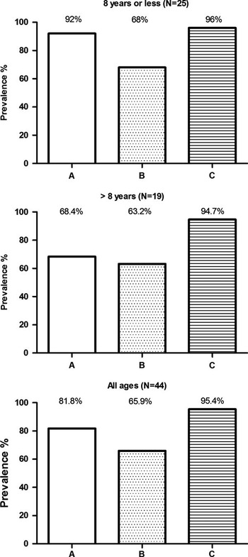
Percentage of the prevalence of radiological ( a ), electrocardiographic ( b ) and echocardiographic ( c ) alterations according to age in sickle cell anaemia patients.

Average z-scores and confidence intervals from the measurements of cardiac structures according to the presence of sickle cell anaemia, Rio Branco, Acre, Brazil, January, 2011 to January, 2012.

CI=confidence interval; LA=left atrium; LV=left ventricle diastolic diameter; PW=posterior wall; RV=right ventricle diastolic diameter; SCA=sickle cell anaemia
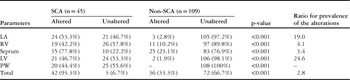
The number of echocardiographic alterations in the sickle cell anaemia group compared with the non-sickle cell anaemia group was 19 times greater for the left atrium, 4.1 times greater for the right ventricle, 3.4 times greater for the septum and 24.6 times greater for the left ventricle (Table 2). For the posterior wall, alterations were only observed in the sickle cell anaemia group (44.4%).
Prevalence of alterations in the echocardiographic measurements expressed as z-score for sickle cell anaemia and non-sickle cell anaemia patients, Rio Branco, Acre, Brazil, January, 2011 to January, 2012.

LA=left atrium; LV=left ventricle; PW=posterior wall; RV=right ventricle; SCA=sickle cell anaemia
As age showed distinct distributions in both groups, the echocardiographic measurements were analysed following age stratification using the average age of the sickle cell anaemia group (8 years) as cutpoint. A statistically significant decrease was observed for the left ventricle z-score according to age in the non-sickle cell anaemia group, 0.03 at age ≤8 years and −0.69 in >8 years (p=0.001), and an increase for the other measurements. This increase according to age was statistically significant for the right ventricle, 0.51 and 1.05, respectively (p=0.027), 1.23 and 1.75, respectively, in the septum (p=0.013), 0.28 and 0.71, respectively, in the posterior wall (p=0.041). In the sickle cell anaemia group, a statistically significant increase according to age was observed in the right ventricle z-score, 1.17 in ≤8 years and 2.33 in older children (data not shown).
The measurement of the right ventricular systolic pressure was possible in 80% of patients with sickle cell anaemia. Of these, 19.4% showed some degree of pulmonary hypertension, that is, levels above 30 mmHg. The average right ventricular systolic pressure for the sickle cell anaemia group was 27.1 (standard deviation=4.4) mmHg, which is equivalent to a tricuspid regurgitant jet velocity of 2.07 m/second, and for the group without the disease it was 22.0 (standard deviation=3.59) mmHg, with an equivalent tricuspid regurgitant jet velocity of 1.73 m/second.
The average myocardial performance index was not statistically different in the sickle cell anaemia and non-sickle cell anaemia groups. In this study, the ejection fraction average was 68.0% and 68.3% for the sickle cell anaemia and non-sickle cell anaemia groups, respectively. Only one patient from the sickle cell anaemia group had an ejection fraction of 57%, which is considered to be below normal.
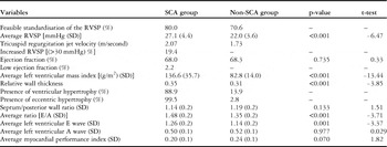
The left ventricular mass index was significantly greater in the sickle cell anaemia group compared with the non-sickle cell anaemia group, respectively, 136.6 g/m2 (standard deviation=35.7) versus 82.8 (standard deviation=14.0), p<0.001 (Table 3). The average in the former indicates ventricular hypertrophy, whereas the average in the latter is considered normal. Ventricular hypertrophy was observed in 88.9% of patients with sickle cell anaemia, with eccentric hypertrophy being responsible for 92.46% of cases. Some other echocardiographic characteristics of patients from the both groups are shown in Table 3.
Echocardiographic parameters, sickle cell anaemia and non-sickle cell anaemia patients, Rio Branco, Acre, Brazil, January, 2011 to January, 2012.

RVSP=right ventricular systolic pressure; SCA=sickle cell anaemia; SD=standard deviation
The comparative analysis of the distinct cardiac assessment methods – chest radiography, echocardiograms, and electrocardiograms – used in exploring the sickle cell anaemia alterations is presented at Figure 1. The highest prevalence of alterations was observed by echocardiogram (95.4%, 95% confidence interval 83.3–99.4), followed by chest radiography (81.8%, 95% confidence interval 66.8–91.3) and electrocardiogram (65.9%, 95% confidence interval 50.0–79.1). In 70.6% of the cases, there was evidence of alterations diagnosis with all three methods.
Interobserver analysis
Cardiomegaly analysed through chest radiography and quantified by the cardiothoracic ratio was observed in 81.8% (n=44) patients. The level of agreement (by the κ coefficient) between the two examiners of the respective radiographs was 90.2%.
As for the analysis of the electrocardiogram, overloading of the cardiac cavities was observed in 64.4% (n=29) patients. The level of interobserver agreement was 94.7%. The most frequent cavity overloading was isolated left ventricular (21 patients, 46.7%), followed by left ventricular overload with left atrial overload (six patients, 13.3%). There was one case with isolated left atrial overload and one case with left atrial, right ventricular and left ventricular overload.
The level of interobserver agreement (by κ coefficient) in the determination of which cavity had been altered was 96.3%. The PR interval was within the normal levels for the age in 88.9% of the patients.
Discussion
Most of the studies involving echocardiographic analysis report dilatations in the left ventricle and hypertrophy.Reference Lamers, Ensing and Pignatelli 9 , Reference Lester, Sodt, Hutcheon and Arcilla 10 However, in some studies, the increase in the thickness of the septum and the posterior wall was not observed.Reference Lamers, Ensing and Pignatelli 9 In a research conducted by HerdyReference Herdy, Aguas and Chedid 16 with 17 Brazilian children, an enlargement of the left atrium and left ventricle in 29.4% of the patients and normal thickness of the posterior wall for all patients was observed.
Lester et alReference Lester, Sodt, Hutcheon and Arcilla 10 observed the echocardiographic alterations present in children up to 18 years of age, taking as normal reference values those included in the range of two standard deviations from the observed measurements within a control group. In that work, the increases encountered were in the left ventricle (60.9%), left atrium (50%), right ventricle (20.3%), posterior wall (20.3%) and septum (12.5%).
In our research, we found a higher frequency of alterations in the septum, both in the sickle cell anaemia group (77.8%) and the non-sickle cell anaemia group (23.1%), probably because of the values used as normal (z-score). Our research also showed a significant increase in the left ventricular mass index (136.6 g/m2), a fact also described in the study by Lester et al (120.4 g/m2).
Research conducted in adolescents and young adults also reported dilatations in the left ventricle, right ventricle, left atrium and septum hypertrophy.Reference Covitz, Esperland, Gallagher, Hellenbrand, Leff and Talner 8 , Reference Martins, Mesquita, Cunha, Pinheiro, Romêo and Pareto 14 However, the difference for wall thickness was not found in all the studies.Reference Covitz, Esperland, Gallagher, Hellenbrand, Leff and Talner 8
It was observed in this investigation that in the non-sickle cell anaemia group, the echocardiographic measurements increased with age, which did not occur for the left ventricle and posterior wall in the sickle cell anaemia group.
The pattern of hypertrophy was essentially eccentric because of the volume overload, as was well documented by GrossmanReference Grossman, Braunwald, Mann, McLaurin and Green 11 in a study on wall stress and hypertrophy, in which the hypertrophies by pressure overloads were predominantly concentric and those by volume overload were of an eccentric pattern. This same pattern of hypertrophy has also been described in other papers.Reference Martins, Mesquita, Cunha, Pinheiro, Romêo and Pareto 14
With the objective of comparing our study with the multi-centre study conducted in the United States of America with 310 patients up to 20 years of age, as described by MinniteReference Minniti, Sable and Campbell 26 , our pulmonary pressure measurements considered as a cut-off point the elevated right ventricular systolic pressure of 30 mmHg or a tricuspid regurgitant jet velocity equal to or above 2.5 m/second. In the study mentioned above, it was observed that 11% of patients had elevated pulmonary arterial pressure – with an average tricuspid regurgitant jet velocity of 2.3 m/second – and that the increase in velocity to ≥2.6 m/second was significantly related to higher risk of haemolysis, hypoxia, acute chest syndrome and the need for transfusions.Reference Minniti, Sable and Campbell 26 In another large multi-centre study conducted with 600 children, described by KatoReference Kato, Onyekwere and Gladwin 27 , a 30% prevalence of pulmonary hypertension was observed. In our groups, 19.4% of the patients had elevated levels of pulmonary pressure and the average jet speed was 2.07 m/second.
The almost normal ejection fraction values in both groups were also consistent with the literature data.Reference Covitz, Esperland, Gallagher, Hellenbrand, Leff and Talner 8 , Reference Lester, Sodt, Hutcheon and Arcilla 10 , Reference Martins, Mesquita, Cunha, Pinheiro, Romêo and Pareto 14 The presence of heart murmur in the auscultation was consistent with the literature data.
The indexation of the echocardiographic measurements according to the body surface, in the form of z-scores, enabled not only the comparison between different groups of patients – sickle cell anaemia or non-sickle cell anaemia – but it also eliminated differences related to gender, age, nutrition and stage of development, which is important when dealing with children who have a chronic disease that has the potential to compromise growth. In addition to being a non-invasive method, in this investigation, when compared with chest radiography and electrocardiograms, the echocardiogram enabled the determination of a higher prevalence of alterations in patients with sickle cell anaemia.
Unfortunately, this study did not allow us to create cut-off values for dilatation of cardiac cavities expressed in z-scores, which would have enabled the definition of danger signals from which clinical intervention procedures could be planned.
Nevertheless, the careful observation of incipient cardiac alterations and the evolution of these alterations greatly assists the cardiologist and haematologist to attend to heart disease and intervene when deemed necessary. There is growing interest in the cardiovascular alterations in sickle cell anaemia, which will lead to the need for further studies to clarify many aspects, especially those related to the therapeutic management and differentiated engagement of the patients according to distinct genetic polymorphism profiles.
Conclusion
This research highlights the importance of echocardiographic measurements being transformed into z-scores for the observation and quantification of the alterations, as many alterations are subtle and pass unnoticed by simple qualitative observation. Owing to the echocardiogram being a diagnostic method dependent on the examiner, this quantification is extremely important for incipient alterations to be properly displayed. In addition, the use of the z-score as a standardised measure will also be extremely useful for comparison purposes and echocardiographic monitoring of patients, especially children, for which the weight and height are variable and, therefore, increased isolated cardiac measurements cannot explain whether the increase is due to a worsening cardiac condition or part of normal heart growth.
By indexing the echocardiographic measurements in the form of z-scores, it was possible to identify alterations in 93.3% of the patients analysed, and quantify the significantly increased dimensions of the left ventricle, right ventricle, left atrium, septum and posterior wall. The presence of left ventricular hypertrophy, essentially eccentric in nature, was also significant, as were the increased levels of pulmonary pressure that were indirectly assessed by tricuspid regurgitation.
Acknowledgements
To the Coordination for Improvement of Higher Education Personnel, Ministry of Education of Brazil, for the Master’s scholarship granted to M.C.V. Ribera and to Marcos Silva for suggestions and support in the organisation of the data. This research was made possible through the academic collaboration established between the Post-graduate Program in Public Health at the Acre Federal University and the Post-graduate Program Environment and Public Health at the Oswaldo Cruz Foundation, supported with resources from the National Council for Scientific and Technological Development.