Significant Outcomes
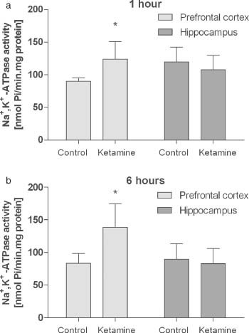
The pathophysiology of schizophrenia is still poorly understood we observed (a) Na+,K+-ATPase activity increased in prefrontal cortex after chronic administration of ketamine; (b) no significant change was found in Na+,K+-ATPase activity in hippocampus; (c) we suggested that chronic administration of 25 mg/kg ketamine provoked imbalance in energetic metabolism by alter the Na+,K+-ATPase activity in prefrontal cortex.
Limitations
This study presented some limitations, first, few brain areas were assessed; second, advanced techniques would be needed to confirm the imbalance of energetic metabolism provoke by ketamine treatment.
Introduction
Schizophrenia is a chronic and prevalent psychiatric disorder with an estimated lifetime prevalence of 1% in the general population. Compared with the general population, life expectancy in patients with schizophrenia is shorter about 20%, attributable to higher rates of suicide and cardiovascular disease, which is already the major mortality cause for schizophrenic patients (Reference Schultz, North and Shields1). The symptoms of schizophrenia can be divided into positive (such as delusions and hallucinations), negative (such as social withdrawal and affective flattening) and cognitive (such as deficits in attention and memory).
The administration of N-methyl-d-aspartate (NMDA) receptor antagonists has been used as an animal model for schizophrenia, generally by the administration of substances such as phencyclidine (PCP) and ketamine. Several reports in literature indicate that sub-anaesthetic doses of ketamine can produce hallucinations and paranoia in humans, similar to positive symptoms characteristics of schizophrenia, and show that this model reproduces positive and negative symptoms, as well as the cognitive impairments of this disease (Reference Hunt, Raynaud and Garcia2,Reference Littlewood, Jones and O'neill3). One of the major hypotheses for the pathophysiology of schizophrenia is that NMDA receptor hypofunctions and therefore glutamatergic dysfunction are primary events leading to hyperdopaminergia (Reference Grace4).
Na+,K+-ATPase is a protein in its role as a maintainer of electrolyte and fluid balance in all mammals. This important enzyme uses the energy from adenosine triphosphate (ATP) hydrolysis to a vectorial transport of sodium and potassium across the plasma membrane with the stoichiometry of 3:2. This creates the electrochemical gradient across the plasma membrane that will facilitate sodium-coupled entry of ions, aminoacids, glucose and many other compounds into the cell (Reference Aperia5).
It is well known that glutamate transporters are sodium-dependent proteins and are related to the normal function of sodium and potassium gradients generated specially by Na+,K+-ATPase, not surprised that alterations in Na+,K+-ATPase activity could cause brain abnormalities such as observed in cerebral ischemia (Reference Wyse, Streck, Worm, Wajner, Ritter and Netto6), schizophrenia (Reference Nanitsos, Nguyen, St'astný and Balcar7), Alzheimer's disease (Reference Hattori, Kitagawa and Higashida8), and in animal models of depression and bipolar disorder (Reference Zugno, Valvassori and Scherer9). In our group, we have already reported, in this animal model of schizophrenia, increased creatine kinase activity, which is an important enzyme from energy metabolism (Reference Canever, Oliveira and De Luca10) and an imbalance of energy metabolism (Reference Oliveira, Fraga and De Luca11).
Considering the fundamental importance of Na+, K+- ATPase in the brain, the aim of our study was investigate the activity of this enzyme in hippocampus and prefrontal cortex of rats submitted to chronic administration of ketamine.
Materials and methods
In this study, we investigated Na+,K+-ATPase activity in the prefrontal cortex and hippocampus of rats submitted to chronic injections of ketamine. We used adult male Wistar rats (250–300 g) obtained from our breeding colony. Animals were housed five per cage with food and water available ad libitum and were maintained on a 12-h light/dark cycle (lights on at 7:00 a.m.) at a temperature of 22 ± 1 °C. Animals were divided in two groups: (a) saline (n = 7) and (b) ketamine (n = 8). All experimental procedures were performed in accordance with the approval of the local Ethics Committee.
Ketamine was used at 25 mg/kg, prepared in saline solution 1 ml/100 g (Reference Imre, Fokkema, Den Boer and Ter Horst12). According to Hunt et al. (Reference Hunt, Raynaud and Garcia2), the characteristic symptoms were induced by chronic sub-anaesthetic doses of 25 mg/kg of ketamine. Control group was injected with same volume with saline solution (i.p).
Sub-anaesthetic doses of ketamine or saline were administrated by 7 days, once a day by i.p. Analyses were performed 1 or 6 h after the last injection. The animals were killed by decapitation, and brain structures as prefrontal cortex and hippocampus were dissected and rapidly frozen to −80 °C until the Na+,K+-ATPase analysis.
The reaction mixture for Na+,K+-ATPase assay contained 5.0 mM MgCl2, 80.0 mM NaCl, 20.0 mM KCl, and 40.0 mM Tris–HCl, pH 7.4, in a final volume of 200 ml. After 10 min of pre-incubation at 37 °C, the reaction was initiated by the addition of ATP to a final concentration of 3.0 mM, and was incubated for 20 min. Controls were carried out under the same conditions with the addition of 1.0 mM ouabain. Na+,K+ -ATPase activity was calculated by the difference between the two assays according to the method of Wyse et al. (Reference Wyse, Streck, Worm, Wajner, Ritter and Netto6). Released inorganic phosphate (Pi) was measured by the method of Chan et al. (Reference Chan, Delfert, Koepnick and McDonald13). Specific activity of the enzyme was expressed as nmol Pi released per min per mg of protein. Data were analysed by Student's t-test, using SPSS program (Statistical Package for the Social Sciences), with significance in p < 0.05.
Results
Our results showed that chronic administration of ketamine was able to increase Na+,K+-ATPase activity in prefrontal cortex at 1 (37%) and 6 h (66%) (after the last administration). However, ketamine has no effect in hippocampus (Fig. 1).
Effect of chronic administration of ketamine on Na+,K+-ATPase activities 1 or 6 h after the last injection in brain of rats. Hippocampus and prefrontal cortex were homogenized and prepared for ATPase assays. Results are expressed as mean ± SD for five independent experiments performed in duplicate; p < 0.01 compared to control (Student's t-test); * different from control.

Discussion
Several lines of evidence suggest that the NMDA system is involved in schizophrenia. This glutamatergic receptor has a complex physiological activity: it controls a variety of functions such as neuronal development and synaptic plasticity and if it is blockade by non-competitive antagonists, such as PCP, MK-801 or ketamine inducing a broad range of behavioural manifestations in animals that closely resemble schizophrenia condition in humans (Reference Oliveira, Fraga and De Luca11). There is also evidence that NMDA receptors are located on brain circuits which regulate dopamine release, suggesting that dopaminergic deficits in schizophrenia may also be secondary to underlying glutamatergic dysfunction. To support this affirmation, ketamine increases cortical dopamine release in in vivo microdialysis studies (Reference Verma and Moghaddam14). It is known that dopamine interacts with proteins, regulating their actions, including Na+,K+-ATPase, it was already speculated that the activation of dopamine D2 receptors increase the activity of Na+,K+-ATPase (Reference Yamaguchi, Walk, Jose and Felder15), while the activation of dopamine D1 receptors inhibited the activity of this enzyme (Reference Snyder, Fienberg, Huganir and Greengard16).
Considering that our model with ketamine is based on glutamatergic hipofunction, and Na+,K+-ATPase plays a special role in the homeostasis of the mammalian brain, any alteration in this enzyme activity can affect many other functions in neurotransmission.
Evidence suggests that the impairment of energy metabolism found in several central nervous system disorders, such as bipolar disorder and schizophrenia could be related to the modulatory effects of glutamate and dopamine in ATPase activity (Reference Ben-Shachar, Zuk, Gazawi and Ljubuncic17). Volz et al. (Reference Volz, Riehemann and Maurer18) also reported reduced ATP levels in the frontal lobe and in left temporal lobe of schizophrenic patients as compared to control. In this context, study showed mitochondrial dysfunction and more specifically, abnormal complex I expression as a pathological characteristic of schizophrenia (Reference Ben-Shachar, Nadri, Karry and Agam19). We also tested energy metabolism and previous studies from our group showed that ketamine administration affects respiratory chain complex in striatum, hippocampus and prefrontal cortex (PFC) in rats (Reference Oliveira, Fraga and De Luca11). In this study, we showed alteration in PFC but not in hippocampus. It could speculate that it could be a compensatory mechanism due to the inhibition of NMDA receptor. It would also be reasonable to suppose that altered binding properties of sodium and potassium to the sodium-pump might be related to the genesis of neuronal dysfunction in schizophrenia (Reference Kurup and Kurup20). A possible explanation for this alteration might be the malfunction of the NMDA receptor system in prefrontal cortex that leads to dysregulation of GABAergic fast-spiking interneurons that stimulated striatal neurons. This mechanism may be involved in the corticolimbic dysfunction associated with the pathophysiology of schizophrenia (Reference Del Arco, Ronzoni and Mora21).
It is well established that the PFC has been shown to have an essential role in cognition, emotion and reward and that PFC dopamine and glutamate neurotransmitter systems play a crucial role in the physiology and pathology of this area of the brain, and an unbalance of this system is related with schizophrenia symptoms (Reference Del Arco and Mora22). To supporting these hypotheses, Corti et al. (Reference Corti, Xuereb, Crepaldi, Corsi, Michielin and Ferraguti23) showed a robust decrease in the protein expression levels of Glu R1 and Glu R2 subunitis in the PFC of schizophrenic patients and showed a significant increase of Na+,K+-ATPase activity in Brodman area 10 in patients with schizophrenia.
The pathophysiology of schizophrenia is still poorly understood; however, the increase in Na+,K+-ATPase activity observed in our animal model indicates that an imbalance of energy metabolism and it could be related to the development of schizophrenia and these findings may explain, at least in part, the mechanisms which underlie this disorder.
Acknowledgements
This research was supported by grants from CNPq, Instituto Cérebro e Mente, INCT and UNESC to AIZ (CNPq Research Fellow). The authors declare that they have no conflict of interest.