Introduction
Cochlear implantation (CI) is effective in restoring hearing in children with profound hearing loss. More recently, the criteria for CI have been expanded to include young children, patients with moderate hearing loss and the elderly.
However, it is generally agreed that CI in patients with long-term bilateral deafness should be considered carefully, because the prognosis is often poor.Reference Blamey, Arndt, Bergeron, Bredberg, Brimacombe and Facer1, Reference Proops, Donaldson, Cooper, Thomas, Burrell and Stoddart2 The main reasons for this poor outcome are: (1) the potentially inadequate number of spiral ganglion neurons present after long-term hearing loss, and (2) poor brain plasticity, which is required for adapting to the new input of information. No formal studies have investigated whether CI is beneficial in patients with long-term monaural deafness. In such patients, CI may be considered in the ear with long-term deafness when the ‘good’ ear suddenly becomes deaf and is unsuitable for any rehabilitation device. Presumably, a better outcome is expected because the auditory system has been working with the input from one ear and thus has not been totally unused, as in patients with bilateral deafness.
Here, we report a patient with long-term profound hearing loss in one ear, who had recently lost all hearing in the other ear after surgery to remove an acoustic neuroma. Post-operatively, the hitherto good ear had become completely deaf, and showed no response on electrical stimulation assessing auditory brainstem responses (ABRs). Thus, CI was performed in the ear with long-term hearing loss, in which ABRs were present. However, CI failed to confer any benefit because the device gave the patient a headache, even with very low stimulation levels.
Case report
Our patient had been identified at the age of two years as having profound right ear hearing loss, and thereafter had depended on her left ear for daily communication.
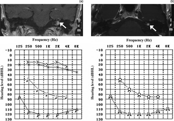
At the age of 58 years, she had been diagnosed with an acoustic neuroma in the left cerebellopontine angle. The tumour had been less than 1 cm in diameter when diagnosed (Figure 1a, upper panel), and the patient had had nearly normal hearing up to 8 kHz (Figure 1a, bottom panel).
Axial magnetic resonance imaging scans and audiograms for the patient, taken before each operation. (a) Scan taken before the initial acoustic neuroma removal surgery via a retrosigmoid approach (upper panel; arrow indicates tumour), and corresponding audiogram (lower panel). (b) Scan taken before removal of acoustic neuroma remnant and resection of the cochlear nerve (upper panel; arrow indicates tumour remnant), and corresponding audiogram (lower panel).

The patient had undergone resection of the acoustic neuroma at another hospital. Although a retrosigmoid approach had been used in an effort to preserve hearing, the ear had become completely deaf post-operatively, and the patient had subsequently developed severe tinnitus.
The patient was referred to our hospital for treatment of her severe tinnitus. Careful examination showed that the acoustic neuroma had not been removed completely (Figure 1b, upper panel), and that the patient had profound hearing loss in both ears (Figure 1b, bottom panel). A definite ABR was found in the right ear (with long-term hearing loss), while there was no response in the left ear (from which the tumour had been removed).
Because there was no ABR in the left ear and tinnitus was a major concern, a second operation was performed via a translabyrinthine approach to ensure total removal of the tumour. During the same procedure, the cochlear nerve was resected to treat the patient's tinnitus, which was greatly mitigated immediately after surgery.
Seven months after the second procedure, the patient returned complaining of recurrent, severe tinnitus in the left ear. She also hoped to gain hearing by using a prosthesis. Because her hearing loss was profound in both ears, and as auditory brainstem implantation is not available in China, the only choice for this patient was CI.
Therefore, a HiRes 90k electrode array (Advanced Bionics, Los Angeles, U.S.) was inserted into the patient's right cochlea. One month later, the implant was turned on and programmed using the HiRes P speech processing strategy.
Unfortunately, the patient reported a headache even when the stimulus current was only 80 current units. She could detect environmental sound at approximately 120 current units, but this perception was accompanied by a moderate headache which the patient could not tolerate.
After comprehensive consultation and several attempts at remapping, the patient still refused to use the cochlear implant.
Discussion
Over the last two decades, CI has gradually been accepted as an effective treatment for many patients with profound deafness, although the outcome varies especially in adults.
Several key factors affect CI outcome. The most significant are age at implantation and duration of deafness before implantation. A long duration of deafness is associated with poorer CI outcomes.Reference Blamey, Pyman, Gordon, Clark, Brown and Dowell3–Reference Waltzman, Fisher, Niparko and Cohen6 Proposed mechanisms for this poor outcome include a reduced number of spiral ganglion neurons surviving after hair cell loss, and a reduced facility for plastic reorganisation in the central auditory system with increasing age and duration of deafness.Reference Blamey, Pyman, Gordon, Clark, Brown and Dowell3, Reference Gantz, Woodworth, Knutson, Abbas and Tyler4, Reference Waltzman, Fisher, Niparko and Cohen6, Reference Summerfield and Marshall7 Although the minimum number of neurons necessary for a satisfactory response to electrical CI stimulation is unknown, there is some concern that prolonged deafness arising from hair cell loss might decrease the neuron population to such an extent that CI is not beneficial.Reference Linthicum and Fayad8
The presented patient had suffered 56 years of deafness in the ear chosen for CI. It is certainly possible that her residual spiral ganglion neurons would function inadequately after such a long duration of deafness. However, this appeared not to be a major issue, as the cochlea showed a definite response to electrical stimulation and the patient could detect environment sound. Instead, an unbearable headache prevented our patient from using an adequate electrical stimulus through her implant. These results suggest that the atrophic and degenerative loss of spiral ganglion neurons is not a major concern in such cases, while the functional status of the central auditory pathway may prove to be a greater obstacle to implant usage. Thus, the presence of ABRs should not be considered the only predictor of outcome in such cases.
In contrast, the success of CI is strongly dependent on higher levels of auditory system response, especially in the cortex. In patients with long-term, profound, binaural hearing loss, the activity level in the auditory cortex may be low, as indicated by the low metabolic rate found in this brain region in such patients.Reference Green, Julyan, Hastings and Ramsden9 Such weak auditory cortex activity may be associated with a smaller degree of auditory activation following CI. This has been suggested as being responsible, at least partially, for the poor outcome of CI in such cases.
However, the pathological processes involved in monaural deafness are presumably different from those in bilateral deafness. In patients with monaural deafness, the central auditory system on both sides should be better adapted to auditory stimuli, because it has been activated by the hearing ear, as in our patient. Theoretically, the plasticity adjustment required for adaptation to CI should be easier, compared with patients with binaural deafness.
However, the failure of CI in the presented patient raises an alarm for the future. The underlying reasons for this failure may be multifactorial, and remain to be explored. Certainly, our patient's unreasonably high expectations and lack of resolve to regain hearing, with a consequent absence of speech training post-CI, contributed to CI failure. In addition, speech stimulation during a critical period may play an important role in cortical recognition. Results in bilaterally, postlingually deaf patients indicate that if auditory deprivation occurs after language acquisition, the auditory evoked potentials can re-normalise upon electrical stimulation, even after a very long period of sensory deprivation.Reference Pantev, Dinnesen, Ross, Wollbrink and Knief10, Reference Pelizzone, Kasper, Hari, Karhu and Montandon11 This emphasises the importance of speech stimulation within a critical period, in order to develop plasticity of the central auditory and speech systems. Thus, our patient's absence of speech stimulation to the innervated auditory cortex, due to her very early deafness in the CI ear, may have interfered with cortical plasticity and contributed to her poor CI outcome.
We believe that two errors occurred in the treatment of this patient.
Firstly, an unsuitable strategy was used carelessly in her first treatment. Three strategies are available for the management of acoustic neuroma patients: adoption of a ‘wait and see’ policy; surgical removal of the tumour; and referral for radio-surgery.Reference Aristegui and Denia12 When deciding on acoustic neuroma treatment in an only hearing ear, hearing preservation should be considered carefully. Thus, most surgeons would adopt a policy of watchful waiting, especially if the tumour is smaller than 1 cm and the patient has functional hearing. Unfortunately, in our patient's case tumour removal was chosen as the initial strategy, and her hearing was lost post-operatively.
• Management decisions for acoustic neuroma in an only hearing ear should be cautious; hearing preservation should be the first choice
• Ear selection for cochlear implantation (CI) is difficult when both ears have disadvantages regarding CI outcome, as in the presented case (one ear had long-term deafness with auditory brainstem responses (ABRs) present, while the other had short-term deafness with no ABRs)
• Caution is advised regarding CI in cases of long-term deafness, even if deafness is monaural
The second treatment error was the decision to completely remove the residual tumour and to resect the cochlear nerve, without considering cochlear nerve preservation. We believe that this approach was overly aggressive. The original surgical decision was based on three factors: (1) the residual tumour posed an underlying risk for long-term rehabilitation; (2) the right ear (with long-term deafness) responded to electrical stimulation and may have benefited from CI; and (3) the only hearing ear went deaf after the first surgical procedure and did not respond to electrical stimulation, so would not have been helped by CI. While the first two factors were true, the last does not appear to have been the case. If the cochlear nerve had been preserved during the second operation, CI in the left ear might still have been possible when the implant in the right ear failed. Several authors have reported successful CI in an ear undergoing acoustic neuroma resection.Reference Hoffman, Kohan and Cohen13–Reference Neff, Wiet, Lasak, Cohen, Pillsbury and Ramsden18 No response from the left ear in one ABR test does not rule out the possibility of CI. We believe that this test should have been repeated later, as suggested by the findings of Neff et al., who proposed that the response to promontory stimulation can be negative immediately after surgery but become positive later on.Reference Neff, Wiet, Lasak, Cohen, Pillsbury and Ramsden18
Conclusion
In this patient, CI failed in terms of its outcome. From this, we should learn that ABR testing should not be regarded as the only predictor of the results of CI. The plasticity of the central auditory system affects the outcome of CI, especially in patients with long-term deafness.
Acknowledgements
We thank Wang Jian and Yin ShanKai for their work in preparing the manuscript. This study was supported by a grant from The National Natural Sciences Foundation of China (30925035/H1303).