Introduction
While epidemiological features of late life depression (LLD) are well known, the neurobiological basis of emotional control in LLD remains controversial. Among the brain areas implicated in the control of emotions in LLD, the ventromedial prefrontal cortex (vMPFC), comprising the orbitofrontal cortex, rostral cingulate cortex, and the ventral medial frontal gyrus, is of interest and has been associated with the evaluation and regulation of emotional events and stimuli and the contextual processing of reward (Drevets et al., Reference Drevets, Price and Furey2008). In a study of female, drug-naïve, first episode LLD patients found reduced activation of the vMPFC in LLD to negatively valenced words in a functional magnetic resonance imaging (fMRI) task (Brassen et al., Reference Brassen, Kalisch, Weber-Fahr, Braus and Büchel2008). Structural brain imaging studies suggest ischemic deficiencies, as measured by increased prevalence of white matter hyperintensities (WMH) (Teodorczuk et al., Reference Teodorczuk2007) especially in frontal brain areas, including the vMPFC, could be contributory to emotional dyscontrol.
We wished to extend these findings and hypothesized that in patients of both sexes with mild to moderate LLD currently managed in a routine psychiatry clinic, compared to a similarly aged comparison population, the vMPFC will show (a) reduced activation with negative valenced words under a fMRI task and (b) total brain WMH volume would be greater in LLD participants. Secondary analyses were planned to assess the effect of gender on the results.
Methods
This study was approved by the Western University Research Ethics Board. Fourteen adult individuals, older than 55 years of age of either sex, who identified themselves as having no major illnesses or any mental health issues (comparison group), were compared with 16 individuals of similar age with a major depressive disorder with or without comorbid anxiety, current episode mild to moderate, meeting Diagnostic and Statistic Manual-IV (DSM-IV) (American Psychiatric Association, 2000) criteria as confirmed by a geriatric-psychiatrist (AV) (LLD group). Patients with other Axis I diagnoses were excluded. In the depressed group 9 participants were male and seven female. The comparison group comprised seven male participants and seven female participants. The exclusion criteria included: being non-ambulatory, presence of significant cognitive impairment (Mini Mental State Examination < 23) (Folstein et al., Reference Folstein, Folstein and McHugh1975), other significant neurological conditions, history of fainting, inadequately controlled hypertension or frequent hypotensive episodes, claustrophobia, or any contraindications to Magnetic Resonance Imaging (MRI). After written informed consent was obtained, the depressed participants underwent Geriatric Depression Scale (GDS-15) (Sheikh and Yesavage, Reference Sheikh and Yesavage1986), and Center for Epidemiologic Studies Depression Scale (CES-D) scale (Radloff, Reference Radloff1977), while all other participants underwent an MMSE (Folstein et al., Reference Folstein, Folstein and McHugh1975), the Cumulative Illness Rating Scale-Geriatrics (CIRS-G) (Parmelee et al., Reference Parmelee, Thuras, Katz and Lawton1995), and the Physical Activity Scale for the Elderly (PASE) (Washburn et al., Reference Washburn, Smith, Jette and Janney1993).
All study participants were imaged at Western University, on a 3 Tesla, Siemens Prisma MRI system (Erlangen, Germany) using a 32-channel head coil. Anatomical contrasts included a whole brain sagittal T1-weighted (T1w) 3D MPRAGE: (TI/TE/TR = 900/2.98/2300 ms; 192 slices, 1 mm isotropic voxels), axial 2D multi-slice T2-FLAIR: (TI/TE/TR = 2400/121/8000 ms; 45 slices at 0.4×0.4×3.0 mm voxels.
Functional MRI volumes were collected using a 2D multi-slice EPI acquisition (TE/TR = 30/2520 ms; 3.0 mm isotropic resolution; iPat = 2). Each volume comprised 45 contiguous, axial-oblique slices oriented parallel to the AC–PC line, spanning the entire cortex.
We excluded one participant with LLD due to technical problems on the fMRI scans. T2-FLAIR images were not acquired on one further LLD participant. Thus the fMRI analysis is on 15 LLD participants, and the WMH on 14 LLD. All 14 comparison participants were included in the analysis.
Words from the affective norms for English words (ANEW) standardized list (Bradley and Lang, Reference Bradley and Lang1999) were used. Each participant underwent four runs of the task, with each trial containing ten positive, ten neutral, and ten negative valence words. The words in the three valence categories were matched for word length and arousal. Words were presented for an average of 2 sec (duration was one of 1.5, 2.0, and 2.5 sec, counterbalanced between valences), followed by a 2 sec response screen, and then a blank screen for 6, 7 or 8 sec (see Supplementary Digital Content, SDC, Figure S1, available as supplementary material attached to the electronic version of this paper at https://doi.org/10.1017/S1041610217000461). Stimuli were projected on a screen and viewed by the participants through a mirror. Participants were instructed to view each word for the entire display time, and subsequently indicated their perceived valence by pressing one of three buttons i.e. “positive, negative, or neutral.” Responses were classified as being correct if the responded valence matched the ANEW classification. Participants underwent a familiarization exposure to the experimental procedures on the same day prior to each experiment and were required to be abstinent from alcohol and caffeine for 12 hours prior to testing.
WMH segmentation was carried out using the FLAIR and T1 structural images using a semi-automated threshold-based algorithm employing SPM8 functions (www.fil.ion.ucl.ac.uk/spm) in an in-house written Matlab package (Matlab R2013a, Mathworks, Inc., Natick, MA, USA) as described previously (Firbank, et al., Reference Firbank, Minett and O'Brien2003). The resulting WMH masks were manually edited using ITK-SNAP (www.itksnap.org) to correct for minor errors. We divided the WMH by total brain volume (TBV) to compensate differing head sizes, and since WMH volumes are non-normally distributed, the log of the WMH/TBV ratio was calculated to use in statistical comparisons.
We used SPM8 (http://www.fil.ion.ucl.ac.uk/spm/) for all image analysis. For each participant, the T1 anatomical image was segmented and spatially normalized in SPM. The first two fMRI volumes were excluded to allow the MRI signal to reach steady state, and slice timing correction was performed, followed by motion correction by aligning all functional images for the participant. Runs with > 2 mm or > 2 degrees head motion were processed with the Art Repair Software (http://cibsr.stanford.edu/tools/human-brain-project/artrepair-software.html) to interpolate over volumes with excessive motion. The aligned fMRI data were then coregistered with the participants's T1 image, whose spatial normalization parameters were used to transform the fMRI data, and the normalized images were smoothed with a 8×8×8 mm Gaussian kernel. A high pass filter of 128 seco was used, and serial correlations were removed with SPM's AR(1) model.
The general linear model (GLM) in SPM was used to conduct a whole-brain analysis of the fMRI data. We created a design matrix with events for the three valence types, using the onset and display duration of correctly responded to words. Missed and incorrectly responded to words were combined as an extra column in the design matrix. These events were convolved with the canonical haemodynamic response function (HRF), and the first derivative of the HRF was also included. The six parameters from the motion correction were also included in the design matrix. The regressors were fitted to the fMRI data to produce voxelwise beta estimates for each participant.
Using the beta images from individual participants, second level (random effects) group analysis was performed on predefined regions of interest (ROIs) taken from the Brassen et al. (2008) study, as 10 mm radius spheres around the following centre coordinates: x = −4, y = 58, z = −6 (medial orbitofrontal cortex [mOFC]); −3, 59, 14 (superior medial frontal cortex [sMFC]); −12, 46, 10 and 12, 43, 7 (left and right rostral anterior cingulate cortex [rACC]). Location of ROIs is shown in SDC Figure S2 (available as supplementary material attached to the electronic version of this paper at www.journals.cambridge.org/jid_IPG). The MarsBaR SPM toolbox (http://marsbar.sourceforge.net/) was used to extract mean Blood Oxygen Level Dependent (BOLD) signal and perform group statistics (one tailed t test) for these ROIs.
Results
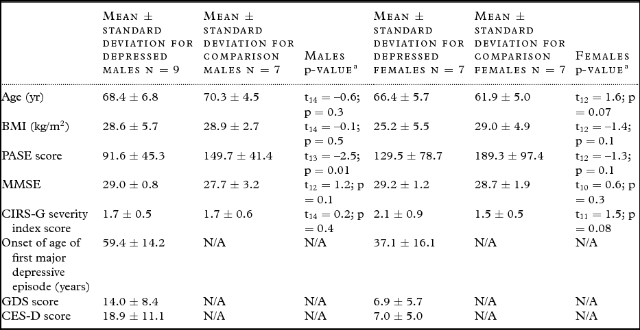
The groups were similar in age, sex, activity levels (PASE), co-morbid medical conditions (CIRS-G), and other demographic features (see Table 1). The mean age of the study participants taken together was 66.9 ± 6.2 years.
Demographic data and questionnaire scores for depressed and comparison participants

aAn independent one-tailed t test was used to calculate p-values, α = 0.01.
Administered scales include the Physical Activity Scale for the Elderly (PASE), Mini-Mental State Examination (MMSE), The Cumulative Illness Rating Scale for Geriatrics (CIRS-G), Geriatric Depression Scale (GDS), and the Center for Epidemiological Studies Depression Scale (CES-D).
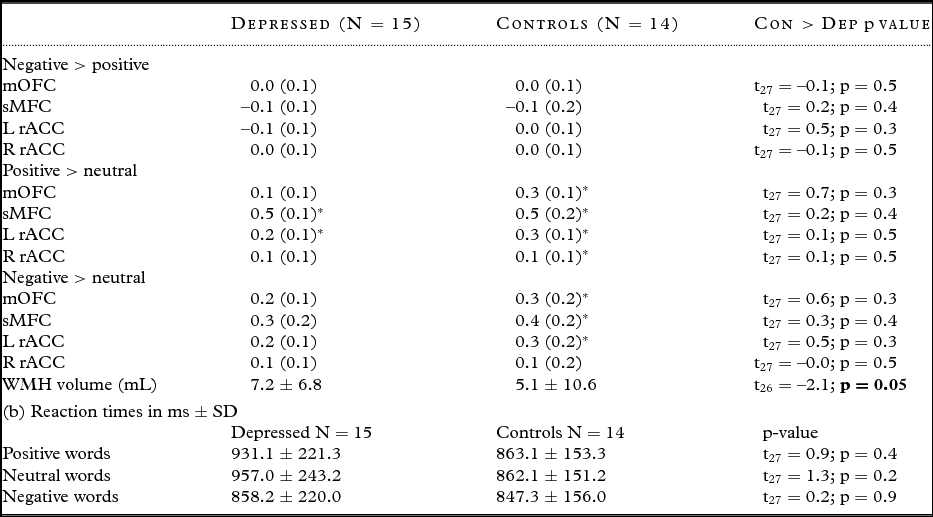
The comparison and depressed participants, across both genders, showed increased BOLD activity to positive versus neutral words, and the comparison participants additionally had increased activation to negative versus neutral words. However, there were no differences in activations between the depressed and comparison participants to any valence contrast (Table 2a). Additionally, there were no significant differences between groups with regards to the reaction times to the words correctly responded to (Table 2b).
(a) Bold values signal mean (SE) in ROIs for the different valence words in the late life depressed (LLD) versus comparison participants. mOFC = medial orbitofrontal cortex; sMFC = superior medial frontal cortex; LrACC and RrACC = left and right rostral anterior cingulate cortex; *indicates within group significant activation (p < 0.05)

The WMH volume was significantly greater in the depressed group compared to comparison participants.
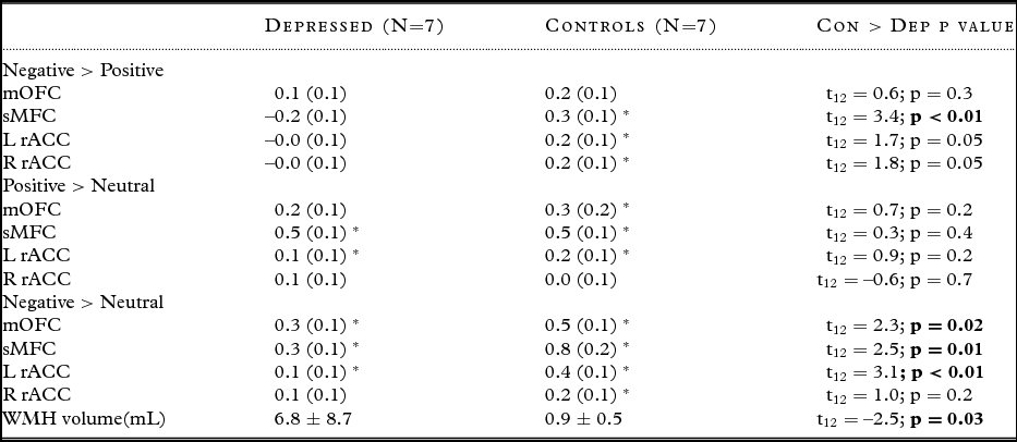
Since the paper by Brassen et al. (2008) showed significant group differences in their female participants, we performed a further exploratory analysis, examining the women only. Compared to female participants with depression, the female comparison participants had significantly greater activation in some areas of the vMPFC for negative versus positive, and negative versus neutral words (Table 3). The WMH remained significantly higher in the female LLD participants relative to comparison participants.
Bold values signal mean (SE) in ROIs for the different valence words in the female participants only. mOFC = medial orbitofrontal cortex; sMFC = superior medial frontal cortex; LrACC and RrACC = left and right rostral anterior cingulate cortex; * indicates within group significant activation (p < 0.05)

We performed additional correlational analysis between WMH and BOLD findings across both genders and groups, and noted lack of statistical significance.
Conclusions
This cross-sectional study in a sample of mixed gender patients with mild to moderate LLD, found a higher total WMH volume in LLD participants compared to comparison participants. No BOLD differences were seen when considering the entire group. However, in the female only subgroup, more marked WMH and BOLD differences were seen. While our positive findings in females are similar to those found by Brassen et al., it is difficult to conceptualize why females had more marked neuroimaging pathology compared to males even though the males scored higher on the depression rating scales (mean GDS score 14 ± 8.4 in males vs. 6.9 ± 5.7 in females). Additionally, though correlational analysis failed to show a relationship between extent of WMH and BOLD activation, it can still be hypothesized that WMH affects the processing of emotional stimuli particularly in the sub-sample of female LLD contributing to impaired emotional evaluation during an emotional word task. A number of studies might like to explore this link fully as this study was not powered enough to assess this. As this study may not have been adequately powered, there is a potential for a type II error. Subsequent larger sample studies might additionally examine relationship to other putative brain functions of the vMPFC, such as autonomic control (Nagai et al., Reference Nagai, Critchley, Featherstone, Trimble and Dolan2004; Critchley, Reference Critchley2005) and decision making (Bechara et al., Reference Bechara, Tranel and Damasio2000), as previously we showed that cognitive dysfunction in patients with LLD correlated with extent of WMHs and autonomic impairment (Vasudev et al., Reference Vasudev2012).
Conflict of interest
AV received a salary support from the Opportunity Fund of the Alternative Funding Plan of the Academic Health Sciences Centres of Ontario to conduct this study.
Description of authors’ roles
A. Vasudev conceptualized the study, applied for research grants, assisted in the study design, screened and recruited participants, assisted in writing the paper, and had overall responsibility for reliability and applicability of the data. M. Firbank assisted in the design of the study, analyzed data, and assisted in writing the paper. J. Gati supervised acquisition of participant MRIs and assisted in writing the paper. E. Ionson assisted in data collection and writing the paper. A. Thomas assisted in the study design and writing the paper.
Acknowledgments
Peer reviewed funding received from: Seed Funding Competition, Department of Psychiatry, Western University; Academic Development Fund, Small Grant Competition, Western University and the Alternative Funding Plan of the Academic Health Sciences Centres of Ontario, Opportunities Fund. The research was also supported by the NIHR Newcastle Biomedical Research Unit based at Newcastle upon Tyne Hospitals NHS Foundation Trust/Newcastle University.
Supplementary material
To view supplementary material for this article, please visit https://doi.org/10.1017/S1041610217000461