Introduction
Cortical development is influenced by both genetic and environmental factors (Giedd et al. Reference Giedd, Schmitt and Neale2007). In schizophrenia, cortical abnormalities may already be present in early life and prenatally (Murray & Lewis, Reference Murray and Lewis1987). Longitudinal studies of patients after their first-episode of psychosis (FEP) have suggested that there are progressive cortical changes. This seems to occur throughout the illness (van Haren et al. Reference van Haren, Schnack, Cahn, van den Heuvel, Lepage, Collins, Evans, Hulshoff Pol and Kahn2011), but is especially prominent during the early years following psychosis onset (van Haren et al. Reference van Haren, Cahn, Hulshoff Pol and Kahn2012). Loss of cortical volume in patients with FEP has been well described in frontal and temporal regions (Ellison-Wright et al. Reference Ellison-Wright, Glahn, Laird, Thelen and Bullmore2008) and systematic reviews of longitudinal voxel-based morphometry (VBM) studies have reported progressive loss of grey matter volume in frontal and temporal regions in patients after their first episode in both adult-onset (Hulshoff Pol & Kahn, Reference Hulshoff Pol and Kahn2008) and childhood-onset schizophrenia (Arango et al. Reference Arango, Moreno, Martinez, Parellada, Desco, Moreno, Fraguas, Gogtay, James and Rapoport2008).
Post-mortem studies have shown that neuropathological changes in patients with schizophrenia are more pronounced in the dorsolateral prefrontal cortex than in other cortical regions (Selemon, Reference Selemon2001) and the loss of brain tissue could be due to a reduction in oligodendrogial cells (Uranova et al. Reference Uranova, Vostrikov, Orlovskaya and Rachmanova2004). Chronic exposure of macaque monkeys to haloperidol or olanzapine was associated with a 10–18% reduction in glial cells in parietal grey matter (Konopaske et al. Reference Konopaske, Dorph-Petersen, Pierri, Wu, Sampson and Lewis2007). It remains to be determined whether these progressive brain changes in humans reflect pathology specific to schizophrenia (Brans et al. Reference Brans, van Haren, van Baal, Schnack, Kahn and Hulshoff Pol2008) or whether they may be determined by environmental factors such as antipsychotic medication (Ho et al. Reference Ho, Andreasen, Ziebell, Pierson and Magnotta2011; Vernon et al. Reference Vernon, Natesan, Modo and Kapur2011), cannabis consumption (Rais et al. Reference Rais, Cahn, Van Haren, Schnack, Caspers, Hulshoff Pol and Kahn2008; Martin-Santos et al. Reference Martin-Santos, Fagundo, Crippa, Atakan, Bhattacharyya, Allen, Fusar-Poli, Borgwardt, Seal, Busatto and McGuire2010) or a combination of both.
Cognitive deficits are also present in patients with FEP (Joyce et al. Reference Joyce, Hutton, Mutsatsa and Barnes2005), but in contrast to those findings of cortical volume loss over time, longitudinal studies (Leeson et al. Reference Leeson, Sharma, Harrison, Ron, Barnes and Joyce2011) suggest that cognitive deficits remain stable over time. A correlation between progressive cortical volume loss and cognitive impairment throughout the illness has been found in some studies (Andreasen et al. Reference Andreasen, Nopoulos, Magnotta, Pierson, Ziebell and Ho2011; Asami et al. Reference Asami, Bouix, Whitford, Shenton, Salisbury and McCarley2012) but not in others (van Haren et al. Reference van Haren, Hulshoff Pol, Schnack, Cahn, Brans, Carati, Rais and Kahn2008; Arango et al. Reference Arango, Rapado-Castro, Reig, Castro-Fornieles, Gonzalez-Pinto, Otero, Baeza, Moreno, Graell, Janssen, Parellada, Moreno, Bargallo and Desco2012).
The area and thickness of the cortex are both high heritability but are determined by different genetic mechanisms (Panizzon et al. Reference Panizzon, Fennema-Notestine, Eyler, Jernigan, Prom-Wormley, Neale, Jacobson, Lyons, Grant, Franz, Xian, Tsuang, Fischl, Seidman, Dale and Kremen2009) and may respond differently to developmental or environmental insults (Du et al. Reference Du, Schuff, Kramer, Rosen, Gorno-Tempini, Rankin, Miller and Weiner2007; Voets et al. Reference Voets, Hough, Douaud, Matthews, James, Winmill, Webster and Smith2008). It would therefore seem appropriate to measure the area and thickness of the cortex separately while investigating the possible effects of developmental and environmental factors on the cortical changes of schizophrenia. Surface-based morphometry (SBM) methods such as Freesurfer (Fischl & Dale, Reference Fischl and Dale2000) are well suited for this purpose and are also capable of detecting longitudinal changes in cortical parameters with small variability of within-subjects measurements at each time point (Reuter et al. Reference Reuter, Schmansky, Rosas and Fischl2012).
Longitudinal studies using SBM in patients with schizophrenia have reported widespread cortical thinning without concomitant cortical area reductions (Cobia et al. Reference Cobia, Smith, Wang and Csernansky2012), with most extensive loss in the frontal and temporal regions (van Haren et al. Reference van Haren, Schnack, Cahn, van den Heuvel, Lepage, Collins, Evans, Hulshoff Pol and Kahn2011; Cobia et al. Reference Cobia, Smith, Wang and Csernansky2012). Long-term antipsychotic treatment has been correlated with thinning of the frontal and cingulate cortex whereas poor social outcome was found to correlate with thinning of the middle temporal cortex (van Haren et al. Reference van Haren, Schnack, Cahn, van den Heuvel, Lepage, Collins, Evans, Hulshoff Pol and Kahn2011), but others failed to find such correlations (Cobia et al. Reference Cobia, Smith, Wang and Csernansky2012; Roiz-Santianez et al. Reference Roiz-Santianez, Tordesillas-Gutierrez, Ortiz-Garcia de la Foz, Ayesa-Arriola, Gutierrez, Tabares-Seisdedos, Vazquez-Barquero and Crespo-Facorro2012). Goghari et al. (Reference Goghari, Smith, Honer, Kopala, Thornton, Su, Macewan and Lang2013) recently reported an increase in prefrontal cortical thickness in FEP patients after short-term treatment with atypical antipsychotics.
Using SBM in a cross-sectional study of FEP patients, we have previously reported reductions in cortical area in the superior temporal gyrus without changes in cortical thickness (Gutiérrez-Galve et al. Reference Gutiérrez-Galve, Wheeler-Kingshott, Altmann, Price, Chu, Leeson, Lobo, Barker, Barnes, Joyce and Ron2010). These cortical changes were associated with premorbid and current IQ.
We report here an exploratory longitudinal study using SBM (Freesurfer) of patients followed up 2 years after their first psychotic episode. The sample of patients included in this longitudinal study overlaps with that of our cross-sectional study (Gutiérrez-Galve et al. Reference Gutiérrez-Galve, Wheeler-Kingshott, Altmann, Price, Chu, Leeson, Lobo, Barker, Barnes, Joyce and Ron2010). We hypothesized that progressive reductions in cortical thickness would be more prominent than those in cortical area in patients compared to healthy controls. We also explored the relationship between cortical changes and changes in symptoms and cognition over the same time points. To our knowledge this is the first longitudinal study to explore the association between changes in area and cortical thickness and cognitive measures in patients with FEP.
Method
Subjects
The patients presented with a psychotic illness for the first time, and had received no more than 12 weeks of antipsychotic medication. As part of the West London Longitudinal First-Episode Psychosis Study (Huddy et al. Reference Huddy, Hodgson, Kapasi, Mutsatsa, Harrison, Barnes and Joyce2007), diagnostic assessments were conducted at presentation and approximately 1 year later. Diagnosis was ascertained using the diagnostic module of the Diagnostic Interview for Psychosis (DIP-DM; Jablensky et al. Reference Jablensky, McGrath, Herrman, Castle, Gureje, Evans, Carr, Morgan, Korten and Harvey2000). Two nurses trained by an experienced psychiatrist (T.R.E.B.) conducted the interviews.
At recruitment, the patients were between 16 and 43 years of age. Twenty-seven FEP patients (17 males) were followed up for 23.8 months after the initial magnetic resonance imaging (MRI) and neuropsychological assessment; at baseline, 24 patients were prescribed antipsychotics (23 atypical, one typical) and at follow-up 18 patients (17 atypical, one typical). Of the 27 patients, 20 had a diagnosis of schizophrenia and seven of schizo-affective disorder. Twenty-five healthy participants (14 males) matched for age and gender who were followed up after a mean of 20.5 months after the initial MRI and neuropsychological assessment served as controls. Sixteen patients and 15 controls who participated in this study had also been included in our previous cross-sectional study (Gutiérrez-Galve et al. Reference Gutiérrez-Galve, Wheeler-Kingshott, Altmann, Price, Chu, Leeson, Lobo, Barker, Barnes, Joyce and Ron2010). Exclusion criteria for all participants were the presence of a medical or neurological illness, including head injury leading to unconsciousness. Controls with prior or family history of psychiatric illness were excluded.
Ethical permission was obtained from the local Ethics Committees. Participants gave written informed consent according to the Declaration of Helsinki.
Demographic details are given in Table 1.
Demographic and total brain volume measures in patients and controls

n.a., Not applicable.
Data shown are mean (standard deviation) [range] with p values of differences between patients (P) and controls (C).
Procedures
Clinical and neuropsychological assessment, MRI acquisition and processing have been described previously (Gutiérrez-Galve et al. Reference Gutiérrez-Galve, Wheeler-Kingshott, Altmann, Price, Chu, Leeson, Lobo, Barker, Barnes, Joyce and Ron2010). Clinical and neuropsychological assessment and MRI were performed at baseline and follow-up. The maximum interval between clinical and neuropsychological assessment and MRI on both occasions was 1 month.
Clinical measures
These included the Scales for the Assessment of Negative and Positive Symptoms (SANS and SAPS; Andreasen, Reference Andreasen1983, Reference Andreasen1984), the Young Mania Rating Scale (YMRS; Young et al. Reference Young, Biggs, Ziegler and Meyer1978), the Hamilton Rating Scale for Depression (HAMD; Hamilton, Reference Hamilton1960), age of onset and duration of untreated psychosis (DUP; Perkins et al. Reference Perkins, Leserman, Jarskog, Graham, Kazmer and Lieberman2000), alcohol and drug use scales (Drake et al. Reference Drake, Osher, Noordsy, Hurlbut, Teague and Beaudett1990), handedness (Annett, Reference Annett1970) and social function. Duration of treatment was calculated as the number of days patients were prescribed antipsychotic medication from the first time, as ascertained from their clinical notes.
Neuropsychological measures
We used the National Adult Reading Test (NART; Nelson & Willson, Reference Nelson and Willson1991) to measure premorbid IQ, the short Wechsler Adult Intelligence Scale – 3rd Edition (WAIS-III; Wechsler, Reference Wechsler1997) to measure current IQ, the Cambridge Neuropsychological Test Automated Battery (CANTAB: working memory span, working memory manipulation and Stockings of Cambridge; Sahakian & Owen, Reference Sahakian and Owen1992) to measure executive function, and the Rey Auditory Verbal Learning Test (RAVLT; Lezak, Reference Lezak1995) to measure verbal memory. Cognitive measures were assessed at baseline and follow-up in patients.
Image processing
Freesurfer 4.5.0 (http://surfer.nmr.mgh.harvard.edu/fswiki/LongitudinalProcessing) was used to generate longitudinal maps of surface area and cortical thickness (Dale et al. Reference Dale, Fischl and Sereno1999; Fischl et al. Reference Fischl, Sereno and Dale1999). The longitudinal process is designed to be unbiased at all time points and a template volume is created instead of initializing the template with information from a specific time point. Using the processed results from the unbiased template, the random variation in the processing procedure is reduced and the robustness and sensitivity of the overall longitudinal analysis is improved (Reuter & Fischl, Reference Reuter and Fischl2011; Reuter et al. Reference Reuter, Schmansky, Rosas and Fischl2012).
Total brain volume was estimated using Freesurfer (Buckner et al. Reference Buckner, Head, Parker, Fotenos, Marcus, Morris and Snyder2004). Cortical parameters were measured by L.G.-G., blind to participant status.
Analysis of cortical parameters
Twenty-two cortical parcellations in each hemisphere were selected from the Desikan template (Desikan et al. Reference Desikan, Segonne, Fischl, Quinn, Dickerson, Blacker, Buckner, Dale, Maguire, Hyman, Albert and Killiany2006): nine frontal (superior, rostral middle, caudal middle, pars opercularis, pars triangularis, pars orbitalis, frontal pole, lateral and medial orbitofrontal), eight temporal (transverse, superior, middle, inferior, temporal pole, fusiform, entorhinal and parahippocampal), three parietal (superior, inferior and precuneus), three occipital (lateral, cuneus and lingual) and four cingulate (rostral anterior, caudal anterior, posterior and isthmus). The average cortical thickness, total surface area and total volume of the cortex for frontal, temporal, parietal, occipital and cingulate regions in each hemisphere were calculated from the individual parcellations at baseline and follow-up. The following comparisons were made for each region: (1) follow-up versus baseline in patients; (2) follow-up versus baseline in controls; (3) patients versus controls at baseline and (4) patients versus controls at follow-up.
Statistical analysis
Baseline and follow-up cortical parameter measures were used for patients and controls. In patients, baseline and follow-up cognitive measures were used to explore associations between cortical parameters and cognitive measures, and baseline cognitive measures were used to predict cortical changes. The statistical package Stata version 10 (Stata Corporation, USA) was used for analyses. Age, gender, duration of follow-up and handedness were compared using t and χ 2 tests.
Linear regression models were used to estimate longitudinal differences for the clinical and cognitive measures in patients.
Linear mixed models were used to estimate longitudinal differences in whole brain cortical parameters (average cortical thickness, total surface area and total volume) of the parcellations in each of the five brain regions, with side (right/left) and assessment time (baseline/follow-up) as within-subject factors, and diagnosis (control/patient) and gender as between-subject factors. Differences in whole brain cortical parameters and total brain volume were estimated with two-way interactions (diagnosis by assessment time). When significant interactions were present, the same model was repeated separately for each brain region. When significant interactions were present for one region, the model was repeated for the parcellations within that region with three-way interactions (parcellation by diagnosis by assessment time).
Linear mixed models were also used to explore whether changes in cortical parameters were associated with changes in clinical and cognitive variables over time in patients. Two-way interactions were included (clinical variable by assessment time, cognitive score by assessment time). When significant interactions were present for whole brain cortical parameters, the model was repeated for each brain region and for the parcellations within that brain region if these were significant. Age, gender and duration of follow-up were covariates in all models.
As in previous studies (Kuperberg et al. Reference Kuperberg, Broome, McGuire, David, Eddy, Ozawa, Goff, West, Williams, van der Kouwe, Salat, Dale and Fischl2003; Gutiérrez-Galve et al. Reference Gutiérrez-Galve, Wheeler-Kingshott, Altmann, Price, Chu, Leeson, Lobo, Barker, Barnes, Joyce and Ron2010), we did not control for whole brain volume, as this is a schizophrenia-related variable that would have obscured possible group differences. Adjustment for multiple comparisons was performed for each model using false discovery rate (FDR; Genovese et al. Reference Genovese, Lazar and Nichols2002) correction and the level of significance was set at 0.05.
To determine whether cognitive functions at baseline predicted changes in cortical parameters at follow-up in patients, linear regression analyses were performed with cortical parameters as the dependent variable. All neuropsychological measures were entered as potential predictors using stepwise entry criteria set at p = 0.10.
Results
Table 1 shows that there were no differences in age, gender, handedness, time to follow-up or total brain volume between patients and controls. Current IQ differed significantly between patients and controls (p = 0.006). Total brain volume between patients and controls was greater at follow-up than at baseline at trend level (mean differences between baseline and follow-up by diagnosis = −6251.8 mm3, p = 0.086).
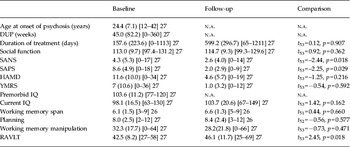
Table 2 shows that positive and negative symptoms improved significantly over time (p < 0.05). There were few significant differences between baseline and follow-up in the cognitive measures. Only verbal learning changed significantly (p = 0.018) and this was seen in the direction of improvement. In a post-hoc analysis, symptoms of depression were not associated with cognitive measures over time (see online Supplementary Table S1).
Clinical and cognitive measures in patients

DUP, Duration of untreated psychosis; SANS, Scales for the Assessment of Negative Symptoms; SAPS, Scales for the Assessment of Positive Symptoms; HAMD, Hamilton Rating Scale for Depression; YMRS, Young Mania Rating Scale; RAVLT, Rey Auditory Verbal Learning Test; n.a., not applicable.
Data shown are mean (standard deviation) [range] n tested with p values of differences between baseline and follow-up in patients.
The analyses were repeated without the schizo-affective subgroup (n = 20) and the results remained unchanged.
Cortical thickness
Whole brain
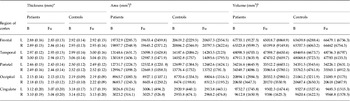
The difference in average cortical thickness across all regions between patients and controls was greater at follow-up than at baseline (mean differences between baseline and follow-up by diagnosis = −0.03402 mm, p = 0.010). In patients, average whole-brain cortical thickness was reduced at baseline at a trend level of significance (patients–controls, mean = −0.05126 mm, p = 0.072) and was significantly reduced at follow-up [patients–controls, mean = −0.08528 mm, 95% confidence interval (CI) −0.14167 to −0.02887, p = 0.003]. Regional cortical parameters in patients and controls unadjusted by age or gender are given in Table 3.
Regional cortical parameters in patients and controls unadjusted by age or gender

B, Baseline; Fu, follow-up; L, left; R, right.
a Values given are mean (standard deviation).
b Values given are mean (standard deviation) of the sums of six parcellations each.
Frontal cortex
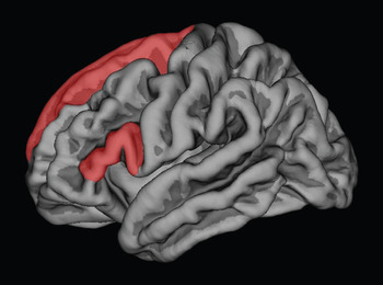
The difference in cortical thickness between the groups was greater at follow-up than at baseline (mean differences between baseline and follow-up by diagnosis = −0.05065 mm, p = 0.008). Cortical thickness did not differ between the groups at baseline (patients–controls, mean = −0.05561 mm, p = 0.112) but it was reduced in patients at follow-up (patients–controls, mean = −0.10626 mm, 95% CI −0.17588 to −0.03665, p = 0.003). This difference was accounted for by thickness reduction in the superior frontal (−0.08569 mm, 95% CI −0.16547 to −0.00593, p = 0.035), pars opercularis (−0.13267 mm, 95% CI −0.21067 to −0.05466, p = 0.001) and pars triangularis (−0.13297 mm, 95% CI −0.21254 to −0.05341, p = 0.001) parcellations (see Fig. 1).
Parcellations where cortical thickness was significantly different between patients and controls followed up for 2 years: superior frontal, pars opercularis and pars triangularis.

The rate of cortical thinning over time was faster in patients (cortical thinning in patients–controls = −0.00255 mm/month, 95% CI −0.00413 to −0.00096, p = 0.002, with cortical thinning of −0.00190 mm/month, 95% CI −0.00298 to −0.00081, p = 0.001 in patients).
Temporal cortex
The difference in cortical thickness between groups was greater at follow-up than at baseline at trend level of significance (mean differences between baseline and follow-up by diagnosis = −0.04391, p = 0.052). Cortical thickness did not differ between groups at baseline (patients–controls, mean = −0.04281 mm, p = 0.251) but was reduced in patients at follow-up (patients–controls, mean = −0.08671 mm, 95% CI −0.16117 to −0.01227, p = 0.022). Cortical thinning in the superior temporal (−0.15801 mm, 95% CI −0.24930 to −0.06672, p = 0.001) parcellation in the patient group accounted for the difference.
The rate of thinning over time was faster in patients (cortical thinning in patients–controls = −0.00197 mm/month, 95% CI −0.00390 to −0.00003, p = 0.046, with a thickness reduction of −0.00235 mm/month, 95% CI −0.00364 to −0.00105, p < 0.001 in patients).
No significant differences between the groups were seen in the thickness of the parietal, occipital or cingulate cortex.
Cortical area
There were no differences in cortical area between groups at baseline or follow-up (mean differences between baseline and follow-up by diagnosis = –324.76 mm2, p = 0.648), indicating that the cortical area had remained unchanged in both groups during the follow-up.
We performed a post-hoc analysis in the patient group, looking for changes in cortical area in those parcellations (superior frontal, pars opercularis, pars triangularis and superior temporal) showing longitudinal reductions in cortical thickness. The cortical area in the superior frontal parcellation was smaller in patients than in controls at both baseline and follow-up but there was no change in area over time (follow-up–baseline mean by diagnosis = –1.99 mm2, p = 0.801; baseline: patients–controls, mean = –269.98 mm2, p = 0.045; follow-up: patients–controls, mean = –271.97 mm2, p = 0.040) (see Supplementary Table S2).
There were no differences in cortical volume between groups at baseline or follow-up (mean differences between baseline and follow-up by diagnosis = –3864.54 mm3, p = 0.193).
Associations with duration of treatment
The association of duration of antipsychotic treatment with average cortical thickness was stronger at follow-up than at baseline (cortical thinning over time = −0.00012 mm/day of treatment, 95% CI −0.00022 to −0.00002, p = 0.020, with a thickness reduction of −0.00009 mm/day, 95% CI −0.00018 to −0.000001, p = 0.048 at follow-up).
The association of frontal cortical thickness with duration of treatment was stronger at follow-up than at baseline (cortical thinning over time = −0.00019 mm/day of treatment, 95% CI −0.00033 to −0.00005, p = 0.007, with thinning of −0.00010 mm/day, 95% CI −0.00018 to −0.000001, p = 0.050 at follow-up). This was accounted for by thinning in the superior frontal (−0.00014 mm/day, 95% CI −0.00025 to −0.00002, p = 0.020) and caudal middle frontal (−0.00015 mm/day, 95% CI −0.00027 to −0.00002, p = 0.019) parcellations.
The association of duration of treatment with frontal cortical volume was stronger at follow-up than at baseline (cortical volume changes over time = −5.05 mm3/day of treatment, 95% CI −9.41 to −0.68, p = 0.023, with a volume reduction of −4.09 mm3/day, 95% CI −7.11 to −1.07, p = 0.008 at follow-up).
Associations with cognitive variables
Premorbid IQ was associated with total cortical area at baseline and follow-up (p < 0.05) and the strength of this association was similar on both occasions (over time mean difference in cortical area per IQ point = 0.94 mm2, p = 0.958). This was explained by the association with frontal area (baseline: increase of 82.75 mm2, 95% CI 13.30–152.20, p = 0.020 per IQ point; follow-up: increase of 89.86 mm2, 95% CI 20.26–159.44, p = 0.011 per IQ point).
Premorbid IQ was associated with total cortical volume at baseline and follow-up (p < 0.05) and the strength of this association was similar on both occasions (over time mean difference in cortical area per IQ point 57.08 mm3, p = 0.442). This was explained by the association with frontal (baseline: increase of 280.59 mm3, 95% CI 34.19–526.98, p = 0.026 per IQ point; follow-up: increase of 293.44 mm3, 95% CI 47.67–539.22, p = 0.019 per IQ point) and parietal volume (baseline: increase of 158.47 mm3, 95% CI 38.36–278.59, p = 0.010 per IQ point; follow-up: increase of 168.25 mm3, 95% CI 49.29–287.21, p = 0.006 per IQ point).
No associations were found between changes in clinical or cognitive variables and cortical parameters over time (see Supplementary Tables S3–S5).
Prediction of cortical parameters from cognitive performance at baseline
Premorbid and current IQ at baseline predicted reduction in the thickness of the parietal cortex (premorbid IQ: r 2 adj = 0.292, t = 2.47, p = 0.022; current IQ: r 2 adj = 0.183, t = 2.59, p = 0.016) at follow-up. Working memory span at baseline predicted thickness reductions of the frontal (r 2 adj = 0.182, t = 2.23, p = 0.045) and parietal (r 2 adj = 0.247, t = 2.54, p = 0.019) cortices at follow-up.
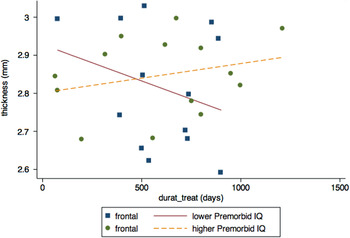
We performed a post-hoc analysis to examine the associations between duration of treatment, frontal cortical thickness and premorbid IQ. A binary variable was created with one group of patients with IQ lower than the median score of 103 (n = 13) and another of patients with IQ higher than or equal to the median score (n = 14). The association of frontal cortical thickness with duration of treatment was stronger in the group of patients with lower premorbid IQ (three-way interaction p = 0.052, with cortical thinning of −0.00039 mm/day, 95% CI −0.00062 to −0.00016, p = 0.004; see Fig. 2).
Scatter plot of the association between duration of treatment and frontal cortical thickness in patients with lower and higher premorbid IQ.

Discussion
The main finding of this study was the thinning of the frontal (superior frontal, pars opercularis, pars triangularis) and, to a lesser extent, superior temporal cortex in patients with FEP followed up for 2 years. By contrast, there were no changes in cortical area over the same period, although in the regions where cortical thinning occurred, reductions in the cortical area were already detectable at baseline in the patient group. We included patients with schizo-affective disorder and schizophrenia in our sample as recent evidence suggests that there are no distinguishing features in the brain structure (Ivleva et al. Reference Ivleva, Bidesi, Keshavan, Pearlson, Meda, Dodig, Moates, Lu, Francis, Tandon, Schretlen, Sweeney, Clementz and Tamminga2013) or cognitive performance (Reilly & Sweeney, Reference Reilly and Sweeney2014) between these two groups. Our findings remained unchanged when patients with schizo-affective disorder were excluded.
Our findings are in keeping with those of others that have reported progressive frontal and temporal cortical thinning in patients with childhood-onset (Thompson et al. Reference Thompson, Vidal, Giedd, Gochman, Blumenthal, Nicolson, Toga and Rapoport2001; Vidal et al. Reference Vidal, Rapoport, Hayashi, Geaga, Sui, McLemore, Alaghband, Giedd, Gochman, Blumenthal, Gogtay, Nicolson, Toga and Thompson2006) and adult-onset schizophrenia (van Haren et al. Reference van Haren, Schnack, Cahn, van den Heuvel, Lepage, Collins, Evans, Hulshoff Pol and Kahn2011; Cobia et al. Reference Cobia, Smith, Wang and Csernansky2012) using SBM. The distribution of cortical thinning in our patients involving the superior and inferior frontal gyrus and, to a lesser extent, the superior temporal cortex is also in keeping with the findings of others in chronic (Kuperberg et al. Reference Kuperberg, Broome, McGuire, David, Eddy, Ozawa, Goff, West, Williams, van der Kouwe, Salat, Dale and Fischl2003) and FEP patients (Narr et al. Reference Narr, Bilder, Toga, Woods, Rex, Szeszko, Robinson, Sevy, Gunduz-Bruce, Wang, DeLuca and Thompson2005; Wisco et al. Reference Wisco, Kuperberg, Manoach, Quinn, Busa, Fischl, Heckers and Sorensen2007; Minatogawa-Chang et al. Reference Minatogawa-Chang, Schaufelberger, Ayres, Duran, Gutt, Murray, Rushe, McGuire, Menezes, Scazufca and Busatto2009; Schultz et al. Reference Schultz, Koch, Wagner, Roebel, Schachtzabel, Gaser, Nenadic, Reichenbach, Sauer and Schlosser2010). We have also reported cortical thinning in the inferior frontal gyrus in patients with temporal lobe epilepsy and interictal psychosis (Gutiérrez-Galve et al. Reference Gutiérrez-Galve, Flugel, Thompson, Koepp, Symms, Ron and Foong2012).
Longitudinal studies using VBM have also found loss of frontotemporal grey matter in patients with first-episode early-onset (Arango et al. Reference Arango, Rapado-Castro, Reig, Castro-Fornieles, Gonzalez-Pinto, Otero, Baeza, Moreno, Graell, Janssen, Parellada, Moreno, Bargallo and Desco2012) and adult-onset (van Haren et al. Reference van Haren, Hulshoff Pol, Schnack, Cahn, Brans, Carati, Rais and Kahn2008) schizophrenia followed up over time. In our study, cortical thinning was not large enough to translate into group differences in cortical volume, although progressive loss of cortical volume may have been detected in a larger sample. Our findings are similar to those of Cobia et al. (Reference Cobia, Smith, Wang and Csernansky2012), who reported thinning of the frontotemporal cortex in 20 patients with adult-onset schizophrenia followed up for 2 years in the absence of changes in cortical area.
Our earlier observations in an overlapping cohort of patients with FEP (Gutiérrez-Galve et al. Reference Gutiérrez-Galve, Wheeler-Kingshott, Altmann, Price, Chu, Leeson, Lobo, Barker, Barnes, Joyce and Ron2010) and those of Rais et al. (2012) in a medication-naive cohort suggest that reductions in cortical area without cortical thinning may be present at or before disease onset, whereas cortical thinning may become more prominent over time, pointing to mechanisms operating later in the disease (van Haren et al. Reference van Haren, Hulshoff Pol, Schnack, Cahn, Brans, Carati, Rais and Kahn2008; Sun et al. Reference Sun, Stuart, Jenkinson, Wood, McGorry, Velakoulis, van Erp, Thompson, Toga, Smith, Cannon and Pantelis2009). The pathological processes underlying these progressive cortical changes have not been fully elucidated, although reduction of the interneuronal neuropil through exaggerated physiological pruning (Boksa, Reference Boksa2012) has been put forward as a possible mechanism.
A study looking at the heritability of grey matter volume in schizophrenia (Owens et al. Reference Owens, Picchioni, Ettinger, McDonald, Walshe, Schmechtig, Murray, Rijsdijk and Toulopoulou2012) has reported a small or non-significant heritability for the grey matter of the superior, middle and inferior frontal cortex, suggesting that cortical changes in these regions are not an endophenotype for schizophrenia and that grey matter loss may be due to illness-related biological changes including, inter alia, exposure to medication and substance abuse.
A recent meta-analysis has reported a correlation between reduction in grey matter volume and cumulative exposure to antipsychotic treatment in patients with schizophrenia (Fusar-Poli et al. Reference Fusar-Poli, Smieskova, Kempton, Ho, Andreasen and Borgwardt2013). A systematic review (Moncrieff & Leo, Reference Moncrieff and Leo2010) has reported loss of frontal cortical grey matter in 14 out of 26 longitudinal MRI studies of medicated patients whereas grey matter loss was only present in five out of 21 studies of medication-naive patients. van Haren et al. (Reference van Haren, Schnack, Cahn, van den Heuvel, Lepage, Collins, Evans, Hulshoff Pol and Kahn2011) have also reported an association between antipsychotic exposure and progressive cortical thinning, particularly in those on typical antipsychotics. Our results also suggest that cortical thinning in those frontal areas of low grey matter heritability (superior and middle frontal gyri) may be due in part to medication exposure, even if most of our patients had only received atypical antipsychotics. However, it should be noted that our measure of exposure to medication was based on duration of treatment, as we did not consider that the available dosage information was accurate enough to explore the association between dose of medication and cortical thinning over time.
The clinical relevance of cortical thinning remains uncertain. Progressive cortical thinning was not associated with the severity of clinical symptoms in our patients, in keeping with the findings of some studies (Cobia et al. Reference Cobia, Smith, Wang and Csernansky2012; Roiz-Santianez et al. Reference Roiz-Santianez, Tordesillas-Gutierrez, Ortiz-Garcia de la Foz, Ayesa-Arriola, Gutierrez, Tabares-Seisdedos, Vazquez-Barquero and Crespo-Facorro2012), but not others (van Haren et al. Reference van Haren, Schnack, Cahn, van den Heuvel, Lepage, Collins, Evans, Hulshoff Pol and Kahn2011). Similarly, the cognitive performance of our patients did not deteriorate over time despite progressive cortical thinning. A similar finding has also been reported by Cobia et al. (Reference Cobia, Smith, Wang and Csernansky2012) in a small group of patients who remained stable clinically and cognitively over a 2-year follow-up. We did find, however, that cortical thinning was predicted by low premorbid IQ and poor working memory present at the time of the first assessment.
Functional imaging studies (Murray et al. Reference Murray, Corlett and Fletcher2010) have suggested that cognitive function in the early stages of schizophrenia may be preserved in the presence of cortical abnormalities by engaging alternative or additional neural networks, a phenomenon that may reflect cognitive reserve (Barnett et al. 2006). More extensive longitudinal studies are required to determine whether these compensatory mechanisms eventually become ineffective.
Our finding that the relationship between duration of treatment and frontal cortex thinning was greater in those with lower premorbid IQ is intriguing. One possible explanation is that patients with lower cognitive reserve (i.e. premorbid IQ) are more vulnerable to medication effects on grey matter. Another is that measures of medication duration or cumulative dose are a proxy for illness severity, which is also reflected in the premorbid IQ (Khandaker et al. Reference Khandaker, Barnett, White and Jones2011) and the severity of cortical changes.
Our findings have to be considered as exploratory, given the small sample size of our study, although SBM is sensitive enough to reliably detect longitudinal changes in cortical parameters in small samples (Reuter et al. Reference Reuter, Schmansky, Rosas and Fischl2012). It is also relevant to mention that we were not able to replicate fully the results of our earlier cross-sectional study performed in an overlapping sample of patients with FEP (Gutiérrez-Galve et al. Reference Gutiérrez-Galve, Wheeler-Kingshott, Altmann, Price, Chu, Leeson, Lobo, Barker, Barnes, Joyce and Ron2010), as only subtle reductions in cortical area were present in the study reported here whereas in our cross-sectional study widespread area reductions were present in the frontotemporal cortex. It remains likely that we may be able to replicate our previous findings fully in a larger patient sample. Another limitation is that we did not have suitable data to explore whether cannabis consumption may have also been related to cortical changes in our patients (Rais et al. Reference Rais, Cahn, Van Haren, Schnack, Caspers, Hulshoff Pol and Kahn2008). Finally, the duration of follow-up was variable in patients and healthy controls, although linear mixed models used in this study cover overdispersion bias (Breslow & Clayton, Reference Breslow and Clayton1993).
Supplementary material
For supplementary material accompanying this paper visit http://dx.doi.org/10.1017/S0033291714001433.
Acknowledgements
We thank I. Harrison and S. Mutsatsa for recruiting patients for the West London study and Dr M. Harrison for the neuropsychological testing. We also thank Professor D. H. Miller and other members of the NMR Unit of the UCL Institute of Neurology. We are grateful to all the subjects who participated in the study.
This study was funded by a programme grant from the Wellcome Trust (Number 064607). E.M.J. was supported by the UCLH/UCL Biomedical Research Centre.
Declaration of Interest
None.