I. INTRODUCTION
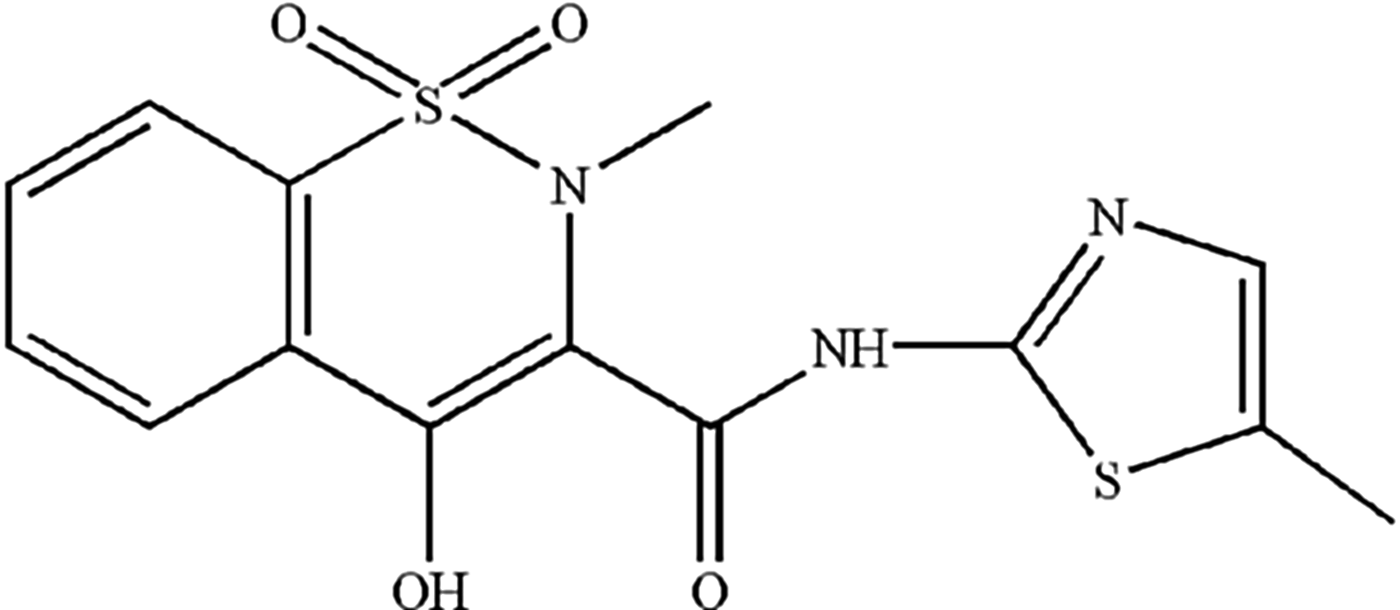
Meloxicam (Figure 1), is a non-steroidal anti-inflammatory drug of the enolic acid class of compounds, used to treat chronic rheumatic diseases and mastitis (Fitzpatrick et al., Reference Fitzpatrick, Chapinal, Petersson-Wolfe, DeVries, Kelton, Duffield and Leslie2013). Meanwhile, it shows a high potency in animal tests for chronic musculoskeletal disorders (Johnston, Reference Johnston2013).
Structural formula of meloxicam.

The single crystallographic data of meloxicam were reported by Fabiola et al. (Reference Fabiola, Pattabhi, Manjunatha, Rao and Nagarajan1998) and Luger et al. (Reference Luger, Daneck, Engel, Trummlitz and Wagner1996), recorded separately in the Cambridge Structural Database with the reference code SEDZOQ and SEDZOQ01. The average values of cell parameters were a = 6.999(6) Å, b = 8.114(1) Å, c = 13.607(6) Å, α = 85.711(1)°, β = 88.336(1)°, γ = 74.871(1)°, unit-cell volume V = 743.908 Å3, Z = 2, and space group P-1. To date, detailed X-ray powder diffraction data for meloxicam have not been reported in the literature.
II. EXPERIMENTAL
A. Sample
Meloxicam, in powder form of more than 99% purity was purchased from Sichuan Sunnyhope Pharmaceutical Co., Ltd., China. The meloxicam sample was also characterized by melting point at 255.1 °C and IR measurements.
B. Powder diffraction data collection and reduction
The X-ray powder diffraction measurements were performed on an X'Pert PRO diffractometer (PANalytical Co., Ltd., Netherlands) equipped with a PIXcel 1D detection system and CuKα 1 radiation (λ = 1.540 56 Å, generator setting: 40 kV and 40 mA). The diffraction data were recorded at room temperature with a step size of 0.013 13° 2θ within 5–50° in 2θ. The structural calculations were mostly conducted using the Reflex module in the software package Material Studio 4.2 (Accelrys Co., Ltd., USA) in the State Key Laboratory of Polymer Materials Engineering (Sichuan University, China).
The unit-cell dimensions can be determined by indexing and analyzing the peak positions in the powder diffraction patterns using X-Cell method (David et al., Reference David, Shankland, McCusker and Baerlocher2002). The indexing result was then refined Pawley refinement to fit the experimental powder pattern (Harris, Reference Harris2012; Wu et al., Reference Wu, Tang, Pan, Cheng and Li2013). After Pawley refinement of the unit cell, the final R wp of the structure was converged at 3.49%.
III. RESULTS
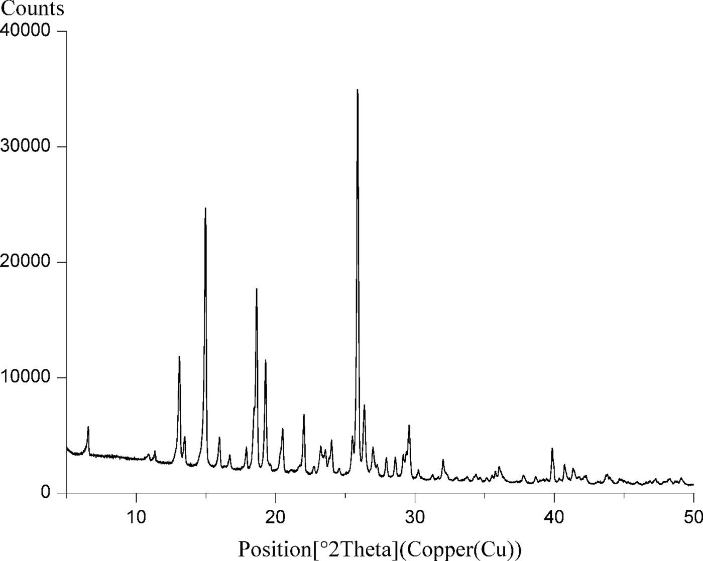
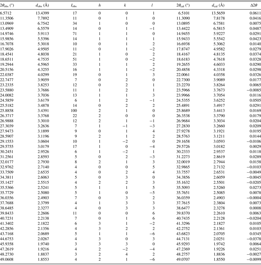
The experimental powder diffraction pattern is shown in Figure 2. Pawley refinement results show that meloxicam is triclinic with space group P-1 and unit-cell parameters: a = 6.997(2) Å, b = 8.113(2) Å, c = 13.604(4) Å, α = 85.774(2)°, β = 88.311(1)°, γ = 74.994(1)°, unit-cell volume V = 743.821 Å3, and Z = 2 (Table I). The results are in good agreement with the single-crystal data and the deviations of the two methods were between 0.01 and 1.60%. All lines of powder and single-crystal data were indexed and are consistent with the P-1 space group.
X-ray powder diffraction pattern of the meloxicam, using CuKα 1 radiation (λ = 1.540 56 Å).
Indexed X-ray powder diffraction data of meloxicam, C14H13N3O4S2. Only the peaks with relative I obs of 1 or greater are reported [a = 6.997(2) Å, b = 8.113(2) Å, c = 13.604(4) Å, α = 85.774(2)°, β = 88.311(1)°, γ = 74.994(1)°, unit-cell volume V = 743.821 Å3, Z = 2, and space group P-1]. All measured lines were indexed and are consistent with the P-1 space group. The d-values were calculated using CuKα 1 radiation (λ = 1.540 56 Å).