Various neurological disorders are characterised by relatively selective degeneration of a subset of neurons, despite the presence of generalised environmental stressors. Examples include the vulnerability of Purkinje cells to hyperthermia, hippocampal neurons to hypoxia or basal ganglia neurons to manganese poisoning. Similarly, disease-related variants in ubiquitously expressed genes can lead to highly selective neuronal loss. Reduction in the level of the ubiquitously expressed survival motor neuron protein (SMN), for example, leads to loss of lower motor neurons from the anterior horn of the spinal cord, causing autosomal recessive spinal muscular atrophy (SMA), the prototypical motor neuron disease of childhood (Ref. Reference Lefebvre1). The most common motor neuron disorder of adulthood, amyotrophic lateral sclerosis (ALS), occurs without a family history in 95% of cases, but mutations in several ubiquitously expressed genes, including SOD1 (superoxide dismutase) and TARDBP (TAR DNA-binding protein 43) cause autosomal dominant forms of disease.
SOD1 was the first familial ALS gene to be identified (Ref. Reference Rosen2), and a vast amount of research has been devoted to understanding how SOD1 mutations cause ALS. Consequently, our view of ALS pathophysiology has, to a large extent, been based on studies utilising SOD1 mouse models (Ref. Reference Turner and Talbot3), which implicate diverse processes in either causation or propagation of ALS, including oxidative stress, excitotoxicity, mitochondrial dysfunction, protein aggregation, defects in axonal transport and impaired cell signalling (reviewed in Refs Reference Cleveland and Rothstein4, Reference Shaw5). None of these insights have yet led to effective therapies. The focus of motor neuron disease research has shifted with the recent discovery of the RNA-binding protein TDP-43 as a key neuropathological marker in the majority cases of ALS (Refs Reference Neumann6, Reference Mackenzie7) and additionally in frontotemporal lobar degeneration with ubiquitinated inclusions (FTLD-U). The subsequent identification of mutations in TARDBP and in a gene encoding a similar RNA-binding protein FUS/TLS (fused in sarcoma/translocated in liposarcoma) (Refs Reference Sreedharan8, Reference Vance9) confirmed the direct role of RNA-associated proteins in the pathogenesis of ALS. At the same time, progress in basic science has led to a rapid expansion of our knowledge of RNA biology (Ref. Reference Licatalosi and Darnell10), including the various functions of SMN in splicing and RNA transport. Abnormal RNA processing provides a possible link between SMA, ALS and other motor neuron disorders. Here, we review the clinical features and molecular pathophysiology of motor neuron disorders and the evidence of altered motor neuron RNA handling from transcription to translation.
Motor neuron anatomy
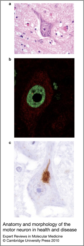
Motor neuron disorders are often classified according to the relative involvement of upper or lower motor neurons. Lower motor neurons comprise those in the ventral horn of the spinal cord and motor nuclei of the brainstem that synapse at the neuromuscular junction (alpha motor neurons) and muscle spindles (gamma motor neurons) forming the final common pathway for voluntary movement. On spinal cord sections, lower motor neurons are conspicuous by their large size, with somata up to 75 µm in diameter, and abundant Nissl substance composed of ribosomal RNA associated with the rough endoplasmatic reticulum (Fig. 1a).
Lower motor neurons are multipolar cells that innervate 2–1000 individual muscle fibres, with axons up to one metre long in humans. The neurotransmitter at the neuromuscular junction is acetylcholine. Each cell has an extensive network of branching dendrites, which are estimated to form 97% of the surface area of the cell, excluding the axon (Ref. Reference Ulfhake and Kellerth11), and harbour up to 10,000 synaptic terminals. These terminals receive input from spinal interneurons predominantly mediated through glycine and γ-aminobutyric acid (GABA), as well as from descending upper motor neurons using excitatory aspartate and glutamate receptors.
Upper motor neurons in the cerebral cortex control lower motor neurons in the spinal cord, by either synapsing directly, or more commonly via interneurons. The large pyramidal Betz cells in layer V of the motor cortex are the archetypal upper motor neurons. The smaller cortical pyramidal cells are more numerous in both the motor and premotor cortex where they are in direct cortical control of movement. Descending fibres travel in the corticospinal tract, along with fibres from more widespread areas of the cortex, including the premotor cortices, the supplementary motor area as well as various parts of the parietal lobe, including the primary somatosensory cortex. Most fibres cross in the lower brainstem (medulla) to synapse onto the contralateral spinal cord, although some spinal cord areas have bilateral innervation via fibres running in the anterior corticospinal tract, which remain ispsilateral or cross only at the relevant spinal segment (Ref. Reference Lemon12). Direct corticomotorneuronal connections to anterior horn cells appear to be a recent evolutionary development that is unique to higher primates (Ref. Reference Eisen13) and appear to have their origin almost exclusively in the primary motor cortex (M1) (Ref. Reference Lemon12). Upper and lower motor neurons share the qualities of extreme polarity, long axons and extensive dendritic networks.
Motor neuron disorders
Lower motor neurons are selectively lost in SMA, which affects 1 in 10,000 live births (Ref. Reference Pearn14) and is characterised by neurogenic muscle atrophy with weakness and wasting, often leading to death from neuromuscular respiratory failure in infancy. SMA is caused by disruption of the SMN1 gene (Ref. Reference Lefebvre1) with subsequent reduction in levels of the SMN protein, which is normally found in discrete nuclear foci and also is also diffusely localised in the cytoplasm (Fig. 1b) (Ref. Reference Lefebvre15). SMN is highly conserved in evolution and ubiquitously expressed. Complete loss of SMN, which is incompatible with life (Ref. Reference Schrank16), is prevented by production of low levels of SMN from the SMN2 gene, a near-identical paralogue of SMN1 which has arisen from a duplication event in recent evolution. The presence of a translationally silent C-T transition in SMN2 exon 7, however, leads to disruption of an exonic splicing enhancer (ESE) element (Ref. Reference Cartegni, Chew and Krainer17), or creation of an exonic splicing silencer (ESS) (Ref. Reference Kashima and Manley18), with subsequent exon 7 skipping in the majority of SMN2-derived transcripts and reduces the level of expression of full-length SMN to about 10% of the normal levels derived from the SMN1 gene (Refs Reference Monani19, Reference Lorson20). Disease severity is broadly proportional to residual SMN levels, which is a function of SMN2 copy number, although other modifying factors are involved in some cases (Refs Reference Wirth21, Reference Oprea22).
The most common adult-onset motor neuron disorder is ALS (also known as Lou Gehrig's disease or simply as ‘motor neuron disease’), which has an incidence of 1–2 per 100,000 population per year and affects both upper and lower motor neurons, although to variable degrees in individual patients. In addition to the lower motor neuron features of muscle atrophy and wasting, upper motor neuron dysfunction is evident clinically by increased muscle tone, exaggerated tendon reflexes and loss of fine motor control. Primary lateral sclerosis (PLS) and progressive muscular atrophy (PMA) are terms used to describe conditions with only upper motor neuron or lower motor neuron clinical features respectively, but which are generally considered part of the aetiopathological spectrum of ALS (Ref Reference McDermott and Shaw23, Reference Wijesekera and Leigh24). In addition to motor dysfunction, many patients show more widespread involvement of the CNS both clinically and pathologically, commonly in the form of mild cognitive dysfunction and less frequently in the form of frank dementia (Ref. Reference Strong25). There is now increasing genetic and neuropathological evidence to suggest that ALS and FTLD-U might be part of the same spectrum of disease (Refs Reference Mackenzie and Feldman26, Reference Neary, Snowden and Mann27). Both share characteristic features of TDP-43-positive neuropathology (Refs Reference Neumann6, Reference Arai28) and mutations in TARDBP can cause not only ALS (Refs Reference Sreedharan8, Reference Gitcho29), but an ALS-determining mutation has recently also been described in a single patient with FTD (Ref. Reference Borroni30). FTLD-U has therefore now been renamed FTLD-TDP-43.
About 5% of ALS is familial, with an autosomal dominant pattern of inheritance (Refs Reference Chiò31, Reference O'Toole32). Mutations in SOD1 constitute up to 20% of familial ALS (Ref. Reference Rosen2), whereas mutations in TARDBP (Ref. Reference Sreedharan8), FUS/TLS (Refs Reference Vance9, Reference Ayala, Misteli and Baralle60) and ANG, which encodes angiogenin (Ref. Reference Greenway33), can also cause classical ALS. The vast majority of ALS occurs sporadically, but is also thought to have a partially genetic basis (Ref. Reference Schymick, Talbot and Traynor34). Despite the marked genetic heterogeneity, there are no clinical features that allow prediction of the underlying aetiology or the distinction between familial and sporadic ALS in individual patients, suggesting that different genetic defects share common pathophysiological mechanisms (Ref. Reference Ravits and La Spada35).
In addition to SMA and ALS, a heterogeneous group of individually rare genetic disorders cause selective loss of upper or lower motor neurons, which are collectively called hereditary spastic paraplegias and distal spinal muscular atrophies (also known as hereditary motor neuropathies), respectively (Ref. Reference James and Talbot36). Despite phenotypic differences across the broader spectrum of motor neuron diseases, it is tempting to speculate that there are common pathways that lead to motor neuron degeneration.
RNA metabolism and motor neuron disorders
Regulation of pre-mRNA splicing
Post-transcriptional processing of mRNA by exon skipping and the use of alternative 3′ or 5′ splice sites, 5′and 3′ terminal exons, and polyadenylation sites can produce numerous mRNA isoforms per gene, thus increasing the cellular protein repertoire. This process is particularly prominent in the nervous system (Ref. Reference Lee and Irizarry37), and there are numerous examples of a role for alternative splicing events in the physiological regulation of neuronal development and maintenance (Ref. Reference Li, Lee and Black38). A switch between alternative mRNA isoforms is essential for neurite outgrowth (Refs Reference Wojtowicz39, Reference Matthews40), synapse formation (Refs Reference Boucard41, Reference Chih, Engelman and Scheiffele42, Reference Chih, Gollan and Scheiffele43) and membrane function (Ref. Reference Raingo, Castiglioni and Lipscombe44) during neuronal development and postnatal maturation.
Splicing is a high-fidelity process that relies on the interaction of specific sequences of the pre-mRNA molecule (cis-acting elements) with a complex array of trans-acting factors, i.e factors that act on the RNA species of interest, including various RNA-binding proteins and the spliceosome. Cis-acting elements include not only the 3′ and 5′ splice site consensus sequences, but also less well characterised splicing regulatory elements in both introns and exons, which can enhance or inhibit exon recognition (exonic or intronic splice enhancers and silencers). The latter sequences are binding sites for protein factors that promote (mainly SR proteins) or inhibit (hnRNPs) exon recognition and assembly of the machinery that removes introns and joins exons – the spliceosome. The major spliceosome consists of five small nuclear ribonucleoprotein complexes (snRNPs), which in turn, consist of snRNAs (U1, U2, U4/6, U5) and hundreds of proteins including a core of Sm proteins (Ref. Reference Wahl, Will and Luhrmann45). A far less abundant minor spliceosome utilises different U snRNAs (U11, U12, U4atac/U6atac, U5) and has different splice-site and branch-point consensus sequences (Ref. Reference Will and Luhrmann46).
A crucial step in the biogenesis of snRNPs, the coupling of the Sm core to the correct snRNA, is catalysed in the cytoplasm by a protein complex consisting of SMN and several other proteins, including gemin2–gemin8 and unrip (STRAP) (Refs Reference Meister47, Reference Pellizzoni, Yong and Dreyfuss48). Although snRNP biogenesis is a ubiquitous process, the stoichiometry of many other splicing factors is specific for the developmental stage and tissue type. Several factors that regulate splicing are specifically expressed in the nervous system (Ref. Reference Li, Lee and Black38). Particularly well characterised are the RNA-binding proteins NOVA1 and NOVA2 (Refs Reference Yang, Yin and Darnell49, Reference Jensen50). NOVA1 and NOVA2 bind to YCAY clusters (Y = pyrimidine base, U or C) that, depending on their position in introns or exons, lead to exon skipping or exon inclusion (Ref. Reference Ule51). NOVA protein targets were identified in an exon-specific microarray screen of Nova-knockout mice, and were shown to be enriched for genes involved in synapse biogenesis, including agrin (AGRN), a factor with an alternatively spliced (Z+) isoform important for acetylcholine receptor clustering at the neuromuscular junction (Ref. Reference Ule52). Nova double-knockout mice are born alive but have markedly reduced levels of Z+ agrin in the spinal cord, and virtually no intact neuromuscular junction. Functional neuromuscular junctions could be re-established by crossing the Nova-knockout mice with mice overexpressing agrin Z+ , but only to uncover a more proximal, as-yet-unidentified defect in motor neuron physiology (Ref. Reference Ruggiu53), suggesting that NOVA1 and NOVA2 are vital for motor neuronal physiology at several levels. The NOVA proteins are examples of trans-acting RNA-binding proteins, deficiency of which can cause specific motor neuronal defects in mice associated with splicing alterations. A rare human disorder, paraneoplastic opsoclonus-myoclonus ataxia (POMA), has a complex phenotype with some motor and cognitive features and autoantibodies against NOVA1 and NOVA2, although a direct pathogenic effect of these antibodies is unlikely (Ref. Reference Yang, Yin and Darnell49).
Two proteins recently implicated in the aetiopathogenesis of ALS, TDP-43 and FUS/TLS, are RNA-binding proteins with a function in splicing regulation. TDP-43 has strong affinity for sequences containing (UG)m repeats (Ref. Reference Buratti and Baralle54) and has been extensively characterised in its role in CFTR exon 9 skipping (Refs Reference Buratti55, Reference Buratti56, Reference Ayala, Pagani and Baralle57) and splicing of the APOA2 gene (Ref. Reference Mercado58), which is probably mediated via an interaction with hnRNPA2 (Ref. Reference D'Ambrogio59). Other pre-mRNA (i.e. splicing) targets of TDP-43 are not yet known, although the top hit in a microarray analysis of TDP-43 knockdown in HeLa cells was Cdk6, which contains several TDP-43 target sequences in its pre-mRNA (Ref. Reference Ayala, Misteli and Baralle60). In addition, there is one report that suggests TDP-43 modifies the splicing of the SMN2 pre-mRNA (Ref. Reference Bose61), which adds an intriguing twist to the relationship between ALS and SMA, but results are based on an artificial minigene system and the significance in vivo is unknown.
FUS/TLS encodes a 526 amino acid, 53 kDa nuclear protein containing a RNA-recognition motif (RRM) surrounded by Arg-Gly-Gly (RGG) repeats. In addition, it contains a zinc-finger domain with RNA-binding properties (Ref. Reference Iko62). FUS interacts with and modulates the splicing function of several SR proteins (Ref. Reference Yang63) and other splicing factors (Refs Reference Meissner64, Reference Sato65), which it binds via its C-terminal domain (Ref. Reference Yang, Embree and Hickstein66). One possibility is that FUS is necessary to direct SR proteins to the correct splice sites (Ref. Reference Meissner64). FUS preferentially binds RNA sequences containing GGUG motifs (Ref. Reference Lerga67), but little is known about specific human FUS RNA targets.
In most cases of ALS and FTLD-U, TDP-43 forms ubiquitinated cytoplasmic or nuclear inclusions, whereas the physiological nuclear protein is translocated to the cytoplasm (Fig. 1b,c) (Refs Reference Neumann6, Reference Mackenzie7, Reference Davidson68, Reference Cairns69). FUS has been shown to form ubiquitinated inclusions in the few autopsy cases of ALS with FUS mutations (Refs Reference Vance9, Reference Kwiatkowski70), as well as in the uncommon form of FTLD-U without TDP-43 pathology (Ref. Reference Neumann71). FUS mutants show increased cytoplasmic and reduced nuclear distribution in cell culture (Refs Reference Vance9, Reference Kwiatkowski70), and there appears to be a degree of nuclear loss of FUS in motor neurons postmortem, albeit to a lesser degree than that seen with TDP-43 (Ref. Reference Neumann71). It is therefore conceivable that loss of nuclear splicing factors in cells affected by ALS leads to a shift in splicing pattern of one or several genes that are crucial for (motor) neuronal function, and that this, rather than a primary toxic effect of neuronal inclusions, is the determinant of disease. In agreement with this, loss of TDP-43 in Drosophila leads to defective neuromuscular junctions (Ref. Reference Feiguin72).
Anatomy and morphology of the motor neuron in health and disease. (a) Cresyl violet stain of a spinal motor neuron showing the large cell body with large nucleus, prominent nucleolus and Nissl substance corresponding to the rough endoplasmatic reticulum. (b) Antibodies against the SMN protein reveal intense staining (red) of nuclear structures associated with ribonucleoprotein assembly in mouse spinal cord. The SMN protein, however, is also present in the cytoplasm and axonal processes. TDP-43 (green) is a predominantly nuclear protein with diffusely granular staining pattern sparing the nucleolus. (c) Motor neurons in ALS are characterised by nuclear depletion of TDP-43, which forms cytoplasmic aggregates.

In contrast to NOVA, both TDP-43 and FUS are ubiquitously expressed proteins (Refs Reference Buratti55, Reference Andersson73, Reference Aman74), whereas ALS and FTLD are diseases in which pathology is limited to the spinal cord and brain. However, loss of a ubiquitous splicing factor could in theory cause tissue-specific alterations by interacting with other cellular factors. Unlike TDP-43 or FUS, SMN does not regulate splicing by binding specific RNA sequences, but has indirect effects through the regulation of spliceosome assembly. This assembly capacity is markedly reduced in models of SMA, although steady state levels of spliceosome components seem to be only very mildly reduced (Refs Reference Gabanella75, Reference Gabanella76). Endstage SMA mice have been shown to have differentially spliced transcripts in several tissues, including the spinal cord (Ref. Reference Zhang77), but the selective loss of lower motor neurons in response to SMN deficiency implies that, if splicing alterations are at the core of SMA pathophysiology, motor neurons are either particularly vulnerable to a global change in the cellular mRNA, or a crucial subset of motor-neuron-specific mRNAs are mis-spliced. We have recently shown that widespread differential exon usage is only a late feature in mouse models of SMA, which argues against a widespread breakdown of splicing fidelity as the cause of SMA (Ref. Reference Baumer78). The lesson learned from NOVA and agrin, however, is that alternative splicing of even a single mRNA can have deleterious consequences for the cell, and a splicing alteration of one or a few genes could be relevant for SMA. So far, no target mRNA has been identified that could explain the SMA phenotype.
An abnormal, intron-retaining splice variant of the intermediate filament protein peripherin has been described to occur in human ALS (Ref. Reference Xiao79). This isoform is prone to aggregation in vitro, and isoform-specific antibodies have been shown to label cytosolic inclusions in ALS motor neurons.
In an unbiased approach, a recent study used exon-specific microarrays to analyse mRNA from laser-capture microdissected residual motor neurons in postmortem spinal cord samples of patients with TDP-43-positive ALS, and identified a large number of differentially expressed exons compared with control samples (Ref. Reference Rabin80). No changes were obvious in the remaining tissue of the anterior horn after the motor neurons had been removed. Although it remains unclear whether or not the observed changes are primary or secondary, this study highlights the importance of considering alternative splicing events in addition to crude levels of transcript in diseases such as ALS. Whether the changes demonstrated reflect a failure of splicing fidelity or a shift in expression to known physiological isoforms appropriate to the ‘stressed’ cell requires further study.
mRNA transport
Motor neurons are the extreme example of a polarised cell, and not only have different protein requirements in different cellular compartments, but also rapidly changing protein needs in response to synaptic stimulation. In addition to the complexity of gene expression brought about by tissue-specific alternative splicing, there is mounting evidence that neurons regulate gene expression both spatially and temporally by localised protein translation in dendrites and axons. Local translation does not seem to be an exceptional mechanism that is important for only a few transcripts, but may involve hundreds of mRNA species (Ref. Reference Martin and Zukin81). Since several proteins implicated in motor neuron disorders have roles in mRNA transport and translation, these pathways constitute points of motor neuron vulnerability beyond pre-mRNA splicing.
Targeting of mRNAs to specific sites in dendrites or axons is a function of cis-acting elements consisting of specific nucleotide sequences on the nascent mRNA that are often, but not exclusively, located in the 3′UTR (localisation elements or ‘zipcodes’), as well as trans-acting protein factors that bind to these sequences. This association often begins during transcription and splicing in the nucleus (Refs Reference Giorgi and Moore82, Reference Oleynikov and Singer83, Reference di Penta84, Reference Gu85). One of the best studied examples of this is the association of the zipcode-binding protein 1 (ZBP1) with a corresponding 54 nucleotide localisation sequence (the zipcode) in the 3'UTR of β-actin mRNA (Ref. Reference Ross86), which is essential for correct asymmetrical targeting of this mRNA in a variety of cell types including neurons (Refs Reference Condeelis and Singer87, Reference Tiruchinapalli88, Reference Zhang89). Upon nuclear export, mRNAs and their associated binding proteins are found in granules that contain a variety of other proteins and facilitate transport or further processing. RNA-transport granules are present in neurons (Ref. Reference Knowles90) and contain part of the machinery needed for later translation of the mRNA (Refs Reference Krichevsky and Kosik91, Reference Kiebler and Bassell92), but there is probably a heterogeneous population of different macromolecular complexes that contain distinct mRNAs and mRNA-binding proteins (Refs Reference Martin and Ephrussi93, Reference Elvira94). Although transport of RNPs can take place by passive diffusion, it is often actively mediated by microtubules and associated kinesin and dynein motors (Refs Reference Knowles90, Reference Kiebler95, Reference Kanai, Dohmae and Hirokawa96).
Several mutations in kinesins are associated with disorders of both upper and lower motor neurons (Ref. Reference James and Talbot36), and there is one report suggesting that variation in kinesin-associated protein 3 (KIFAP3) modulates survival in ALS (Ref. Reference Landers97). In addition, mutations in dynactin have been reported in a few cases of ALS or motor neuropathy (Refs Reference Munch98, Reference Munch99, Reference Puls100). Although there is no direct evidence that mutations lead to ALS through disruption of RNA-transport granules, RNA transport does appear to be dependent on the integrity of axonal-transport motors.
Both TDP-43 and FUS have been identified outside the nucleus in association with protein complexes likely to have a role in RNA transport. FUS was found to be a constituent of kinesin-associated RNA-transport granules in hippocampal neuronal dendrites that contained mRNAs encoding CamKIIa (CAMK2A) and Arc (ARPC3) (Ref. Reference Kanai, Dohmae and Hirokawa96). Translocation of FUS to dendrites was shown to be dependent on intact microtubules, as well as on mGluR5 stimulation. Hippocampal pyramidal neurons from mice deficient in FUS were characterised by abnormal dendritic spine morphology (Refs Reference Fujii101, Reference Fujii and Takumi102). FUS was also shown to be transported by a myosin-Va motor, which is actin based (Ref. Reference Yoshimura103), and binds not only β-actin mRNA, but also mRNA encoding an actin-stabilising protein, Nd1L (Ref. Reference Fujii and Takumi102).
In addition to its cytoplasmic and nuclear role in snRNP assembly and maturation, SMN has been shown to be present in axons and growth cones in cultured cells (Refs Reference Fan and Simard104, Reference Rossoll105, Reference Jablonka106), in rat motor neuronal dendrites (Refs Reference Bechade107, Reference Pagliardini108) and axons (Ref. Reference Pagliardini108), as well as in proximal axons in human neurons (Refs Reference Briese109, Reference Giavazzi110). A number of studies report that SMN interacts with proteins involved in the transport and post-transcriptional regulation of various transcripts, including β-actin. Examples include KSRP/ZBP2/MARTA1, FBP, hnRNP R and hnRNP Q (reviewed in Ref. Reference Rossoll and Bassell111). Reduction of SMN levels induces altered axonal morphology in zebrafish (Ref. Reference McWhorter112) and cultured mouse motor neurons (Ref. Reference Rossoll113), and altered axonal growth is associated with reduced β-actin mRNA transport (Ref. Reference Rossoll113), which is mediated by an SMN-binding partner, hnRNP R (Ref. Reference Rossoll105), but apparently independently of Sm proteins, indicating that the SMN function in axons is distinct from its role in snRNP biogenesis (Ref. Reference Zhang114). hnRNP R was shown to bind to the β-actin mRNA zipcode (Ref. Reference McWhorter112), and although it cannot be ruled out that this association is dependent on the zipcode-binding proteins ZBP1 or ZBP2, the absence of these proteins in several proteomic studies on SMN-binding partners (Ref. Reference Meister, Eggert and Fischer115) makes it possible that the SMN–hnRNP complex is unique in motor neuronal mRNA transport. β-actin is important in axonal and dendritic growth, as well as in synapse formation (Ref. Reference Luo116) and an actin-stabilising protein, plastin 3 (PLS3), was recently shown to be a modifier of disease severity in SMA (Ref. Reference Oprea22), but it is unknown whether β-actin mRNA, or other, as yet unidentified mRNAs are relevant to disease pathogenesis, although locally synthesised dendritic and axonal proteins are known to function in synaptic plasticity and neural repair.
TDP-43 has been shown to bind to the 3′UTR of low molecular weight neurofilament (NFL) mRNA (Ref. Reference Strong117) and forms part of RNA-transport granules enriched for mRNA encoding β-actin (Ref. Reference Elvira94). TDP-43 is also a component of hippocampal dendritic RNA granules, which also contain β-actin and CaMKIIα mRNA (Ref. Reference Wang118). This implicates TDP-43 in mRNA stability and transport, which could be disrupted by pathological TDP-43 aggregate formation in ALS.
Although much of the research on SOD1-related ALS has focussed on the toxicity of SOD1 aggregates, oxidative stress and mitochondrial dysfunction, there are recent reports that mutant SOD1 binds to the VEGF 3′UTR (Refs Reference Lu119, Reference Li120), as well as the NFL 3′UTR (Refs Reference Ge121, Reference Volkening122), implying a possible dysregulation of post-transcriptional mRNA regulation as the disease mechanism.
Local mRNA translation
Neuronal transport granules are generally not translationally competent (Ref. Reference Krichevsky and Kosik91) and the transported mRNAs are translationally repressed until released to polysomes. Translational repressors can act by various means, including blocking the recruitment of ribosomal components, shortening of the polyA tail of mRNAs, with resulting reduced affinity for polyA-binding protein (PABP), and, most commonly, by inhibition of the cap-dependent translation-initiation process (Ref. Reference Besse and Ephrussi123). For example, one of the best known components of RNA granules, FMRP, disrupts translation initiation by targeting the eIF4F complex through a protein-binding partner (Ref. Reference Napoli124), modulating dendritic maturation and synaptic plasticity. FMRP has been reported to bind to SMN, providing indirect evidence that SMN could have a neuron-specific function by regulating local translation, in addition to modulating RNA transport. As well as a role in mRNA transport, alteration of local translation could thus be an alternative explanation for the finding of reduced β-actin protein levels in primary motoneuronal axons taken from SMA mice (Ref. Reference Rossoll113).
The release of mRNAs from translational repressors and subsequent translation has been well studied in dendritic compartments, where it is often the consequence of synaptic-activity-dependent depolarisation and spatially restricted phosphorylation of RNA-binding proteins. For example, β-actin mRNA remains translationally repressed through binding of ZBP1, but phosphorylation of ZBP1 by a Src-kinase releases β-actin mRNA onto polysomes (Ref. Reference Huttelmaier125). Phosphorylation of the translational repressor CPEB in response to NMDA receptor-mediated aurora kinase activation leads to stimulation of polyadenylation of CPEB target mRNAs such as CaMKIIα (Ref. Reference Huang126). In turn, phosphorylation of general translation-initiation factors, such as eIF4e, will lead to global changes of mRNA translational activity.
Although local translation is currently best characterised for the dendritic compartment in hippocampal cell cultures, motor neurons are likely to use similar mechanisms in their extensive dendritic tree. In addition, there is increasing evidence that despite the absence of easily identifiable ribosomes beyond the axonal hillock, functional components of the protein-synthesis machinery are present in the axon (Refs Reference Koenig and Martin127, Reference Koenig128, Reference Merianda129) and that local mRNA translation occurs in axons as in dendrites (reviewed in Refs Reference Lin and Holt130, Reference Giuditta131). For instance, the local translation of β-actin mRNA in the axonal growth cone takes place in response to external guidance cues (Refs Reference Leung132, Reference Lin and Holt133) during development, but local mRNA transport and translation is also thought to be a feature in mature neurons (Refs Reference Hanz134, Reference Yudin135, Reference Taylor136). A recent study systematically examining the axonal compartment of a dorsal root ganglion model system for locally translated mRNA content using a combination of proteomics and RT-PCR, identified more than a hundred proteins of diverse function (Ref. Reference Willis137), many of which overlapped with mRNAs identified in axonal preparations of mature cortical neurons (Ref. Reference Taylor136). In parallel to what is known from dendrites, axonal protein translation is activity dependent (Ref. Reference Jimenez-Diaz138), and changes in the repertoire of axonally synthesised protein are part of a neuronal-injury-response programme (Refs Reference Taylor136, Reference Yoo139).
Mutations in tRNA synthetases GARS and YARS, enzymes that charge tRNAs with the cognate amino acid glycine and tyrosine, respectively, cause a motor-predominant form of peripheral neuropathy, (Refs Reference Antonellis140, Reference Jordanova141). GARS is present in axons, suggesting a function in supporting local protein translation. Mutations in GARS and YARS do not consistently lead to loss of enzyme function or altered cellular localisation. The presence of these proteins in axons implies a potential role in axonal protein synthesis, failure of which could underlie the neuropathy (Refs Reference Antonellis and Green142, Reference Motley, Talbot and Fischbeck143), a view that is supported by findings in Drosophila (Ref. Reference Chihara, Luginbuhl and Luo144).
Mutations in IGHMBP2, the gene encoding the immunoglobulin-µ-binding protein 2, cause a more severe type of distal spinal muscular atrophy (Ref. Reference Grohmann145). IGHMBP2 is a DNA/RNA helicase that was initially thought to be involved in transcription or pre-mRNA splicing, but there is now evidence that it is in fact an RNA helicase associated with ribosomes in the neuronal cytoplasm that is also present in axons and growth cones (Ref. Reference Grohmann146). IGHMBP2 mutants lose their enzymatic activity in vitro, suggesting that dysfunction of mRNA translation underlies distal SMA (Ref. Reference Guenther147). Senataxin (SETX), the gene mutated in a juvenile form of slowly progressive ALS (ALS4) (Ref. Reference Chen148), is a member of the same helicase superfamily. Although functions in transcription, DNA repair and pre-mRNA splicing have been described (Refs Reference Suraweera149, Reference Suraweera150), senataxin shows strong cytoplasmic staining in mature murine neurons (Ref. Reference Chen151), and might have a function in translation regulation, similarly to IGHMBP2. Interacting partners identified by mass spectrometry include PABP and SMN, in addition to several hnRNPs (Ref. Reference Suraweera150).
Translation control: stress granules, P bodies and microRNAs
Whereas local mRNA translation contributes to fine spatial resolution of protein synthesis, temporal control of translation after nuclear export of mRNAs is achieved by temporary storage of mRNAs in ribonucleoprotein granules that can be classified according to their key protein constituents into stress granules or processing bodies (P bodies), although a considerable degree of overlap exists.
Cells, including neurons (Refs Reference Tsai, Tsui and Wei152, Reference DeGracia153), respond to a variety of stressors with a change in gene expression partially mediated by translational arrest of some mRNAs, while specific proteins, such as heat shock proteins, continue to be synthesised (Refs Reference Anderson and Kedersha154, Reference Anderson and Kedersha155, Reference Kawai156). In the stereotypical stress response, cap-dependent translation initiation is inhibited by phosphorylation of eIF2α (EIF2S1) and disassembly of polysomes from translating mRNAs, the majority of which are then stabilised and stored temporarily in stress granules (Refs Reference Anderson and Kedersha154, Reference Anderson and Kedersha155).
The phosphorylation of eIF2α is mediated by a number of kinases that respond to specific stressors, including heat and viral infections, but also factors implicated in the pathogenesis of ALS, such as oxidative stress and endoplasmatic reticulum stress induced by the unfolded protein response (Refs Reference Kawai156, Reference Harding157, Reference Harding158).
Stress granules are characterised by the presence of stalled initiation complexes containing small ribosomal subunits, eIF3, eIF4e and PABP (Ref. Reference Kedersha159). In addition, they contain a variety of proteins involved in various aspects of RNA metabolism, including stabilisation, translational repression and localisation. Several of the proteins involved in motor neuron disorders appear to be present in stress granules. TDP-43 and FUS are nuclear–cytosolic shuttling proteins (Refs Reference Ayala160, Reference Zinszner161), a function shared by many stress granule components (Refs Reference Kedersha162, Reference Taupin163, Reference Gallouzi164). FUS has been shown to be part of stress granules induced by oxidative stress in cell culture (Ref. Reference Andersson73). Cytosolic redistribution of TDP-43 is a key feature of ALS (Ref. Reference Neumann6), and transient TDP-43 relocalisation is observed in motor neurons after axotomy (Refs Reference Moisse165, Reference Moisse166), indicating that cytoplasmic TDP-43 might be a physiological response to injury. In addition, TDP-43 was shown to localise to stress granules in a motor neuronal cell line under oxidative stress (Ref. Reference Colombrita167). Although the cytoplasmic TDP-43 in ALS motor neurons was not found to colocalise with the known stress granule markers TIAR or HuR in autopsy material (Ref. Reference Colombrita167), another study found increased expression of stress granule marker TIA1 and colocalisation with cytosolic TDP-43 in ALS motor neurons (Ref. Reference Volkening122). Stress granule proteins TIA1 and PABP are key components of basophilic inclusions in a rare form of juvenile ALS (Ref. Reference Fujita168). In our experience, these PABP-positive inclusions are also strongly immunoreactive for FUS, which is in keeping with a recent observation of FUS-positive basophilic inclusions in FTLD-U (Ref. Reference Munoz169). Basophilic inclusions, although they contain some stress granule components and FUS, are negative for TDP-43, which suggests that aggregation of various RNA-binding proteins occurs in a disease-specific manner in motor neuron disorders.
Stress-induced translational repression can also occur independently of eIF2α phosphorylation (Refs Reference McEwen170, Reference Mazroui171) in a pathway involving cleaved tRNAs (Ref. Reference Yamasaki172). This pathway depends on the ribonuclease activity of angiogenin, mutations of which are a rare cause of familial and sporadic ALS (Refs Reference Greenway33, Reference Gellera173). The ribonuclease activity of angiogenin is reduced by mutations (Ref. Reference Crabtree174), suggesting that a disturbance of protein translation repression or faulty stress response might underlie the pathogenesis of these types of ALS. In keeping with this, wild-type angiogenin has been shown to protect motor neurons from hypoxia-induced stress, whereas mutant angiogenin lacks this function (Ref. Reference Subramanian, Crabtree and Acharya175). The SMN protein has been identified in stress granules (Refs Reference Hua and Zhou176, Reference Hua and Zhou177), although little is known about its function in the stress-response pathway.
P-bodies are RNA granules that are related to stress granules. Similarly to stress granules, they are present in neurons (Refs Reference Cougot178, Reference Zeitelhofer, Macchi and Dahm179, Reference Zeitelhofer180) and have a role in translation control by storing untranslated mRNAs (Ref. Reference Brengues, Teixeira and Parker181), but also function in 5′–3′ decay (Ref. Reference Cougot, Babajko and Seraphin182) and nonsense-mediated decay of mRNAs harbouring a premature stop codon (Refs Reference Cougot, Babajko and Seraphin182, Reference Sheth and Parker183). mRNAs contained in P-bodies are generally linearised and deadenylated, and not found in association with PABP (Ref. Reference Anderson and Kedersha184). They contain proteins necessary for deadenylating and decapping mRNAs well as a 5'–3' exonuclease, XRN1. There is evidence that mRNA recognition by the general machinery for mRNA decay is mediated by mRNA-sequence-specific binding of microRNAs, which, in turn, recruit specific regulatory proteins (Ref. Reference Parker and Sheth185).
Research on translational regulation through microRNAs is in its early stages. MicroRNAs are the result of a two-step processing mechanism comprising cleavage of pre-microRNA in the nucleus by the RNAse Drosha (RNASEN). Both TDP-43 and FUS have recently been identified as two of several Drosha-associated proteins in human HEK293T cells (Ref. Reference Gregory186). The resulting pre-microRNAs are then exported to the cytoplasm, where further cleavage by another RNAse, Dicer, occurs. The resulting microRNA is a ~21-nucleotide-long RNA duplex that is unwound and incorporated into the ‘RNA-induced silencing complex’, or RISC, which acts on complementary regions in the 3′UTR of target mRNAs, which, depending on the degree of complementarity, are either degraded or translationally silenced (Refs Reference Bicker and Schratt187, Reference Carthew and Sontheimer188).
MicroRNAs are abundant the nervous system and have an important role in both neuronal development and in the adult brain, where they are thought to regulate synaptic plasticity (Ref. Reference Kosik189). In mice, conditional ablation of Dicer in Purkinje cells or the retina leads to cerebellar degeneration or retinal cell loss, respectively (Refs Reference Schaefer190, Reference Damiani191), underlining the general importance of an intact microRNA system for neuronal survival. Altered microRNAs have been implicated in Parkinson disease (Ref. Reference Kim192), fragile-X mental retardation syndrome (Ref. Reference Li and Jin193), Tourette syndrome (Ref. Reference Abelson194) and Alzheimer disease (Ref. Reference Lukiw195), and altered levels of microRNA can modify the disease phenotype of polyglutamine disease models (Refs Reference Bilen, Liu and Bonini196, Reference Bilen197). Despite this, a causal relationship between altered microRNA expression and disease pathogenesis remains hypothetical. However, there is some evidence that genetic variation in cis can alter microRNA binding and lead to altered gene expression in the absence of coding mutations. For example, loss-of-function mutations in the progranulin gene (GRN) cause FTLD-U (Ref. Reference Gass198), but do not explain all cases of disease. The binding of microRNA659 to the progranulin 3′UTR is increased by a single nucleotide polymorphism (SNP) in the progranulin 3′UTR, leading to reduced progranulin expression in vitro (Ref. Reference Rademakers199). The SNP rs5848 is strongly associated with FTLD-U, adding another genetic mechanism to the spectrum of GRN mutations. No direct role for microRNAs has been demonstrated in motor neuron disorders to date, but with increasing interest in this rapidly evolving field, studies are probably under way.
RNA modifications
To add another layer of gene expression regulation, post-transcriptional mRNA modifications in the form of base-pair change can occur in the cytoplasm. For example, the adenosine deaminase acting on RNA (ADAR) enzyme family catalyses the exchange of selected adenosine residues to inosine on mRNAs, which is then translated as a guanosine residue (Ref. Reference Bass200). Glutamate receptor subunit GluR-2 (GRIA2) mRNA is edited in such a way at a specific site, leading to an amino acid change that alters channel properties (Refs Reference Greger201, Reference Burnashev202). There are some reports suggesting that this physiological editing function is almost completely lost in spinal motor neurons from patients with ALS, producing channels that are pathologically permeable for Ca2+, which could explain the glutamate toxicity observed in ALS (Refs Reference Kawahara203, Reference Kwak and Kawahara204).
As opposed to the physiological modifications of RNA introduced by A-I editing, oxidation of bases, in particular guanine to 8-hydroxy-guanosine (8-OHG) (Ref. Reference Wamer, Yin and Wei205), is a pathological event that leads to disruption of translation (Refs Reference Tanaka, Chock and Stadtman206, Reference Shan, Chang and Lin207). An increase in oxidised RNA has been detected in a variety of neurodegenerative conditions including Alzheimer disease (Ref. Reference Nunomura208). RNA oxidation, as assessed by RNA immunoprecipitation with an antibody specific to 8-OHG, was also a feature in affected postmortem brain regions of patients with ALS, as well as in spinal cords from mutant SOD1 transgenic mice, even at the presymptomatic stage (Ref. Reference Chang209).
Summary and Conclusions
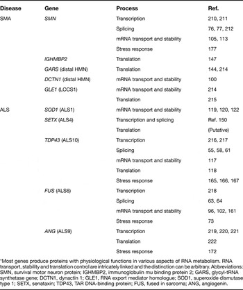
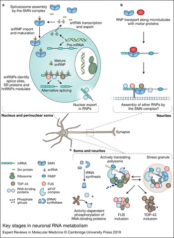
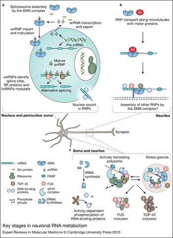
Many proteins implicated in motor neuron disorders have a physiological function in one or several RNA-processing steps (see Table 1) from transcription to translation (Fig. 2). Loss of this function can occur by genetic deletion or pathological sequestration of protein. However, specific disease-causing RNA-handling deficits have yet to be identified for the majority of motor neuron disorders. With growing understanding of gene expression in neurons, several of the diverse mechanisms previously implicated in the pathogenesis of motor neuron disease can be integrated into a concept of shared vulnerability. For instance, axonal-transport deficits are likely to impair transport of RNA granules; glutamate toxicity could lead to altered local translation of mRNAs in dendrites by altering the phosphorylation status of RNA-binding proteins; oxidative stress can lead to oxidised RNA species and faulty translation; and the unfolded protein response can trigger a stress response involving RNA-binding proteins.
Major genes implicated in forms of spinal muscular atrophy and amyotrophic lateral sclerosis*

* Most genes produce proteins with physiological functions in various aspects of RNA metabolism. RNA transport, stability and translation control are intricately linked and the distinction can be arbitrary. Abbreviations: SMN, survival motor neuron protein; IGHMBP2, immunoglobulin mu binding protein 2; GARS, glycyl-tRNA synthetase gene; DCTN1, dynactin 1; GLE1, RNA export mediator homologue; SOD1, superoxide dismutase type 1; SETX, senataxin; TDP43, TAR DNA-binding protein; FUS, fused in sarcoma; ANG, angiogenin.
Key stages in neuronal RNA metabolism. (a) Many alternative splice isoforms, some neuron specific, result from RNA processing. Small nuclear RNAs are transcribed and exported to the cytoplasm, where they are coupled with Sm proteins to form snRNPs, a reaction controlled by SMN, the protein depleted in spinal muscular atrophy. Nuclear export of mRNAs involves various mRNA-binding proteins as well as GLE1, mutations in which cause LCCS1. (b) SMN is hypothesised to have a second, possibly motor neuron specific, function in neurites in complex with RNA binding proteins to target, stabilise and translationally repress the RNA cargo. (c) Protein translation involves the complex interplay of mRNA, RNA-binding proteins, ribosomes and tRNAs. Translation can occur in the soma, dendrites or axons and is dependent on the phosphorylation of RNA-binding proteins and by local signalling. A variety of stressors can lead to disassembly of the polysomes and aggregation of RNA, ribosomal subunits and various RNA-binding proteins into stress granules that serve to temporarily store mRNA. Both TDP-43 and FUS form aggregates in ALS and an active area of research is whether aggregation leads to loss of function. Abbreviations: Sm, Smith proteins; snRNPs, small nuclear ribonucleoproteins; GLE1, RNA export mediator homologue; tRNA, transfer RNA; TDP43, TAR DNA-binding protein; FUS, fused in sarcoma.
Future work will focus on identifying target RNAs for the RNA-binding proteins known to be involved in ALS, as well as identifying novel disease-related proteins by targeting RNA metabolism in genetic and proteomic screens. Although this growing understanding of motor neuron disease pathophysiology does not immediately translate into therapies, it seems unlikely that a cure for these devastating disorders can be developed without a thorough understanding of the basic disease mechanisms. In particular, if processing of RNA or RNA–protein complexes emerges as a common feature of motor neuron disease pathology and as a proximal step in disease initiation and propagation, this would suggest that drugs targeting these pathways might be effective in several diseases. For ALS, at least, which is largely a sporadic age-related condition that presents to specialist clinics after a median delay of one year from symptom onset, applying such therapies will require dramatic improvements in our ability to make an early diagnosis.
Acknowledgements and funding
We are very grateful to the reviewers of this article, who made a number of suggestions that significantly improved its clarity. Our work on motor neuron disorders is generously supported by grants from the SMA Trust, Motor Neuron Disease Association and the BDF Newlife Foundation.