I. INTRODUCTION
In this era of exploring alternative and sustainable energy resources, catalyst aided water splitting plays a substantial role. However, basic electrochemical water splitting is associated with substantial energy loss, fundamentally due to the high overpotentials required at the anode corresponding to the oxygen evolution reaction (OER). Although traditionally precious metal oxides such as RuO x and IrO x were viewed as efficient water oxidation catalysts, Reference McCrory, Jung, Peters and Jaramillo1–Reference Lee, Suntivich, May, Perry and Horn3 scarcity of the raw materials and their high cost has been prohibitive for wide scale practical applications of these catalysts. To circumvent this issue, over the past several years, researchers have delved deeper into transition metal oxides as OER electrocatalyst with performance comparable to the precious metal oxides. Reference Gong and Dai4–Reference Yan, Tian and Chen23 More recently, new families of OER electrocatalysts has been identified comprising of transition metal chalcogenides M x E y (M = Ni, Fe, Co; E = S, Se), Reference Pu, Luo, Asiri and Sun24–Reference Chen, Ma, Liu, Li, Su, Davey and Qiao28 some of which outperform even the transition metal oxides. Reference Swesi, Masud and Nath26,Reference Masud, Swesi, Liyanage and Nath27 Some of the major advantages of using chalcogenides over oxides is that the electronegativity of S, Se, and Te being smaller than O, metal-chalcogen bonds show a higher degree of covalency, which leads to decreasing band gap energy, as well as raising the valence band edge to be more closer to the water oxidation level. In fact theoretical band structure calculations have shown that the band gap and band edge energies does show a progressive change (decreasing band gap and increasing valence band edge energy) from oxides to tellurides. Reference Schuster, Gatti and Rubio29,Reference Rasmussen and Thygesen30 While most of the transition metal oxides are wide band gap semiconductors, the selenides and tellurides are mostly semimetal or metallic. Additionally, the transition metal chalcogenides are known to form a wide range of stoichiometries stabilizing the metal in different oxidation states, which leads to lower redox potential of the transition metal center. Cumulative effects of all these are expected to result in much better electrocatalytic activity in the chalcogenides. In fact, our recent report showed that a simple binary nickel selenide, Ni3Se2, can outperform some of the best OER electrocatalyst and produced 10 mA cm−2 exchange current density at an overpotential of 290 mV. Reference Swesi, Masud and Nath26 Following this report several other selenides were shown to be active for OER including diselenides, Reference Pu, Luo, Asiri and Sun24,Reference Masud, Swesi, Liyanage and Nath27,Reference Kwak, Im, Jang, Kim, Park, Lim, Cha and Park31,Reference Kong, Cha, Wang, Lee and Cui32 monoselenides, Reference Tang, Cheng, Pu, Xing and Sun33 and sulfides such as Ni3S2. Reference Zhu, Yue, Zhang, Yu, Zhang, Wang and Wang34 Although the OER overpotential has been reduced substantially in these new families of electrocatalysts, further improvement of overpotentials is still desirable for the development of energy efficient water splitting devices.
Apart from higher efficiency, another important aspect of these transition metal oxides and chalcogenides electrocatalyst is that a good proportion of them are active for both OER and HER (hydrogen evolution reaction) in alkaline medium. Over the past couple of years, several bifunctional water splitting catalysts have been reported such as NiFe layered double hydroxide on nickel foam, Reference Luo, Im, Mayer, Schreier, Nazeeruddin, Park, Tilley, Fan and Grätzel35 cobalt–cobalt oxide/N-doped carbon hybrids, Reference Jin, Wang, Su, Wei, Pang and Wang36 porous cobalt phosphide/cobalt phosphate thin film, Reference Yang, Fei, Ruan and Tour37 cobalt-phosphorous-derived film, Reference Jiang, You, Sheng and Sun38 Ni2P nanoparticles, Reference Stern, Feng, Song and Hu39 Ni5P4 film, Reference Ledendecker, Calderon, Papp, Steinruck, Antonietti and Shalom40 CoSe, Reference Liu, Liu, Asiri, Luo and Sun41 and NiSe nanowires film on Ni-foam. Reference Tang, Cheng, Pu, Xing and Sun33 A bifunctional catalyst active for both OER and HER in the same electrolytic solution can offer full water splitting and is even more attractive for practical applications. However, such bifunctional electrocatalysts with reasonably good activity for both OER and HER is very challenging to design.
One of the possible ways to increase the catalytic activity is by increasing the active surface area which can be done though nanostructuring and increasing the surface roughness. Reference Xu, Su, Wu, Cheng, Guo, Li and Liu22 Along with this creating a porous surface is also advantageous for catalytic activities since reactants can diffuse into the catalyst layer easily on such structures and reduce the liquid sealing effect, hence improving the catalyst performance. Reference Lu, Yu, Wang, Zhai, Xie, Ling, Tong and Li42 One of the favored methods to create a porous surface is through etching. In electrodeposited films, electrochemical etching can lead to partial dissolution of the film thereby forming porous rough films with higher surface area with well-preserved conductivity of the film. Herein, we have demonstrated how the bifunctional electrocatalytic activity of Ni3Se2 film can be improved through electrochemical etching induced nanostructuring. The OER activity especially shows a significant improvement achieving 10 mA cm−2 at an overpotential of 190 mV in 1 M NaOH which is one of the lowest overpotential reported till date. The HER also shows a low overpotential of 230 mV at 10 mA cm−2, making this bifunctional catalyst active for full water splitting at 1.65 V (versus RHE). The catalyst showed exceptional stability under conditions of continuous O2 and H2 generation for extended periods of time exceeding 18 h with low Tafel slope values. Detailed structural and morphological characterization revealed that the films were compositionally stable after OER and HER activity.
II. EXPERIMENTAL AND METHODS
A. Preparation of as-deposited Ni3Se2 film and roughened Ni3Se2 film
Electrodeposition of Ni3Se2 films was performed in a conventional three-electrode cell, using Ag|AgCl as the reference electrode while working and counter electrodes were made from Au-coated glass [Deposition Research Lab Incorporated (DRLI)] and platinum mesh, respectively. The details of the Ni3Se2 electrodeposition procedure has been published elsewhere. Reference Swesi, Masud and Nath26 The Au substrates were cleaned by ultrasonic treatment in micro-90 detergent followed by isopropanol and deionized (DI) water. Reference Swesi, Masud and Nath26 The clean Au substrates were masked with a scotch tape, leaving an exposed area for the electrodeposition. The solutions were prepared using analytical grade reagents and DI water (resistivity ∼18 MΩ cm). Prior to mixing, the solutions were purged with N2 gas to remove any dissolved O2. Dilute HCl was added to maintain the pH at 2.5 and to prevent the formation of hydroxyl species. The deposition potential of Ni3Se2 was controlled at −0.80 V (versus Ag|AgCl) for 300 s from an aqueous solution containing 10 mM NiSO4·6H2O, 10 mM Na2SeO3, and 25 mM LiCl at 25 °C. An IviumStat potentiostat was used to control the electrodeposition process and to monitor the current and voltage profiles. Immediately after deposition, the deposits were washed with deionized water to remove impurities and adsorbents from the surface.
Surface-roughened Ni3Se2 film was prepared by further electrochemical etching of the as-deposited Ni3Se2 film in 0.05 M HCl. Typically, Ni3Se2 film was connected as the anode while Pt was the cathode in the electrochemical cell, and a current density of 10 mA cm−2 was maintained between the two electrodes for 15 and 30 s. Such treatment led to partial dissolution of the Ni3Se2 film. After electrochemical dissolution, products were washed in deionized water, and then air-dried. The as-deposited Ni3Se2 film was silvery black and the surface of the coating was very bright. After electrochemical dissolution, the surface color remained same and no discolored patches were observed.
III. METHODS OF CHARACTERIZATION
A. Powder X-ray diffraction (XRD)
The electrodeposited substrates were studied as such without any further treatment. Philips X-Pert X-ray diffractometer (PANalytical, Almelo, The Netherlands) was used to obtain the characteristic XRD pattern of the catalysts where the radiation source was Cu Kα (1.5418 Ǻ). As the deposited layer of catalyst on the substrate was very thin, the pxrd was collected at grazing angles in thin film geometry (GI mode with Göbel mirrors).
B. Scanning electron microscopy (SEM)
FEI Helios NanoLab 600 FIB/FESEM (FEI Corporation, Hillsboro, Oregon) at an accelarating voltage of 10 kV and a working distance of 4.8 mm was used to check the morphology of catalyst. The energy dispersive spectroscopy (EDS) analysis of the films were performed from the same SEM microscope. Cu tapes were used to connect the sample surfaces to the stubs to provide a good “path to ground” to reduce charging effect. ImageJ software was used for the measurement of the particles to determine the average particle size.
C. Transmission electron microscopy (TEM)
A high-resolution TEM imaging (HRTEM) and selected area electron diffraction (SAED) patterns were obtained from a FEI Tecnai F20 microscope operating at 300 kV accelerating voltage (FEI Corporation). The sample for TEM analysis was prepared by scratching little bit of the powder from the Au-glass substrate (after dissolution) and dispersing it in methanol, followed by dropping it on the formvar-coated Cu TEM grids.
D. X-ray photoelectron spectroscopy (XPS)
KRATOS AXIS 165 x-ray photoelectron spectrometer (Kratos Analytical Limited, Manchester, United Kingdom) with a source of monochromatic Al X-ray source was used to perform the XPS measurements of the electrodeposited films. The spectra were collected after 2 min sputtering with Ar to remove the immediate oxide layer from the surface of catalyst. All XPS binding energies were corrected by using the reference C1s signal at 284.5 eV.
E. Electrochemical performance study
Linear sweep voltammetry (LSV), cyclic voltammetry (CV), and chronoamerometry techniques were used for the catalytic evaluation of the deposited films. Typical three-electrode electrochemical cell coupled with an IviumStat potentiostat was used for the study where Ni3Se2 modified Au, Ag|AgCl|KCl(sat.) and Pt mesh were used as working, reference and counter electrodes, respectively. All potentials obtained with Ag|AgCl|KCl(sat.) reference electrode were converted to the reversible hydrogen electrode (RHE) by using the following equation.
 $${E_{{\rm{RHE}}}} = {E_{{\rm{Ag}}|{\rm{AgCl}}|{\rm{KC}}{{\rm{l}}_{{\rm{(sat}}{\rm{.)}}}}}} + 0.059\;{\rm{pH}} + E_{{\rm{Ag}}|{\rm{AgCl}}|{\rm{KC}}{{\rm{l}}_{{\rm{(sat}}{\rm{.)}}}}}^0\quad ,$$
where E
RHE is the calculated potential as RHE,
 ${E_{{\rm{Ag}}|{\rm{AgCl}}|{\rm{KC}}{{\rm{l}}_{{\rm{(sat}}{\rm{.)}}}}}}$
is the experimentally measured potential, and
 $E_{{\rm{Ag}}|{\rm{AgCl}}|{\rm{KC}}{{\rm{l}}_{{\rm{(sat}}{\rm{.)}}}}}^0$
is the thermodynamic potential at 25 °C (0.197 V).
IV. RESULTS AND DISCUSSION
A. Structure and morphology of Ni3Se2 films
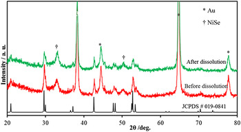
The pxrd patterns of as-synthesized films deposited on Au-coated glass substrates showed peaks corresponding to Ni3Se2 phase (PDF # 019–0841) along with peaks characteristic of Au arising from the underlying substrate as shown in Fig. 1. Ni3Se2 has an interesting crystal structure (shown as inset of Fig. 1) built up of anion close packing where Ni occupies 3/4th of the tetrahedral holes specifically showing a distorted tetrahedral coordination. Se on the other hand, shows a distorted trigonal prismatic coordination with the neighboring Ni atoms. Interestingly, energy density studies have suggested that Ni3Se2 may have Ni–Ni bonding influenced by the closeness of the edge-shared Ni polyhedra. Reference Agarwala and Sinha43 The diffraction pattern obtained after electrochemical etching (as shown in Fig. 1), still clearly shows the presence of crystalline Ni3Se2 with no other discernible impurity peaks, indicating that the film obtained after etching was indeed predominantly Ni3Se2. This confirmed that Ni3Se2 phase was not lost in electrochemical etching progress. However, the intensity of the diffraction peaks of Ni3Se2 phase became stronger after electrochemical dissolution.
PXRD patterns of Ni3Se2 catalysts before and after dissolution (acid leaching) showing the presence of Ni3Se2 (PDF # 019–0841) along with Au.

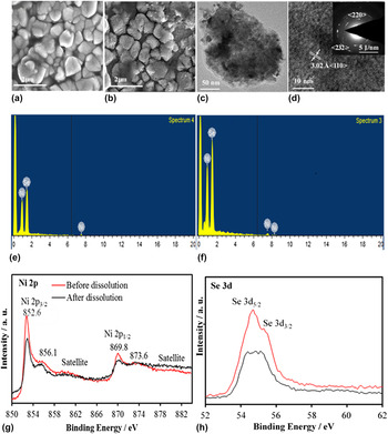
Figures 2(a) and 2(b) show morphology of the as-deposited Ni3Se2 film and the etched Ni3Se2 film, respectively. Detailed SEM study of the as-deposited film showed that the film was mainly granular and composed of micron sized grains with a broad size distribution (0.21–1.2 μm) and an average particle diameter of 0.74 μm. From the SEM [Fig. 2(a)] it was also apparent that the as-deposited micron-sized grains had very smooth surfaces before electrochemical dissolution. After electrochemical etching SEM of the Ni3Se2 film showed a distinct difference as can be seen in Fig. 2(b). The film became noticeably porous with very rough surface of the grains which seemed to be covered with thin nanoflakes. Nevertheless, representative SEM images [Figs. 2(a) and 2(b)] clearly showed that even after dissolution, the Au-glass substrate was still uniformly coated with the interconnected nanograins network structure without developing any fragmentation or obvious breakage. Particle size analysis revealed that the dimensions of the grains varied from 0.072 to 0.77 μm, where the lower limit was for the thickness of the flakes while the upper limit was the grain size. Average grain size was obtained to be around 0.31 μm.
SEM images of Ni3Se2 (a) before and (b) after dissolution. (c) TEM and (d) HRTEM images of Ni3Se2 catalyst after dissolution. Inset in (d) shows the SAED pattern. (e) and (f) are the EDS spectra of Ni3Se2 before and after dissolution, respectively. (g) Ni 2p and (h) Se 3d XPS spectra where red and black lines indicate the before and after dissolution of catalysts.

The nanostructured film after dissolution was also studied by TEM and HRTEM imaging as shown in Figs. 2(c) and 2(d). Low-magnification TEM image [Fig. 2(c)] shows the rough surface texture of the granules. Higher magnification HRTEM image on the other hand, shows the crystalline nature of the granules with lattice fringes corresponding to 〈110〉 lattice planes of Ni3Se2 shown in Fig. 2(d). The crystalline nature of the nanostructured grains were also confirmed by SAED which showed diffraction rings corresponding to 〈220〉 and 〈232〉 lattice planes. Thus it was further confirmed with TEM and SAED analysis that the nanostructured grains were indeed crystalline Ni3Se2 after the dissolution step.
The composition of the film was further confirmed through quantitative EDS, elemental mapping, and XPS. While the elemental mapping clearly showed that Ni and Se were homogeneously distributed in nanograins, quantitative analysis showed that the relative ratio of Ni:Se was close to 3:2, which confirmed the film composition was indeed Ni3Se2 [Fig. 2(e)]. The Ni:Se ratio did not change after electrochemical dissolution confirming the stability of the film [Fig. 2(f)]. XPS spectra showed peaks corresponding to Ni 2p 3/2 and 2p 1/2 at 855 and 869 eV as shown in Fig. 2(g), while Se 3d peaks [Fig. 2(h)] were observed at 54.5 and 55.6 eV (3d 5/2 and 3d 3/2, respectively). These XPS peaks agrees very well with those reported for Ni3Se2. Reference Swesi, Masud and Nath26 Interestingly XPS did not show any evidence for the presence of oxidic impurities on the surface even after the electrochemical etching step. All of the above characterization confirmed that the composition of the film did not show a drastic change after electrochemical etching. In fact, this etching step can be possibly used as a surface cleaning technique where the acid removes soluble impurities from the surface (including oxides) thereby yielding a more pristine surface. Since this surface cleaning is achieved through electrochemical etching induced dissolution, this step has been simply referred to as dissolution hereafter.
B. Electrochemical performance of Ni3Se2 films
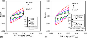
One of the very obvious changes observed after the dissolution was that the morphology of the film changed from smooth to rough and porous, which is accompanied by an increased surface area. Specifically, as the electrodeposited Ni3Se2 was aged as an anode in an acidic solution, it led to sporadic dissolution from the film surface thus creating a rough surface. This expectedly led to an increase of the electrode surface area which was further confirmed through measuring the electrochemically active surface area (ECSA) and roughness factor (RF) of the film after dissolution and comparing it with the as deposited film. ECSA was estimated by measuring the double layer charging current at different scan speeds. The probed electrode was potentiostatically cycled (−0.25 to −0.1 V versus Ag|AgCl) with scan rates ranging between 10 and 160 mV s−1 while the charging current was collected [Figs. 3(a) and 3(b)]. The electrochemical double layer capacitance, C DL, is proportional to the collected charging current, i c, and inversely proportional to the scan rate, ν, as shown in the following equation,
 $${i_{{\rm{DL}}}} = {C_{{\rm{DL}}}} * \nu \quad .$$
Cyclic voltammograms measured at (a) before and (b) after dissolution of Ni3Se2 catalysts in N2 saturated 1.0 M NaOH solution at different scan rate from 10 to 160 mV s−1. Inset shows plot of anodic and cathodic current measured at −0.19 V as function of scan rate.

The C DL was estimated as the slop of the plot of i c as a function of ν. The ECSA of the catalysts can be calculated based on the following equation,
 $${\rm{ECSA}} = {{{C_{{\rm{DL}}}}} \mathord{\left/ {\vphantom {{{C_{{\rm{DL}}}}} {{C_{\rm{s}}}}}} \right. \kern-\nulldelimiterspace} {{C_{\rm{s}}}}}\quad .$$
Where C s is the specific capacitance (0.040 mF cm−2) in 1 M NaOH. Reference Swesi, Masud and Nath26 The roughness factor (RF) of each electrode was then calculated by dividing obtained ECSAs by the geometric area (0.14 and 0.283 cm2 for the catalyst with and without dissolution, respectively). It was observed that the ECSA increased more than 3-fold after dissolution for 30 s (1.5 cm2 in pristine Ni3Se2 to 4.9 cm2 after dissolution), while the roughness factor showed a more dramatic change, increasing almost 7-fold following the dissolution step (5.3–35, respectively).
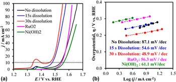
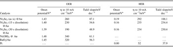
The electrocatalytic performance for OER of the electrodeposited films was investigated through linear sweep voltammetry (LSV) in alkaline solution (1 M NaOH) near pH 14. For comparison, we had also checked electrodeposited RuO2, and Ni(OH)2 for OER catalytic activity. Figure 4(a) shows the LSVs of electrochemical oxygen evolution with Ni3Se2 catalysts, before and after dissolution, compared with that of RuO2 and Ni(OH)2. As observed, after electrodeposition of Ni3Se2 the substrates showed high activity for OER with onset overpotential of about 200 mV for the as-deposited Ni3Se2 electrode and 160 mV (170 mV) for the treated Ni3Se2 electrode for 15 s (30 s), respectively [all potentials have been reported with respect to RHE]. This 30–40 mV shift in onset overpotential is expected as more catalytically active sites were exposed to the electrolyte resulting from the increased surface area and higher roughness factor. Specifically, the dissolution step creates high density of exposed active metal sites on the surface which in turn increases the electrocatalytic performance by reducing the overpotential and increasing current density. Conventionally, the effectiveness of OER catalysts are estimated by listing the overpotential (η) required to reach a current density of 10 mA cm−2 (per geometric surface area), which is considered to be equivalent to 10% solar energy conversion efficiency. Reference Li, Hasin and Wu6 The η at 10 mA cm−2 per geometric area of all films are reported in Table I. It can be clearly found that the surface-etched Ni3Se2 catalyst exhibits higher catalytic activity toward OER with a much lower η (190 mV). It should be mentioned here that this is one of the lowest overpotential that has been reported for OER electrocatalyst.
(a) LSVs measured at catalysts in N2 saturated 1.0 M NaOH solution at a scan rate of 10 mV s−1. Dotted line shows the current density of 10 mA cm−2. (b) Tafel plots of catalysts.

Electrochemical parameters of the catalysts measured in 1 M NaOH.

a Potential versus RHE.
The kinetic parameters corresponding to the OER activities were estimated from the measured Tafel slope collected near the onset potential for OER. The Tafel plots for the as-prepared Ni3Se2 and surface-modified Ni3Se2 catalysts (after dissolution) in 1 M NaOH solution are shown in Fig. 4(b). The observed Tafel slope of the surface-modified Ni3Se2 catalyst were about 56.4 and 48.9 mV dec−1 for 15 and 30 s of dissolution, respectively which were much smaller than that of as-prepared Ni3Se2 catalysts (87.1 mV dec−1), which can be attributed to the facilitated diffusion through the highly porous and rough surface of the modified electrode. The smaller Tafel slope of the modified electrode compared to that of the pristine electrode also suggest faster reaction kinetics.
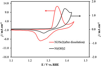
In addition to reporting enhanced electrocatalytic activity for OER, it is also important to understand what causes such improvement of the onset and overpotential in the transition metal selenides, as compared to the oxides. It was observed from the LSV plots that in both Ni3Se2 as well as Ni(OH)2, the OER activity was preceded by Ni2+ → Ni3+ oxidation peak and it has been widely accepted that Ni3+ is the actual catalytically active species. Reference Zhang44 We looked more closely at the Ni2+ → Ni3+ oxidation peak by measuring cyclic voltammograms (CV) across a short potential range and compared the CVs obtained from the Ni3Se2 with that obtained from Ni(OH)2. This comparison very clearly showed that the preoxidation peak itself was shifted to lower potential (by about 50 mV) in the selenide compared to the oxide as shown in Fig. 5. This indicates that the catalytically active species (Ni3+) is generated at a much lower potential in the selenide. This observation coupled with the smaller band gap and higher conductivity of the selenide matrix may be instrumental in enhancing their OER catalytic activity compared to the oxides.
CVs measured at Ni3Se2 after 30 s dissolution and Ni(OH)2 catalysts in N2 saturated 1.0 M NaOH at 10 mV s−1.

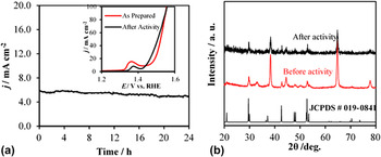
In addition to the outstanding catalytic activity, the modified Ni3Se2 catalyst also showed good stability for OER (Fig. 6). The chronoamperometry (current versus time) plot for porous Ni3Se2 electrode maintained at a constant potential of 0.42 V versus Ag/AgCl exhibits stable current for over 24 h of continuous operation suggesting that the modified Ni3Se2 catalyst displayed good durability for OER in 1 M NaOH. Comparison of LSVs before and after 24 h of electrolysis [inset of Fig. 6(a)] also confirmed the stability of catalyst.
(a) Stability of catalyst under continuous O2 evolution for 24 h studied at constant potential. Inset of (a) is the comparison of LSVs of catalyst in N2 saturated 1.0 M NaOH before and after chronoamperometry for 24 h (b) XRD of catalyst before and after chronoamperometry for 24 h.

The compositional stability of the catalyst after periods of continuous O2 evolution was also studied by XRD collected after 24 h OER activity. The pxrd study revealed that the structural integrity of Ni3Se2 was maintained even after 24 h of continuous O2 evolution as shown in Fig. 6(b) indicating the durability of the catalyst.
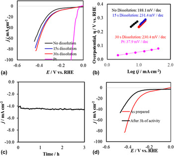
The Ni3Se2 has also been studied for possible HER catalytic activity in the same electrolyte (1 M NaOH) by measuring the LSV between −0.5 and 0.0 V versus RHE using a three-electrode system with a scan rate of 10 mV s−1. Au coated-glass was used as the substrate due to its negligible HER activity. Commercially available Pt was also used for comparison of the HER catalytic activity. Compared to the as-deposited Ni3Se2, the electrochemically treated Ni3Se2 showed better HER performance (Table I), achieving 10 mA cm−2 at an overpotential of 234 mV and showing a Tafel slope of 230.4 mV dec−1 [Fig. 7(b)]. Chronoamperometric measurements at −0.2 V versus RHE revealed that the catalysts retained stable HER activities even after 3 h [Figs. 7(c) and 7(d)], suggesting their potential as an attractive bifunctional electrocatalyst for full water splitting. Accordingly, a cell was created by coating both the cathode and anode with surface modified Ni3Se2, and it was observed that this bifunctional catalyst could effectively split water producing a current density of 10 mA cm−2 at a cell voltage of 1.69 V. The electrolysis energy efficiency was calculated to be 84% following methods as has been reported previously. Reference Kreuter and Hofmann45
(a) Polarization curves for catalysts in N2 saturated 1.0 M NaOH solution at a scan rate of 10 mV s−1. (b) Tafel plots of catalysts. (c) Stability study of catalyst under continuous H2 evolution for 3 h at constant potential and (d) is the LSVs of catalyst in N2 saturated 1.0 M NaOH for comparison.

V. CONCLUSION
Morphology-controllable Ni3Se2 films were successfully prepared by electrodepositing Ni3Se2 film on Au substrate and followed by electrochemical dissolution. In electrochemical dissolution process, Ni3Se2 phase is well-preserved and roughened, porous structure was formed. This technology is a promising way to fabricate other roughened high surface area films. The etched Ni3Se2 film showed an enhanced OER catalytic performance in comparison with the as-deposited Ni3Se2 film. This enhancement in catalytic activity could be explained by the increased surface roughness factor and flake-like morphology of the film which facilitates transfer of charge across the electrolyte–electrode interface. Introduction of pores on the catalyst surface through electrochemical dissolution also improves the catalytic performance. Specifically, the overpotential for OER catalytic activity exhibited by the etched Ni3Se2 is clearly the lowest amongst all other reported OER electrocatalyst (Table I). This etched Ni3Se2 electrocatalyst competes other earth-abundant material-based OER and HER catalysts, and places itself as one of the best bifunctional electrocatalysts to date, with a high current density and excellent stability.
ACKNOWLEDGMENTS
This research was funded through financial support from ACS PRF (grant #54793- ND10) and Energy Research and Development Center (ERDC) at Missouri S&T.