Kawasaki disease is an acute, self-limited febrile vasculitis with a predilection for coronary arteries that predominantly affects children aged 6 months to 5 years. Reference Son and Newburger1 It was first described by Dr Tomikasu Kawasaki, a Japanese paediatrician, as mucocutaneous lymph node syndrome in 1967. Reference Kawasaki2 It is the second most common vasculitis in childhood, behind Immunoglobulin A vasculitis (formerly called Henoch–Schönlein purpura). Reference Younger3 It is also the leading cause of acquired heart disease in children in developed countries, a spot previously occupied by acute rheumatic fever. Reference Uehara and Belay4 Its aetiology is unknown, although there is some evidence to suggest that Kawasaki disease may be secondary to an infection and subsequent activation of the immune system in genetically susceptible children. Reference Rife and Gedalia5
The clinical manifestations include fever, mucosal changes, conjunctivitis, polymorphous rash, extremity changes, and lymphadenopathy. Reference Rife and Gedalia5 One of the most feared complications of Kawasaki disease is coronary artery lesions, which include stenosis, dilation, and aneurysms. If left untreated, they present in approximately 25% of the patients. Reference Suzuki, Kamiya and Kuwahara6 Timely treatment with intravenous gamma globulin and aspirin reduces the incidence of coronary artery lesions. Reference Newburger, Takahashi and Burns7,Reference Furusho, Kamiya and Nakano8 Nevertheless, approximately 10–20% of patients treated have refractory disease defined as persistent or recurrent fever despite treatment with intravenous gamma globulin and aspirin, and thus are at an increased risk of developing coronary artery lesions. Reference Durongpisitkul, Soongswang, Laohaprasitiporn, Nana, Prachuabmoh and Kangkagate9–Reference Wallace, French, Kahn and Sherry11 Therefore, it is important to identify those who are at an increased risk of refractory disease or coronary artery lesions to initiate or restart treatment in a timely fashion.
As inflammation is the leading mechanism in the development of coronary artery lesions and intravenous gamma globulin resistance, the use of biomarkers of inflammation could be useful in diagnosis and prediction. There are multiple studies that have analysed clinical or laboratory characteristics that could predict refractory disease and/or coronary artery lesions, including days of illness at initial treatment c-reactive protein, total bilirubin, aspartate transaminase, neutrophil count, and others. Reference Sano, Kurotobi and Matsuzaki12–Reference Fukunishi, Kikkawa and Hamana14 Similarly, scoring systems have been developed with a various combination of these characteristics and have shown mixed results, Reference Kobayashi, Inoue and Takeuchi15–Reference Tremoulet, Best and Song19 and some have shown not to be effective when applied to other populations. Reference Arane, Mendelsohn and Mimouni20,Reference Davies, Sutton and Blackstock21 Leukocytes and their subpopulations are the primary mediators of inflammation, and their changes classically reflect the immune response. The neutrophil-to-lymphocyte ratio has been shown to express the severity of inflammation and the disease in process in critically ill patients. Reference Zahorec22 Additionally, multiple studies have shown that neutrophil–lymphocyte ratio is a strong predictor of mortality and poor outcomes in patients with different diseases such as acute coronary syndrome, Reference Azab, Zaher and Weiserbs23,Reference Park, Jang and Oh24 with malignancies, Reference Tang, Lu, Li, Li, Xu and Dong25,Reference Cummings, Merone and Keeble26 and bacterial meningitis. Reference Widjaja, Rusmawatiningtyas, Makrufardi and Arguni27 Therefore, the use of neutrophil–lymphocyte ratio has been proposed to be useful in Kawasaki disease and several studies have reported its effectiveness in the prediction of outcomes in these patients. Reference Kawamura, Takeshita, Kanai, Yoshida and Nonoyama28,Reference Ha, Lee and Jang29
The primary objective of this study was to determine if neutrophil–lymphocyte ratio before or after intravenous gamma globulin therapy could predict refractory disease and coronary artery lesions in paediatric patients with Kawasaki disease.
Methods
A systematic review of the literature was performed to identify manuscripts describing comparisons of neutrophil–lymphocyte ratio between those who had refractory disease and those who had non-refractory disease, and between those who had coronary artery lesions and those who did not have coronary artery lesions. The primary variable of interest was the predictive value of neutrophil–lymphocyte ratio in paediatric patient with Kawasaki disease. The reporting of this systematic review was guided by the standards of the Preferred Reporting Items for Systematic Review and Meta-Analysis (PRISMA) Statement. Reference Page, McKenzie and Bossuyt30 This study did not require institutional review board approval as it utilised previously published data that were deidentified. This study is in concordance with the Helsinki declaration.
Inclusion criteria
The following inclusion criteria must have been met for a study to be included in the study: 1) must have had paediatric patients only (less than 18 years of age); 2) studies must have reported neutrophil–lymphocyte ratio values before and/or after intravenous gamma globulin therapy; 3) neutrophil–lymphocyte ratio must have been compared between those who had refractory disease and those who had non-refractory disease, and/or compared between those who had coronary artery lesions and those who did not have coronary artery lesions.
Manuscript search and identification strategy
Manuscripts were identified using electronic databases, including PubMed, EMBASE, Ovid, and Cochrane reviews. These databases were queried using the following search terms individually and in various combinations: “neutrophil-to-lymphocyte ratio”, “neutrophil”, “NLR”, “Kawasaki disease”, “refractory”, “resistance”, “coronary artery lesions”, and “coronary aneurysm”. No specific restriction on the year of publication was used. The final search was conducted on September 29th, 2021. Resulting studies were screened by title and abstract. Those felt to be pertinent to help fulfill the objectives then had their full text retrieved in their entirety. References of these studies were then hand-searched for additional relevant manuscripts. No direct contact with manuscript authors was made to obtain full-text manuscripts or additional data. Published manuscripts available in full text were included in this review if they met the inclusion criteria.
Study identification was conducted separately by two authors (RL and JF). Studies identified for inclusion by these two authors were then reviewed by a third author (SF). Any discrepancies between the two authors were identified by the third author and reviewed by all authors to come to a consensus.
Study quality and bias assessment
These full-text manuscripts were then reviewed by the authors for the presence of bias and overall quality with Newcastle–Ottawa Scale (NOS). Reference Wells G.A., O.'Connell, Peterson, Welch, Losos and Tugwell31 Quality and bias were assessed independently by two authors (JF, RL). Discrepancies between the two authors were then reviewed by a third author (SF), and a consensus was reached.
Endpoints
Studies deemed to be appropriate for inclusion after full-text review, quality, review, and bias review then had all the endpoints reviewed to identify endpoints reported by multiple studies. Endpoints with data from three or more studies were deemed eligible for data extraction.
In all studies, the following endpoint was identified for analysis: neutrophil–lymphocyte ratio value. It was identified in two timepoints: 1) before intravenous gamma globulin therapy and 2) after intravenous gamma globulin therapy. For the second timepoint data, either 1 or 2 days after intravenous gamma globulin therapy was included. In those studies that conducted a receiver operator curve analysis to determine a cut-off value for neutrophil–lymphocyte ratio to refractory disease, the following endpoints were also identified: area under the curve, cut-off, sensitivity, and specificity.
Data extraction
Data regarding baseline patient characteristics and study characteristics were extracted from the manuscripts identified for inclusion. Study-level data were extracted with use of a data collection form that was developed specifically for this review. The data extraction was conducted by two separate authors (JF, EV) to ensure integrity of the resulting data. Differences were then identified by a third author (SF). Discrepancies in the data extraction between the two authors were then reviewed by all authors to come to a consensus.
Study-level data was extracted as mean and standard deviation for each timepoint of neutrophil–lymphocyte ratio. If data were reported as median and range, it was converted to mean and data for data extraction using the methods proposed by Wan et al. Reference Wan, Wang, Liu and Tong32
Data analysis
Continuous data are presented as mean and standard deviation. Categorical data are presented as frequencies with absolute numbers as well as percentages.
The first set of pooled analyses was conducted using Review Manager version 5.4 (Cochrane, London, England). They compared neutrophil–lymphocyte ratio before therapy between those who had refractory disease and those who did not, and between those who had coronary artery lesions and those who did not. Also, they compared neutrophil–lymphocyte ratio after therapy between those who had refractory disease and those who did not. Lastly, they compared neutrophil–lymphocyte ratio variation after therapy in those who experienced refractory disease and those who did not. This was done utilising the mean and standard deviation of the study-level data. A fixed-effects model was run initially for each endpoint. Heterogeneity was assessed using two methods: 1) Q-statistics and its resulting p-value; and 2) I-squared value. Heterogeneity was considered statistically significant if the p-value for the Q-statistic was less than 0.05 or the I-squared value was greater than 50%. For endpoints with statistically significant heterogeneity, a random effect was used for the pooled analyses. Results of these analyses are presented with mean difference and 95% confidence interval.
The second set of pooled analyses were conducted using MedCalc Version 19.2.6 (MedCalc Software Ltd) to pool area under the curve for receiver operator analyses to determine the accuracy of neutrophil–lymphocyte ratio on prediction of refractory disease. Heterogeneity was assessed using two methods: 1) Q-statistics and its resulting p-value; and 2) I-squared value. Heterogeneity was considered statistically significant if the p-value for the Q-statistic was less than 0.05 or the I-squared value was greater than 50%. For endpoints with statistically significant heterogeneity, a random effect was used for the pooled analyses. Results of these analyses are presented with the mean.
Publication bias was assessed qualitatively by a review of funnel plots. Forest plots were created using GraphPad Prism version 9.0.1 (GraphPad Software, San Diego, CA, USA).
Results
Neutrophil–lymphocyte ratio for prediction of refractory Kawasaki disease
Study characteristics
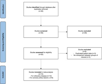
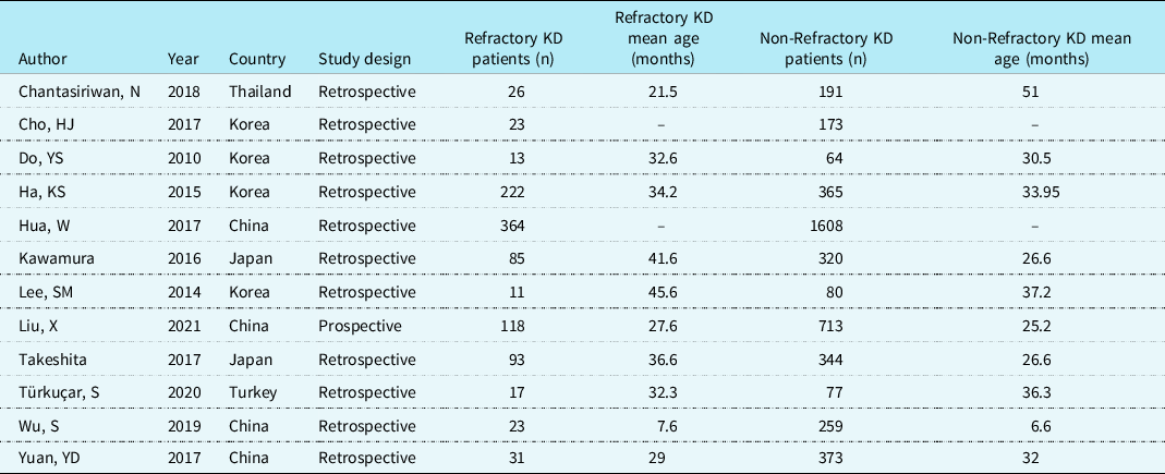
A total of 12 studies with 5593 patients were included in the final analyses of neutrophil–lymphocyte ratio for prediction of refractory Kawasaki disease (Fig 1). Out of these, 1026 (18.3%) experienced refractory Kawasaki disease and 4567 (81.7%) experienced non-refractory Kawasaki disease. The mean age for the refractory group was 32.7 months and 28.4 months for the non-refractory group (Table 1).
PRISMA flowchart.

Characteristics of included studies of NLR for prediction of refractory Kawasaki disease.

KD, Kawasaki disease.
Pooled analysis of neutrophil–lymphocyte ratio before intravenous gamma globulin therapy
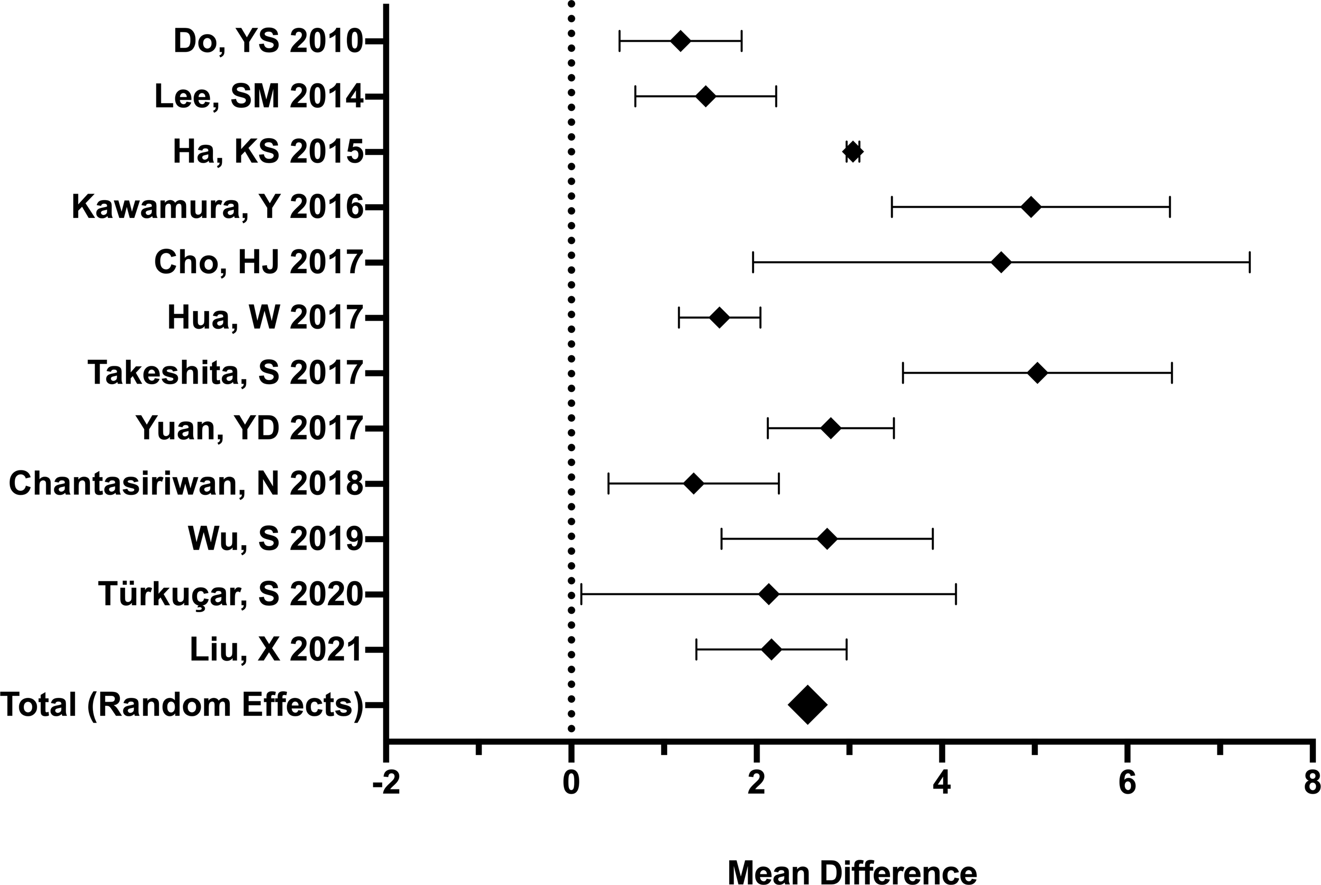
A total of 12 studies with 5593 patients were included in this analysis. Reference Kawamura, Takeshita, Kanai, Yoshida and Nonoyama28,Reference Ha, Lee and Jang29,Reference Do, Kim, Chun, Cha, Namgoong and Lee33–Reference Liu, Shao and Wang42 The Q-statistic for heterogeneity had a p-value of less than 0.01 and the I-squared value was 91%, demonstrating significant heterogeneity. Thus, a random effects model was used. Neutrophil–lymphocyte ratio before intravenous gamma globulin therapy was higher in those who experienced refractory Kawasaki disease (5.7 versus 2.88). This resulted in a mean difference of 2.55 (95% confidence interval 1.93 to 3.17, p-value of less than 0.01) (Fig 2). No publication bias was found by visual assessment of the publication bias funnel plot.
Forest plot showing pooled analysis of mean difference in NLR before intravenous gamma globulin therapy for prediction of refractory disease.

Pooled analysis of receiver operator curves of neutrophil–lymphocyte ratio before intravenous gamma globulin therapy
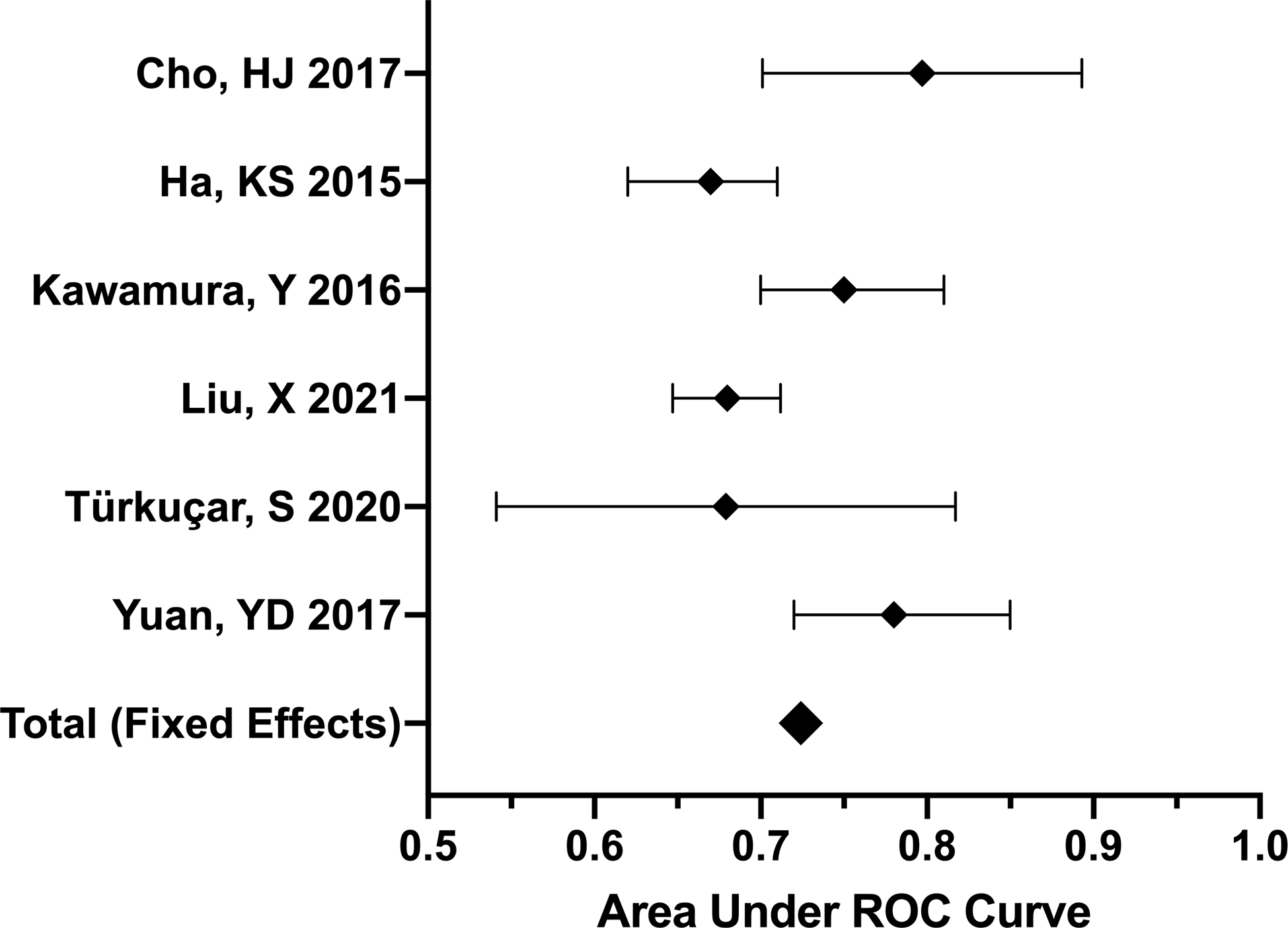
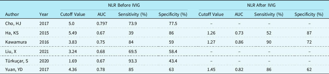
A total of six studies with 2517 patients were included in this analysis Reference Kawamura, Takeshita, Kanai, Yoshida and Nonoyama28,Reference Ha, Lee and Jang29,Reference Cho, Bak and Kim35,Reference Yuan, Sun, Li, Wei and Yu38,Reference Turkucar, Yildiz, Acari, Dundar, Kir and Unsal41,Reference Liu, Shao and Wang42 (Table 2). The Q-statistic for heterogeneity had a p-value of less than 0.05 and the I-squared value was 27%, demonstrating no significant heterogeneity. Thus, a fixed-effects model was used. The pooled area under the curve for neutrophil–lymphocyte ratio before intravenous gamma globulin therapy was 0.724 (Fig 3).
Forest plot showing pooled analyses of receiver operating curves of NLR before intravenous gamma globulin for prediction refractory Kawasaki disease.

Receiver operator curve analyses of included studies for prediction of refractory Kawasaki disease.

IVIG, Intravenous immune globulin; NLR, Neutrophil–Lymphocyte Ratio.
Pooled analysis of neutrophil–lymphocyte ratio after intravenous gamma globulin therapy
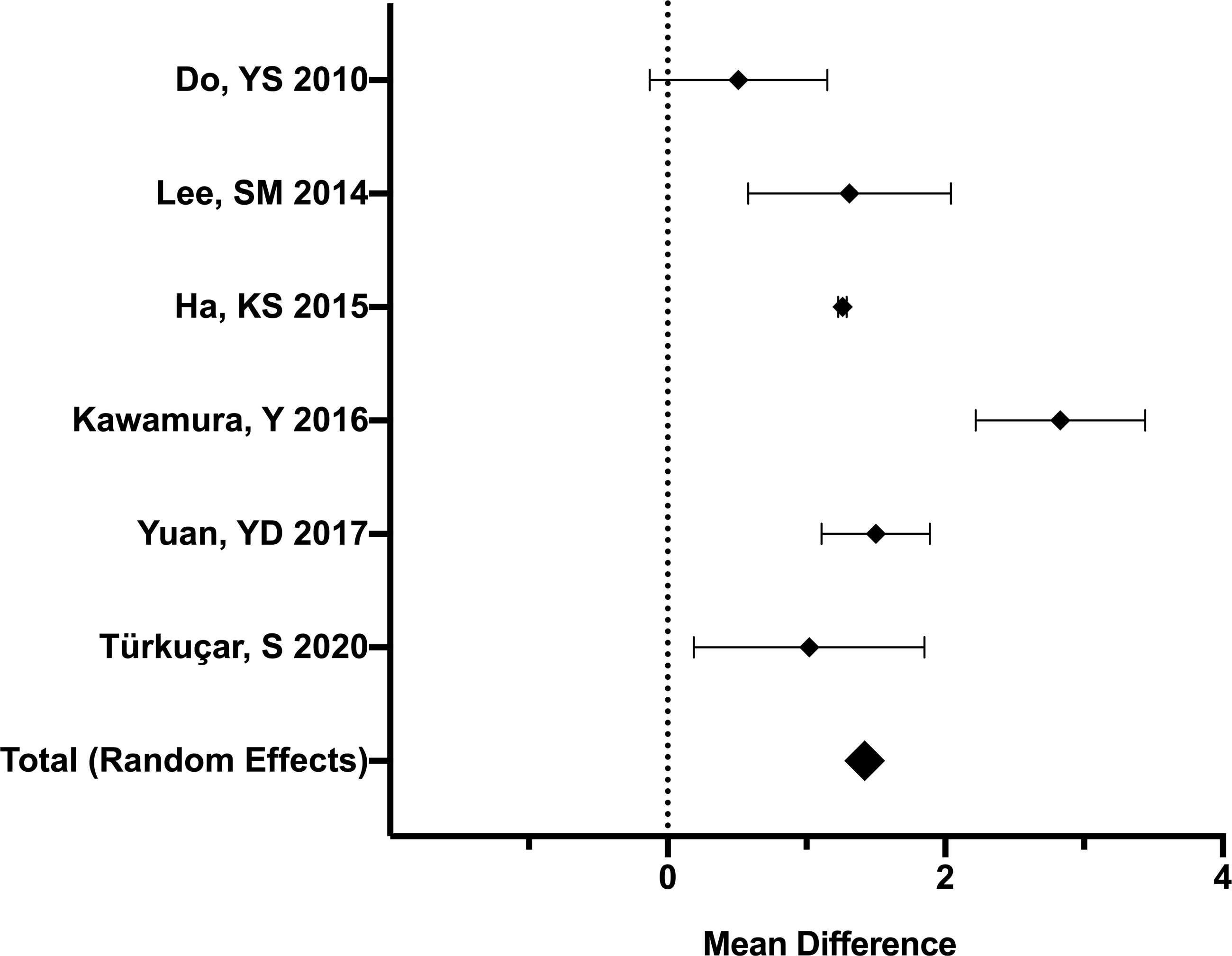
A total of six studies with 1658 patients were included in this analysis. Reference Kawamura, Takeshita, Kanai, Yoshida and Nonoyama28,Reference Ha, Lee and Jang29,Reference Do, Kim, Chun, Cha, Namgoong and Lee33,Reference Lee, Lee, Go, Song, Lee and Kwak34,Reference Yuan, Sun, Li, Wei and Yu38,Reference Turkucar, Yildiz, Acari, Dundar, Kir and Unsal41 The Q-statistic for heterogeneity had a p-value of less than 0.01 and the I-squared value was 85%, demonstrating significant heterogeneity. Thus, a random effects model was used. Neutrophil–lymphocyte ratio after intravenous gamma globulin therapy was higher in those who experienced refractory Kawasaki disease (1.97 versus 0.7). This resulted in a mean difference of 1.42 (95% confidence interval 0.96 to 1.87, p-value of less than 0.01) (Fig 4). No publication bias was found by visual assessment of the publication bias funnel plot.
Forest plot pooled analysis of mean difference in NLR after intravenous gamma globulin therapy for prediction of refractory disease.

Pooled analysis of receiver operator curves of neutrophil–lymphocyte ratio after intravenous gamma globulin therapy
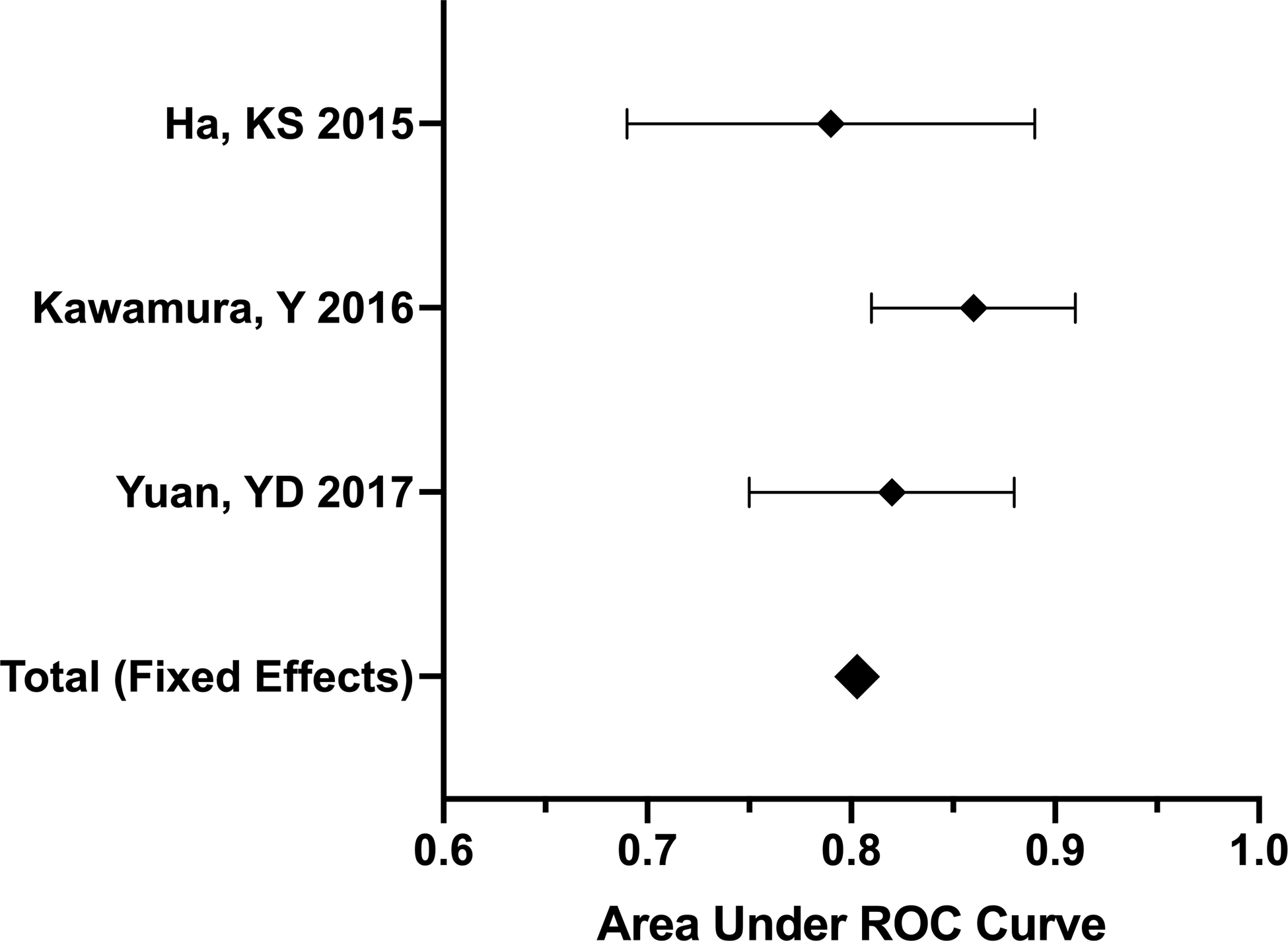
A total of three studies with 1396 patients were included in this analysis Reference Kawamura, Takeshita, Kanai, Yoshida and Nonoyama28,Reference Ha, Lee and Jang29,Reference Yuan, Sun, Li, Wei and Yu38 (Table 2). The Q-statistic for heterogeneity had a p-value of less than 0.05 and the I-squared value was 31%, demonstrating no significant heterogeneity. Thus, a fixed-effects model was used. The pooled area under the curve for neutrophil–lymphocyte ratio after intravenous gamma globulin therapy was 0.803 (Fig 5).
Forest plot showing pooled analyses of receiver operating curves of NLR after intravenous gamma globulin for prediction refractory Kawasaki disease.

Pooled analysis of neutrophil–lymphocyte ratio variation after intravenous gamma globulin therapy in those who experienced refractory Kawasaki disease
A total of six studies with 379 patients were included in this analysis. Reference Kawamura, Takeshita, Kanai, Yoshida and Nonoyama28,Reference Ha, Lee and Jang29,Reference Do, Kim, Chun, Cha, Namgoong and Lee33,Reference Lee, Lee, Go, Song, Lee and Kwak34,Reference Yuan, Sun, Li, Wei and Yu38,Reference Turkucar, Yildiz, Acari, Dundar, Kir and Unsal41 The Q-statistic for heterogeneity had a p-value of less than 0.01 and the I-squared value was 95%, demonstrating significant heterogeneity. Thus, a random effects model was used. The neutrophil–lymphocyte ratio was lower after intravenous gamma globulin therapy compared to before therapy (1.97 versus 5.97). This resulted in a mean difference of −3.02 (95% confidence interval −4.40 to −1.65, p-value of less than 0.01). No publication bias was found by visual assessment of the publication bias funnel plot.
Pooled analysis of neutrophil–lymphocyte ratio variation after intravenous gamma globulin therapy in those who did not experience refractory Kawasaki disease
A total of six studies with 1279 patients were included in this analysis. Reference Kawamura, Takeshita, Kanai, Yoshida and Nonoyama28,Reference Ha, Lee and Jang29,Reference Do, Kim, Chun, Cha, Namgoong and Lee33,Reference Lee, Lee, Go, Song, Lee and Kwak34,Reference Yuan, Sun, Li, Wei and Yu38,Reference Turkucar, Yildiz, Acari, Dundar, Kir and Unsal41 The Q-statistic for heterogeneity had a p-value of less than 0.01 and the I-squared value was 98%, demonstrating significant heterogeneity. Thus, a random effects model was used. The neutrophil–lymphocyte ratio was lower after intravenous gamma globulin therapy compared to before therapy (0.7 versus 2.88). This resulted in a mean difference of −1.82 (95% confidence interval −2.54 to −1.09, p-value of less than 0.01). No publication bias was found by visual assessment of the publication bias funnel plot.
NLR for prediction of coronary artery lesions
Study characteristics
A total of five studies with 1690 patients were included in the final analyses of neutrophil–lymphocyte ratio for the prediction of coronary artery lesions (Fig 1). Out of these, 300 (17.8%) developed coronary artery lesions and 1390 (82.2%) did not develop coronary lesions (Table 3).
Characteristics of included studies of NLR for prediction of coronary artery lesions.

CAL, coronary artery lesions.
Pooled analysis of neutrophil–lymphocyte ratio before intravenous gamma globulin therapy
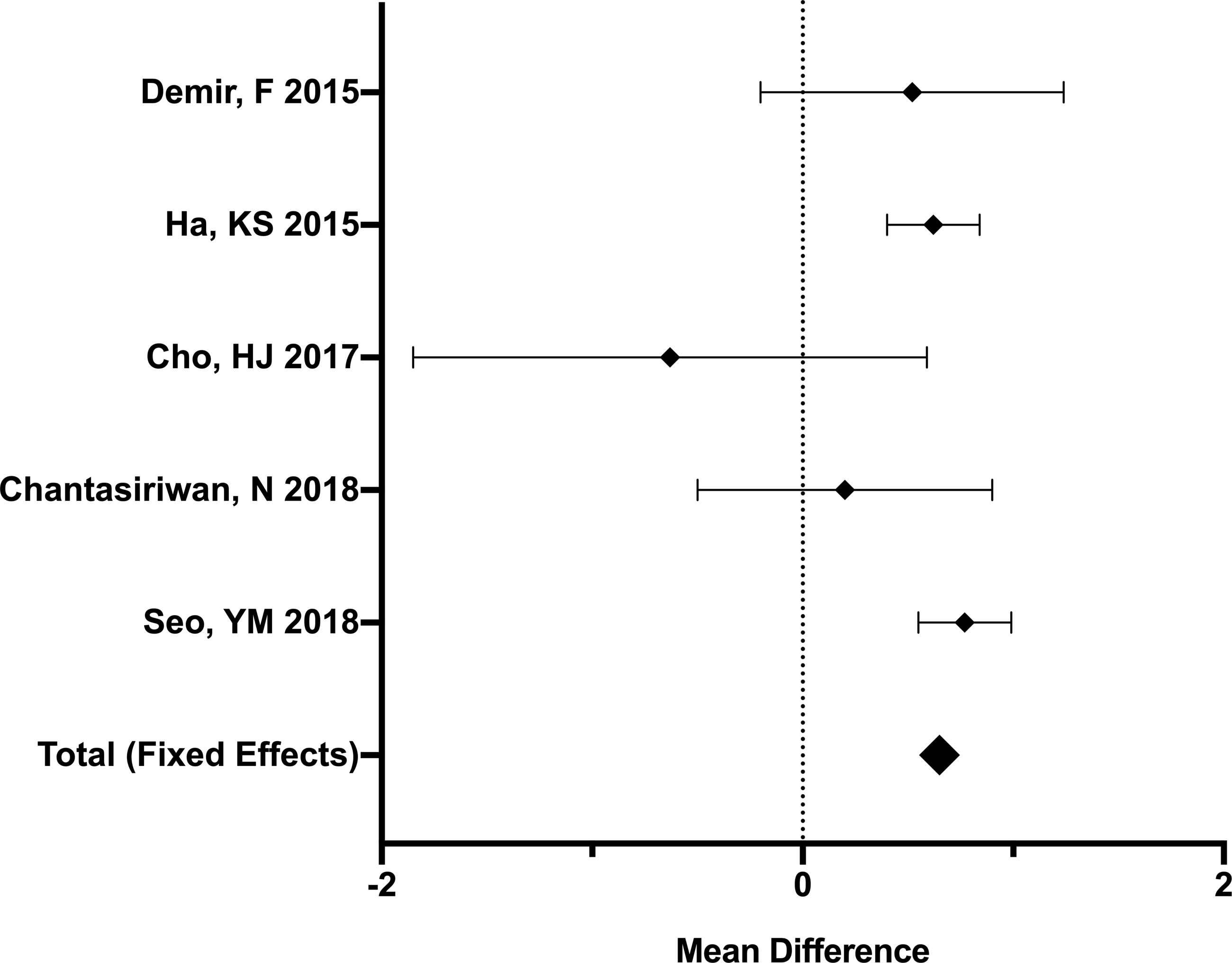
A total of five studies with 1690 patients were included in this analysis. Reference Ha, Lee and Jang29,Reference Cho, Bak and Kim35,Reference Chantasiriwan, Silvilairat, Makonkawkeyoon, Pongprot and Sittiwangkul39,Reference Demir, Karadeniz and Ozdemir43,Reference Seo, Kang and Lee44 The Q-statistic for heterogeneity had a p-value of 0.3 and the I-squared value was 44%, demonstrating no significant heterogeneity. Thus, a fixed-effects model was used. Neutrophil–lymphocyte ratio before intravenous gamma globulin therapy was higher in those who experienced coronary artery lesions (3.85 versus 3.74). This resulted in a mean difference of 0.65 (95% confidence interval 0.5 to 0.79, p-value of less than 0.01) (Fig 6). No publication bias was found by visual assessment of the publication bias funnel plot.
Forest plot pooled analysis of mean difference in NLR before intravenous gamma globulin therapy for prediction of coronary artery lesions.

Discussion
As coronary artery lesions are one of the most feared complications of Kawasaki disease, and resistance to intravenous gamma globulin therapy has been associated with their development, the identification of patients at increased risk of this complication is very important. Thus, several scoring systems have been proposed for both the prediction of refractory disease and coronary artery lesions with mixed results. Reference Kobayashi, Inoue and Takeuchi15,Reference Egami, Muta and Ishii17–Reference Tremoulet, Best and Song19,Reference Hua, Ma and Wang45–Reference Tang, Yan and Sun48 These systems generally use a combination of white blood cells, CRP, TB, AST, ALT, platelets, age, and days of illness at treatment. Unfortunately, some of them have failed to be effective across populations. Reference Arane, Mendelsohn and Mimouni20,Reference Davies, Sutton and Blackstock49
Previous studies have reported the relationship between inflammatory markers and cardiovascular disease; some of the most important are C-reactive protein and white blood cells. Reference Danesh, Collins, Appleby and Peto50,Reference Ates, Canpolat and Yorgun51 More recently, attention has shifted to leukocyte subpopulations. In similar diseases, such as multisystem inflammatory syndrome, absolute lymphocyte count is usually decreased and profound lymphopenia is associated with a more severe course, ICU admission and shock. Reference Whittaker, Bamford and Kenny52,Reference Abrams, Oster and Godfred-Cato53 Nonetheless, a low absolute lymphocyte count is rarely found in patients with Kawasaki disease. Reference Matsubara, Ichiyama and Furukawa54 In comparison to multisystem inflammatory syndrome, patients with Kawasaki disease are less likely to have lymphopenia but are more likely to have leukocytosis and neutrophilia. Reference Rowley55–Reference Godfred-Cato, Abrams and Balachandran58 In Kawasaki disease, absolute lymphocyte count remains stable in the acute, subacute, and convalescent phase of the disease, and it is rather the lymphocyte percentage that increases after ntravenous immune globulin treatment. Reference Tremoulet, Jain, Chandrasekar, Sun, Sato and Burns59 Instead of a specific value of either lymphocytes or neutrophils, studies have looked at the neutrophil-to-lymphocyte ratio in Kawasaki disease.
Neutrophil–lymphocyte ratio has been demonstrated to correlate with disease severity in critically ill patients and predicts mortality in different pathologies, particularly in the coronary artery disease. Reference Zahorec22–Reference Widjaja, Rusmawatiningtyas, Makrufardi and Arguni27 There are two factors that may make the use of neutrophil–lymphocyte ratio more attractive: first, it is not affected by exercise or dehydration, and second, because neutrophils and lymphocytes represent two complementary immune mechanisms. Reference Demir, Karadeniz and Ozdemir43 Neutrophil levels are considered an unspecific inflammatory response marker, and lymphocyte levels are a marker of immune regulation. Thus, the neutrophil–lymphocyte ratio is considered to reflect the balance between inflammation and immune regulation, and it may be more predictive than either parameter alone. Reference Demir, Karadeniz and Ozdemir43
Particularly in Kawasaki disease, the use of neutrophil–lymphocyte ratio would seem beneficial as its pathophysiology involves interactions between neutrophils and lymphocytes, and its most serious complication is cardiovascular. In the acute phase of Kawasaki disease, neutrophils increase and infiltrate coronary arteries which cause necrotising arteritis which may contribute to the development of coronary artery aneurysms. Reference Noval Rivas and Arditi60 In further stages, chronic vasculitis and luminal myofibroblast proliferation contribute to the later stages mediated by CD8+ T cells, IgA+ plasma cells, eosinophils, and macrophages, which may lead to stenosis and thrombosis. Reference Orenstein, Shulman and Fox61
Persistent inflammation after treatment has been reported as an important factor in the development of coronary artery lesions. Reference Kim, Choi and Woo62 Therefore, the current study evaluates the use of neutrophil–lymphocyte ratio in the prediction of refractory Kawasaki disease and coronary artery lesions.
These pooled analyses demonstrate that neutrophil–lymphocyte ratio is useful in the prediction of refractory disease. The mean neutrophil–lymphocyte ratio for those who experienced refractory Kawasaki disease was 5.7 before intravenous gamma globulin therapy and 1.97 after therapy. The pooled area under the curve for neutrophil–lymphocyte ratio before intravenous gamma globulin therapy was 0.724, demonstrating good value in prediction of refractory disease, and the pooled area under the curve for neutrophil–lymphocyte ratio after therapy was 0.803, demonstrating excellent value in predicting resistance to intravenous gamma globulin therapy. In both the refractory and non-refractory groups, the neutrophil–lymphocyte ratio was significantly lower after intravenous gamma globulin therapy compared to before it. Further studies are needed to test if the variation of neutrophil–lymphocyte ratio before and after intravenous immune globulin therapy could help guide clinicians in risk stratification.
Although a pooled area under the curve analysis could not be done for the prediction of coronary artery lesions, these analyses demonstrated that neutrophil–lymphocyte ratio could also be useful in their prediction. Neutrophil–lymphocyte ratio before intravenous gamma globulin therapy was higher in those who experienced coronary artery lesions with a mean difference of 0.65 (p-value of less than 0.01). Further studies are needed to evaluate the effectiveness. While previous studies have demonstrated these findings, the findings from these pooled analyses offer a quantitative summary.
A similar study to this one was performed by Wu et al on the use of neutrophil–lymphocyte ratio in the prediction of refractory Kawasaki disease. Reference Wu, Yue, Ma, Zhang, Zheng and Li63 The current study expanded on the findings of Wu et al, as it included more studies and also examined the mean difference of neutrophil–lymphocyte ratio between groups. Additionally, the current study also includes a pooled analysis of neutrophil–lymphocyte ratio for the prediction of coronary artery lesions, which in the end, it could be considered the more important endpoint.
These analyses, while additive, are not without their limitations. Firstly, data is all study-level data and not patient-level data. Thus, patient-specific confounders cannot be accounted for. Secondly, heterogeneity was present in some of the pooled analyses but this in and of itself is not a limitation. In fact, the presence of heterogeneity speaks to the need for such pooled analyses as a random-effects model helps provide a meaningful, quantitative summary of otherwise heterogeneous data. While the current pooled analyses do not assess the value of neutrophil–lymphocyte ratio in combination with other clinical parameters, neutrophil–lymphocyte ratio in combination with other clinical parameters could have even greater value in such risk stratification of patients at risk of refractory disease and coronary artery lesions.
Conclusion
Neutrophil–lymphocyte ratio values before and after intravenous gamma globulin therapy are useful for the prediction of refractory Kawasaki disease and were higher in those patients who had refractory disease compared to those who did not. It was also higher in patients who developed coronary artery lesions compared to those who did not. The use of – may help physicians in the identification of patients at risk of refractory disease and coronary artery lesions in patients with Kawasaki disease.
Acknowledgements
None.
Financial support
This research did not receive any specific grant from funding agencies in the public, commercial, or not-for-profit sectors.
Conflicts of interest
None.
Ethics approval
These analyses did not require institutional review board approval as they used previously published data that were deidentified. These analyses are in compliance with the Helsinki declaration of 1975 and its subsequent revisions.