Classification of naked dinoflagellates has been arbitrary for a long time (Fensome et al., Reference Fensome, Taylor, Norris, Sarjeant, Wharton and Williams1993; Daugbjerg et al., Reference Daugbjerg, Hansen, Larsen and Moestrup2000) due to insufficient data and they were generally classified in the order Gymnodiniales (Saldarriaga et al., Reference Saldarriaga, Leander, ‘Max’ Taylor and Keeling2003). Recent studies using ultrastructural and molecular data helped to clarify this classification (Shao et al., Reference Shao, Chen, Zhou, Yuan, Qu, Zhao and Lin2004). Consequently, several new genera of naked dinoflagellates (e.g. Akashiwo, Karenia, Karlodinium Daugbjerg et al., Reference Daugbjerg, Hansen, Larsen and Moestrup2000; Takayama De Salas et al., Reference De Salas, Bolch, Botes, Nash, Wright and Hallegraeff2003 and Togula Flø Jørgensen et al., Reference Flø Jørgensen, Murray and Daugbjerg2004) have been created and some species, previously thought as naked, were discovered to be theacate (e.g. Katodinium rotundatum was reclassified to the peridinialean genus Heterocapsa, Hansen, Reference Hansen1995). However, there are still many poorly studied species of dinoflagellates, which appear naked.
Lessardia genus and Lessardia elongata Saldarriaga & Taylor species were for the first time described in the samples taken from Georges Bank (north-west Atlantic, off the coast of Massachusetts, USA (Saldarriaga et al., Reference Saldarriaga, Leander, ‘Max’ Taylor and Keeling2003)). It was suggested by Saldarriaga et al. (Reference Saldarriaga, Leander, ‘Max’ Taylor and Keeling2003) that this species had been identified as ‘Gymnodinium elongatum Hope 1954 or Gymnodinium sp.’ in the past and observed along Norwegian and Danish coasts (Hansen & Larsen, Reference Hansen, Larsen and Thomsen1992), in the north-west Atlantic (Shapiro et al., Reference Shapiro, Haugen and Carpenter1989) and the north-east Pacific (Shapiro et al., Reference Shapiro, Haugen and Carpenter1989; Sherr & Sherr, Reference Sherr and Sherr2002). After revealing their thecal plates, Saldarriaga et al. (Reference Saldarriaga, Leander, ‘Max’ Taylor and Keeling2003) classified this species in the order Peridiniales and the family Podolampaceae, rather than in naked Gymnodiniales. Recently, it was also reported by Throndsen et al. (Reference Throndsen, Hasle and Tangen2007) that this species was sporadically observed in south Norwegian fjord areas in summer.
In the present study Lessardia elongata, which was not mentioned previously in the Black Sea (Ivanov, Reference Ivanov1965; Bodeanu, Reference Bodeanu1987/1988; Eker et al., Reference Eker, Georgieva, Senichkina and Kideys1999; Velikova et al., Reference Velikova, Moncheva and Petrova1999; Moncheva et al., Reference Moncheva, Gotsis-Skretas, Pagou and Krastev2001; Uysal, Reference Uysal2001; Eker-Develi & Kideys, Reference Eker-Develi and Kideys2003; Gomez & Boicenco, Reference Gomez and Boicenco2004), was identified for the first time in this area using scanning electron microscope (SEM) in samples from the 2004 cruise and under epifluorescence microscope in samples from the 2006 cruise.
During the present study samples were collected from the western Black Sea on two different cruises. The first cruise was on board of the RV ‘Poseidon’, along the Romanian and Ukrainian waters (2–7 September 2004); samples were taken from the subsurface chlorophyll-a maximum layer at 22 stations. The second cruise was on board of the RV ‘Akademik’ along Romanian and Bulgarian waters (2–17 June 2006); samples were taken from surface waters at 30 stations. In both sampling periods phytoplankton samples were immediately fixed with buffered formaldehyde to have a final concentration of 2% onboard. For SEM (JEOL 840) examination, phytoplankton samples from the September 2004 cruise were filtered through polycarbonate membrane filters with 0.2 µm pore size. After filtration membranes were placed on a SEM stub and sputter-coated with gold and searched for cells with the SEM.
Phytoplankton samples from the June 2006 cruise were settled to concentrate cells to count under the microscope using a Sedgewick-Rafter cell. Samples were kept immobile ~2 weeks in the laboratory, after this time the samples were concentrated from a total volume of ~250 ml to 20–50 ml by siphoning the supernatant using thin curved tubes (Eker et al., Reference Eker, Georgieva, Senichkina and Kideys1999). Microscopic visualization and image acquisition of the cells stained with 0.01% fluorescent brightener 28 (Sigma) were performed using an F-View digital monochromatic camera and Cell P software coupled to an Olympus IX-81 motorized inverted microscope with an Olympus U-RFL-T epifluorescence attachment. Cell volume, calculated considering a fusiform shape, was 300 ± 250 µm3 on average and ranged from ~200–800 µm3; N = 400. The in situ temperature and salinity measurements were performed using a Seabird-19 CTD.
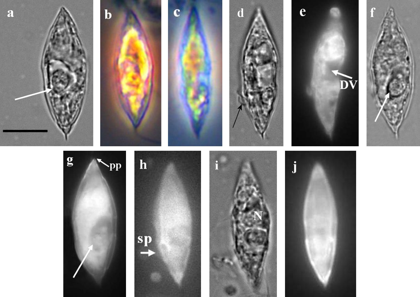
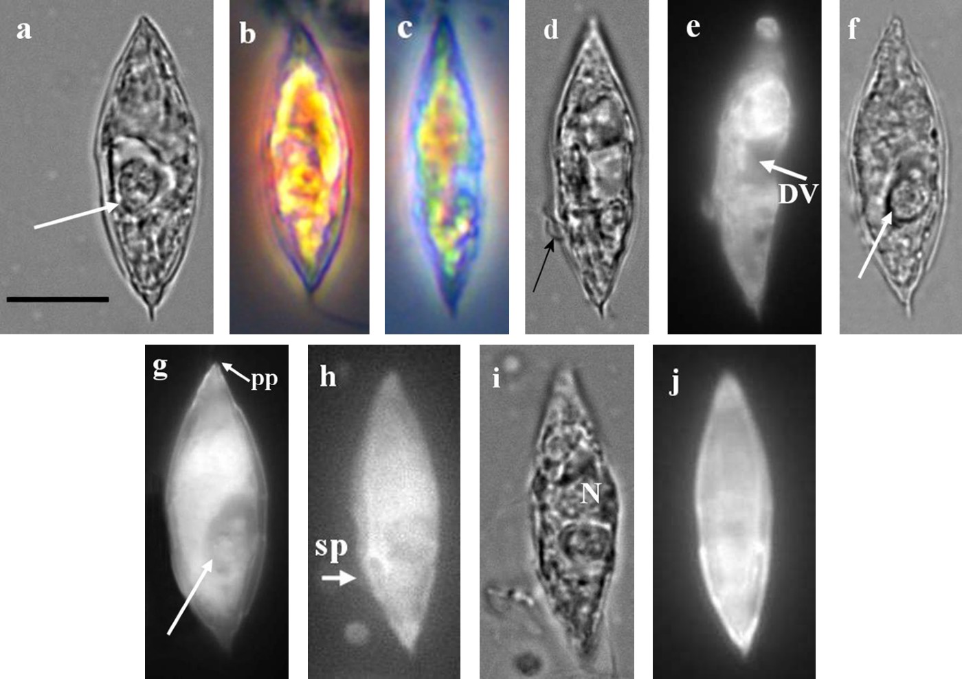
Cells were 22–30 µm long, 5–10 µm wide in formaldehyde fixed samples, spindle shaped, transparent and lack chloroplasts (Figures 1, 2a–g). The epitheca was slightly larger than the hypotheca and had a weakly impressed cingulum (Figure 2c, d). Recently ingested prey could often be seen in the antapical half of the cell within very conspicuous vacuoles (Figure 2a, f, g). Empty vacuoles of cells were sometimes visible (Figure 2d, e). Sulcal plates were also observed (Figure 2h). The nucleus was located in the apical half of the cell (Figure 2i). Faint thecal plates were seen barely on Figure 2j.
SEM micrographs of Lessardia elongata; both are side view.

Morphology of Lessardia elongata. (a) Phase contrast light micrograph showing digestive vacuole with ingested prey (arrow); (b) transparent appearance of another cell; (c) transparent cell in which epitheca clearly seems greater than hypotheca; (d) cell with a broad girdle and a flagellum (arrow); (e) the same cell as Figure 2d stained with fluorescent brightener 28, under UV light, empty digestive vacuole (DV); (f) cell with digestive vacuole with ingested prey; (g) same cell as Figure 2f illuminated with UV light, pore plates (pp) located at the apical side; (h) sulcal plates (sp) shown with UV light; (i) the nucleus (N); (j) faint thecal plates observed with UV light. Scale bar: 10 µm.
Cell abundance of Lessardia elongata ranged from 950–18,400 cells l–1 in September 2004 and from 400–87,000 cells l–1 in June 2006 in the western Black Sea. Abundance of this species was higher at the coastal stations than at the offshore stations.
There was a significant negative correlation between cell abundance of Lessardia elongata and salinity (r2 = 0.39, P < 0.00018). The temperature and salinity ranges were 21.3–24.4°C and 10.4–17.7 psu during September 2004 sampling and 16.7–20.9°C and 4.2–18.5 psu along the stations during June 2006 sampling.
Lessardia elongata Saldarriaga & Taylor, a thecate dinoflagellate, was previously identified as athecate Gymnodinium elongatum Hope 1954 (Saldarriaga et al., Reference Saldarriaga, Leander, ‘Max’ Taylor and Keeling2003) since it may appear like an athecate species under light microscopy and was observed in different regions with this name or as Gymnodinium sp. previously (Shapiro et al., Reference Shapiro, Haugen and Carpenter1989; Hansen & Larsen, Reference Hansen, Larsen and Thomsen1992; Sherr & Sherr, Reference Sherr and Sherr2002). Although neither L. elongata nor G. elongatum has been reported in any peer reviewed publication in the Black Sea so far (Ivanov, Reference Ivanov1965; Bodeanu, 1997/1988; Eker et al., Reference Eker, Georgieva, Senichkina and Kideys1999; Velikova et al., Reference Velikova, Moncheva and Petrova1999; Moncheva et al., Reference Moncheva, Gotsis-Skretas, Pagou and Krastev2001; Uysal, Reference Uysal2001; Eker-Develi & Kideys, Reference Eker-Develi and Kideys2003; Gomez & Boicenco, Reference Gomez and Boicenco2004), it has been observed and included in total phytoplankton abundance and local protocols since the 1980s with different names; Gymnodinium sp., Cryptaulax sp., Gymnodinium elongatum or Gymnodinium fusus (V. Velikova, unpublished; N. Derezyiuk, Odessa University, Ukraine, personal communication). In the present study, based on our fluorescence and SEM observations, we confirm that the spindle shaped dinoflagellate, which was sporadically observed in the Black Sea waters, is Lessardia elongata.
Heterotrophic microplankton temporarily plays a major role in the transfer of food and energy to higher trophic levels within the pelagic system (Hansen, Reference Hansen1991; Tillmann & Hesse, Reference Tillmann and Hesse1998). One of the heterotrophic microplankton species, Lessardia elongata, was recorded for the first time in the western Black Sea in this study.
ACKNOWLEDGEMENTS
This work has been supported by the JRC of the European Commission. I thank J-F. Berthon from JRC for collecting samples, A.C. Cardoso and M. Ghiani from JRC for allowing me to use their microscopes. The colleagues of the Institute of Oceanology of Varna (Bulgary), the Captain and the crew of the RV ‘Akademik’ are duly acknowledged for their support during the measurement campaign.