Implications
This paper reviews current knowledge on the proteins in milk that contribute to host defence, with emphasis on how these proteins contribute to this function, and incorporating some previously unpublished data relating to the function of individual milk proteins as innate immune effectors, or as facilitators of the recognition of pathogens. The understanding at the molecular level of milk as an immune fluid is an emerging area of study, and has not been previously reviewed with this emphasis. Thus, it will provide a valuable guide to future study in this area.
Introduction
It has been well established that the primary function of milk is to provide nutrition for the newborn mammal. Thus, milk contains a high concentration of proteins, dominated by the caseins, which provide a supply of amino acids; fat, in the form of a lipid globule surrounded by a phospholipid bilayer, as well as free fatty acids; carbohydrate, predominantly in the form of lactose, and also oligosaccharides; and many essential minor nutrients, such as minerals, vitamins and nucleotides. It has been suggested that host-defence was in fact the original function of milk in proto-mammals (Goldman, Reference Goldman2002; Vorbach et al., Reference Vorbach, Capecchi and Penninger2006). Specifically, milk may have evolved from a specialised skin secretion that was applied to the surface of soft-shelled eggs during nesting in order to regulate water transport and prevent microbial colonisation of the egg (Oftedal, Reference Oftedal2002). If this was indeed the case, the proto-milk could be assumed to have been rich in antimicrobial substances. In fact, the major whey protein α-lactalbumin has evolved from the bactericidal enzyme, lysozyme (Blackburn et al., Reference Blackburn, Hayssen and Murphy1989).
The milk of mammals contains a wide range of bioactive substances. These include minerals and vitamins such as calcium and vitamins A and D (Hollis et al., Reference Hollis, Roos and Lambert1982; Flynn, Reference Flynn1992), a range of signalling molecules such as cytokines (Goldman et al., Reference Goldman, Chheda, Garofalo and Schmalstieg1996), IGF-1, and growth hormone (Koldovsky and Thornburg, Reference Koldovsky and Thornburg1987), bioactive oligosaccharides (Newburg, Reference Newburg2009), bioactive lipids (Isaacs, Reference Isaacs2001) and a range of proteins and peptides at lower abundance than the caseins, among which are those whose function is associated with host-defence (Clare et al., Reference Clare, Catignani and Swaisgood2003). Many of these bioactivities have been the subject of previous reviews. This review will focus on those proteins present in cows’ milk that contribute to a role in host-defence, using some of our recently produced data to illustrate emerging concepts. The emphasis will be on intact proteins identified through proteomics studies; however, it should be noted that milk also contains a range of peptides derived from proteolytic cleavage of the major milk proteins, and some of these have been shown to have activities consistent with a role in innate immunity. These peptides have been reviewed elsewhere (Clare and Swaisgood, Reference Clare and Swaisgood2000; Politis and Chronopoulou, Reference Politis and Chronopoulou2008). Moreover, this review will focus on bioactive proteins in milk, rather than those in colostrum.
Proteomics
It has long been known that among the minor proteins in cows’ milk, there are some with a function in host-defence. The immunoglobin (Ig)G in colostrum plays an important role in transferring immunity to the newborn, and IgA in milk may play a role in establishing an optimal microfloral population in the gut of the newborn. In fact this property of milk was instrumental in the discovery of antibodies in the late 1800s (Wheeler et al., Reference Wheeler, Hodgkinson, Prosser and Davis2007). Upon ingestion by the calf, the IgG is transferred to the calf's circulation while the IgA, which is present as a complex comprising a dimer linked by secretory component, acts within the intestinal mucosa. The properties and biological roles of the milk Igs have been described in a number of excellent reviews (Sordillo et al., Reference Sordillo, Shafer-Weaver and DeRosa1997; Reiter, Reference Reiter1978; Korhonen et al., Reference Korhonen, Marnila and Gill2000; Kehrli and Harp, Reference Kehrli and Harp2001) and will not be further discussed here. Similarly, lactoferrin, lactoperoxidase and lysozyme have also been long known to be present in milk, and were shown to contribute to the antimicrobial property of milk as long ago as the early 1900s (Wheeler et al., Reference Wheeler, Hodgkinson, Prosser and Davis2007). However, up until recently, the presence of additional host-defence-related proteins in cows’ milk has not been very well documented. This is in part due to the dominance of the six major milk proteins (αS1-casein, αS2-casein, β-casein, κ-casein, β-lactoglobulin and α-lactalbumin), and the unavailability, until recently, of suitable high-resolution protein separation methods, and sensitive identification technologies. Initial studies resolving milk proteins by two-dimensional electrophoresis and identifying them using MS identified only a small number of additional proteins (Hogarth et al., Reference Hogarth, Fitzpatrick, Nolan, Young, Pitt and Eckersall2004). However, with the continued development of proteomics technologies and improvement of bovine protein sequence databases, a hitherto hidden complexity has been revealed in the repertoire of proteins present in milk. Our own work revealed the presence of as many as 95 minor proteins present in cows’ milk, of which approximately a quarter have functions associated with host-defence (Smolenski et al., Reference Smolenski, Haines, Kwan, Bond, Farr, Davis, Stelwagen and Wheeler2007). The low concordance in the list of proteins detected by the two different methods used in our study suggests that the complexity of the milk protein repertoire may be even greater (Smolenski et al., Reference Smolenski, Haines, Kwan, Bond, Farr, Davis, Stelwagen and Wheeler2007), and more recent studies have extended the list of bovine milk proteins even further (Affolter et al., Reference Affolter, Grass, Vanrobaeys, Casado and Kussmann2010; Le et al., Reference Le, Barton, Sanders and Zhang2011). The recently completed sequencing and assembly of the bovine genome has greatly improved the database of bovine protein sequences, thus enabling identification of milk proteins at unprecedented efficiency. We are currently utilising this resource to further investigate the complexity of milk using more powerful proteomics methodologies. Although not yet complete, these studies reveal the existence of over 200 intact proteins in cows’ milk, among which approximately a quarter are associated with host-defence.
Components of the innate immune system
The innate immune system is the first and most important line of defence against pathogens, particularly in mucosal epithelia such as in the oral cavity, digestive tract, lungs or reproductive tract. As implied from the name, these host-defence responses are elicited using either constitutively expressed or induced components that are pre-encoded in the genome, involving no genomic recombination in somatic cells. Typically, the presence of pathogens at these sites initiates a cascade of events that results in the rapid elimination of the threat. The first step in this process is recognition of the pathogen by one or more opsonising proteins, followed by presentation to one or more pathogen-recognition receptors that are present in mucosal epithelial cells as well as immune cells resident in the tissue. This in turn is followed by activation of intracellular signalling pathways that result in expression of signalling proteins (the cytokines and chemokines) as well as effector proteins. The former class of proteins act to recruit additional immune cells to the site of incipient infection while the latter act to neutralise the pathogen directly. These essential features of innate immune pathways have increasingly been elucidated at the molecular level in the past 10 years and have been extensively reviewed (Oviedo-Boyso et al., Reference Oviedo-Boyso, Valdez-Alarcon, Cajero-Juarez, Ochoa-Zarzosa, Lopez-Meza, Bravo-Patino and Baizabal-Aguirre2007; Kawai and Akira, Reference Kawai and Akira2010). The most well-known of these pathways involves a family of structurally related proteins known as the Toll-like receptors (TLRs), which are found on the plasma membrane or on endosomes, each of which is activated by a distinct molecular pattern, which are often, but not always, pathogen-derived. These receptors activate transcription factors such as nuclear factor kappaB and interferon regulatory factor 3 (IRF3) via a range of signalling intermediates, ultimately resulting in expression and secretion of pro-inflammatory cytokines and antimicrobial proteins, the so-called effector-response proteins.
The best characterised of these TLR response pathways is the activation of the inflammatory response by Gram-negative bacteria such as Escherichia coli, which is responsible for the classic toxic shock response that occurs during septicaemia. These bacteria contain an outer envelope containing a complex lipid, lipopolysaccharide (LPS), which is not present in eukaryotes. Mucosal epithelial cells secrete at least two opsonising proteins, LPS-binding protein (LBP) and CD14, which bind LPS and present it to the extracellular domain of TLR4. This causes a conformational change that results in dimerisation of TLR4 and activation of an intracellular-signalling cascade that results in activation of expression of the pro-inflammatory cytokine, tumour necrosis factor-α (TNFα). In some cells, TLR4 activation also triggers secretion of antimicrobial proteins such as bactericidal permeability-increasing protein or β-defensin (Lu et al., Reference Lu, Yeh and Ohashi2008). While the LPS–TLR4 pathway has been well-defined and characterised in human- and murine-immune cells, the molecular mechanisms by which other so-called pathogen-associated molecular patterns elicit particular responses are less well understood. Bovine mammary epithelial cells have been shown to express TLR4 and TLR2 (Goldammer et al., Reference Goldammer, Zerbe, Molenaar, Schuberth, Brunner, Kata and Seyfert2004), and to be functional (Yang et al., Reference Yang, Zerbe, Petzl, Brunner, Gunther, Draing, von Aulock, Schuberth and Seyfert2008b). However, the roles that TLR activation signals might play in the udder to influence its milk production or contribute to host-defence are still not very well understood.
Effector proteins in cows’ milk
Milk has long been known to contain antimicrobial properties, which have been presumed to contribute to suppression of pathogens either in the mammary gland itself or in the digestive tract of the newborn. Lysozyme, lactoferrin and lactoperoxidase each possess the ability to suppress the growth of certain bacteria and were identified as minor milk proteins in the early part of the twentieth century (Wheeler et al., Reference Wheeler, Hodgkinson, Prosser and Davis2007). More recently, it has become apparent that milk also contains a range of additional antimicrobial proteins and peptides, including members of the β-defensin, complement, cathelicidin and S100 calgranulin families, as well as several acute phase proteins (Eckersall et al., Reference Eckersall, Young, McComb, Hogarth, Safi, Weber, McDonald, Nolan and Fitzpatrick2001; Jia et al., Reference Jia, Starner, Ackermann, Kirby, Tack and McCray2001; Rainard, Reference Rainard2003; Swanson et al., Reference Swanson, Gorodetsky, Good, Davis, Musgrave, Stelwagen, Farr and Molenaar2004; Murakami et al., Reference Murakami, Dorschner, Stern, Lin and Gallo2005; Lutzow et al., Reference Lutzow, Donaldson, Gray, Vuocolo, Pearson, Reverter, Byrne, Sheehy, Windon and Tellam2008). The role that two of these effector proteins play in contributing to the host-defence function of milk is described below, drawing on previously published work as well as our own unpublished data.
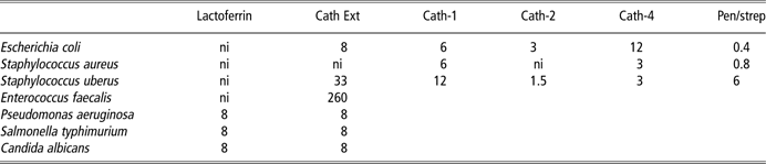
The cathelicidins are potent antimicrobial proteins that are expressed in neutrophils, stored in secretory granules and secreted upon activation in response to pro-inflammatory signalling initiated by a pathogen. During secretion, the full-length protein is cleaved by elastase to release the mature antimicrobial peptide (1.5 to 7 kDa, depending on which cathelicidin) and the remaining portion of the mature protein, known as the cathelin domain (11 kDa). Unlike mice and humans, cattle have multiple cathelecidin genes, each encoding a distinct antimicrobial peptide (Scocchi et al., Reference Scocchi, Wang and Zanetti1997). These peptides have extremely potent and broad-spectrum antimicrobial activity – significantly more potent than that of lactoferrin (Table 1). The cathelin domain is also secreted in response to pathogens; however, the biological role, if any, of this protein domain is much less well understood. The cathelin protein appears to be stable in milk and is easily measured using antibodies. We have found that the abundance of cathelin is elevated in milk from most cows that harbour a mammary infection (Pryor et al., Reference Pryor, Smolenski, Wieliczko, Broadhurst, Stelwagen, Wheeler and Haigh2010; Smolenski et al., Reference Smolenski, Wieliczko, Pernthaner, Broadhurst, Stelwagen, Hein, Wheeler and Haigh2010), and we are currently investigating its utility as a mastitis biomarker in the dairy industry. Intriguingly, we have also obtained evidence that the cathelin domain, rather than being a biologically inert by-product of antimicrobial peptide secretion, may have immunomodulatory properties in its own right. We have purified the cathelin domain from bovine milk and found it binds to LPS with a similar avidity as LBP. Furthermore, adding even quite low concentrations of the purified cathelin domain to the human pathogen Candida albicans caused it to aggregate, and addition of the protein to neutrophils in culture rendered them non-viable (data not shown).
Growth suppression activity of lactoferrin and cathelicidins
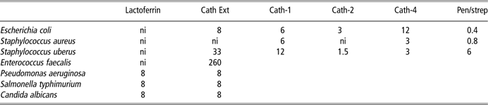
Cath Ext = cathelicidin mixture; ni = no inhibition observed.
Lactoferrin was purified from bulk skimmed cows’ milk after factory processing. Cath Ext was extracted from pooled raw milk from cows with clinical signs of mastitis. Cathelicidin-1, -2 and -4 antimicrobial peptides were chemically synthesised using their known amino acid sequences. The growth suppression was estimated using the serial twofold dilution method in liquid culture using conditions optimal for growth. Values are MIC50 (defined as the concentration of inhibitor that results in a 50% reduction of cfu per unit volume) and are given as μg/ml for all the proteins and peptides. The MIC50 values for the mixture of penicillin and streptomycin is given as μ units/ml. All values are representative of at least three independent analyses carried out on separate occasions, each done in triplicate. No value entered indicates the condition was not tested.
Bovine milk has been shown to contain a number of acute-phase proteins, including haptoglobin and serum amyloid A3 (SAA3). Both of these proteins are increased in abundance in milk from cows with mastitis (Gronlund et al., Reference Gronlund, Hulten, Eckersall, Hogarth and Persson2003) and SAA3 induces mucin secretion in the gut (Larson et al., Reference Larson, Wei, Weber, Mack and McDonald2003). The SAA3 protein, at least, appears to act as an innate immune-effector protein. The structure of SAA3 bears some resemblance to the pentraxin family of proteins. The SAA3 protein appears to form six-membered rings, as opposed to the five-membered pentraxin rings (Wang et al., Reference Wang, Lashuel, Walz and Colon2002). We have shown that the recombinantly produced protein has potent antimicrobial activity as well as liposome-disrupting activity (Molenaar et al., Reference Molenaar, Harris, Rajan, Pearson, Callaghan, Sommer, Farr, Oden, Miles, Petrova, Good, Singh, McLaren, Prosser, Kim, Wieliczko, Dines, Johannessen, Grigor, Davis and Stelwagen2009), properties that are consistent with permeabilisation of bacterial membranes. The SAA3 protein is produced in the liver and is present in the circulation, from where it is transported into milk. However, we have shown that the mammary epithelial cells themselves also express a variant of SAA3, which we term M-SAA3 (Molenaar et al., Reference Molenaar, Harris, Rajan, Pearson, Callaghan, Sommer, Farr, Oden, Miles, Petrova, Good, Singh, McLaren, Prosser, Kim, Wieliczko, Dines, Johannessen, Grigor, Davis and Stelwagen2009). Indeed, the SAA3 in milk has been shown to be a mixture of locally and systemically produced protein (Jacobsen et al., Reference Jacobsen, Niewold, Kornalijnslijper, Toussaint and Gruys2005). Besides its role as an effector protein through its antimicrobial activity, there is also evidence that serum amyloid proteins can act to facilitate pathogen recognition through an opsonising activity. Specifically, it has been shown that SAA1 can bind to the OmpA antigen, which is present in the cell wall of some pathogenic bacteria. This stimulates the phagocytosis of these bacteria by macrophages (Shah et al., Reference Shah, Hari-Dass and Raynes2006). Thus, SAA3 may act as an effector protein as well as facilitating pathogen recognition.
Pathogen-recognition proteins in cows’ milk
The recognition of pathogens is the first and arguably most important step in host-defence. Yet, not much is known about the molecular mechanisms through which this occurs in milk. Although the opsonising proteins for LPS (LBP and CD14) and their subsequent activation of TLR4 are reasonably well characterised in vitro, the existence of this mechanism in milk has not been reported previously. Other pattern-recognition receptors are known to be activated by a range of specific molecules; however, their mechanisms of activation, including the possible involvement of opsonising proteins, is largely unknown. Our proteomics investigation of the complexity of milk has led to the identification of a number of putative pathogen-recognition proteins. Some of these are further discussed below.
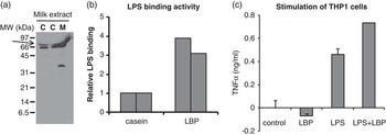
We have found that LBP is present in cows’ milk through our proteomics analysis, and its presence in milk from either healthy cows or cows with mastitis was confirmed by immunoblotting. The protein was purified from milk by selective precipitation followed by cation-exchange chromatography and shown to have potent LPS-binding activity as well as to stimulate inflammatory signalling in the THP1 human macrophage cell line (Figure 1). As milk contains macrophages and other immune cells, these data suggest that milk is primed to detect the presence of Gram-negative bacteria through this mechanism.
Presence and activity of LBP in cows’ milk. (a) Extracts of milk enriched for cationic proteins from (c) uninfected cows or (m) a cow showing clinical signs of mastitis were subjected to SDS-polyacrylamide gel electrophoresis followed by immunoblotting using rabbit IgG raised against bovine LBP. The position of an immunoreactive doublet band at 60 kDa, indicating the presence of LBP, is arrowed. The identity of the 33 kDa reactive band is likely to be a proteolytic cleavage product of LBP. The signal in the mastitis lane has been distorted upwards due to an edge effect. (b) The LPS-binding activity of LBP, which has been purified to >95% from bovine milk. Activity was measured by incubating the LBP preparation or a control protein for non-specific interaction (Casein) in wells of a 96-well plate containing LPS immobilised on its surface. After washing, the bound protein was detected by incubation with a peroxidase-LPS conjugate, followed by visualisation with diaminobenzidine, as described previously (Haigh et al., Reference Haigh, Hood, Broadhurst, Medele, Callaghan, Smolenski, Dines and Wheeler2008). The level of activity relative to that of casein is shown for two separate experiments. (c) Stimulation of secretion of TNFα from the human monocyte cell line THP1 in response to the addition of (control) vehicle alone (LBP = 10 μg/ml purified LBP; LPS = 10 μg/ml LPS; and LBP + LPS = 10 μg/ml of both LBP and LPS). TNFα was measured using an ELISA kit (BD Pharmingen, San Diego, CA, USA) using the manufacturer's instructions. Values are means of four replicates, expressed relative to PBS, which was used as a control. Error bars represent the s.d. (LBP = lipopolysaccharide-binding protein; IgG = immunoglobulin G; LPS = lipopolysaccharide; TNFα = tumour necrosis factor-α; PBS = phosphate-buffred saline).

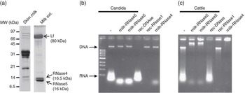
Other possible pathogen-recognition proteins in milk are the RNases. The mammalian RNases are a large family of proteins that are expressed in diverse tissues and secreted into the extracellular space. Some RNases are expressed in mucosal epithelia such as the intestinal tract, and in exocrine secretions such as saliva and milk. These observations, together with the reported antimicrobial and cytotoxic activities of some members of the family have led to the idea that the RNases function in host-defence (Dyer and Rosenberg, Reference Dyer and Rosenberg2006; Boix and Nogues, Reference Boix and Nogues2007). One member of the RNase family, RNase5, has long been known to be present in milk (Maes et al., Reference Maes, Damart, Rommens, Montreuil, Spik and Tartar1988). RNase5 is also known as angiogenin, as it was first discovered through its potent angiogenic activity (Fett et al., Reference Fett, Strydom, Lobb, Alderman, Bethune, Riordan and Vallee1985), a property that it uniquely possesses among the RNases. RNase5 has also been shown to have antimicrobial activity, leading to the suggestion that it plays a role in host-defence in the intestinal tract (Hooper et al., Reference Hooper, Stappenbeck, Hong and Gordon2003). Our proteomics investigations have revealed that bovine milk contains at least one additional RNase, namely RNase4. We have purified both RNases individually from cows’ milk and shown that unlike RNase1, both RNase4 and RNase5 digest RNA in a characteristic manner, leading to oligonucleotides rather than single nucleotides, and that both RNases interact with DNA, leading to a supershift when resolved on agarose gels (Figure 2). Furthermore, RNase5, and to a lesser extent, RNase4, suppress the growth of the human fungal pathogen, C. albicans, particularly in its hyphal form, and this is associated with binding to the surface of the pathogen (Harris et al., Reference Harris, Johannessen, Smolenski, Callaghan, Broadhurst, Kim and Wheeler2010). Other members of the RNase family have been shown to have immunostimulatory activity, further suggesting a role in host-defence. For example, RNase2 has been shown to activate TLR2-mediated pro-inflammatory signalling in dendritic cells (Yang et al., Reference Yang, Chen, Su, Zhang, Kurosaka, Caspi, Michalek, Rosenberg, Zhang and Oppenheim2008a), and an RNase has been shown to stimulate recognition of DNA through the TLR9 pathway in dendritic cells (Miyazato et al., Reference Miyazato, Nakamura, Yamamoto, Mora-Montes, Tanaka, Abe, Tanno, Inden, Gang, Ishii, Takeda, Akira, Saijo, Iwakura, Adachi, Ohno, Mitsutake, Gow, Kaku and Kawakami2009). We have started to investigate whether the RNases in milk also can stimulate pro-inflammatory signalling. Our preliminary data suggest that, indeed, both milk RNases can enhance the stimulation of IL12 and TNFα secretion in mouse macrophages by bacterial DNA, presumably through a TLR9-mediated pathway (S. Gupta, unpublished data).
Presence and activity of RNase4 and RNase5 in cows’ milk. (a) Skim milk (left lane) and an extract of milk enriched for cationic proteins (right lane) were subjected to SDS-polyacrylamide gel electrophoresis followed by staining of proteins with Coomassie blue. The protein bands corresponding to lactoferrin, RNase4 and RNase5 are indicated by arrows. The identity of these proteins has been confirmed by immunoblotting and mass spectrometry (data not shown). (b) Nucleic acids were extracted from Candida albicans by a procedure involving alkali lysis, ethanol precipitation, proteinase K digestion and phenol extraction using established procedures (Ausubel et al., Reference Ausubel, Brent, Kingston, Moore, Seidman, Smith and Struel1995). Stock solutions of RNase were added to aliquots of a 1-μg/ml solution of nucleic acid to bring them to a final concentration of 1 μg/ml for either milk-purified RNase5 (duplicate experiments), recombinant RNaseA (Promega, Madison, WI, USA), recombinant RNase-qualified 1 (RQ1) DNase (Promega) or milk-purified RNase4, and incubated for 60 min at 37°C. The mixtures were then subjected to electrophoresis on Tris-acetate-EDTA-agarose gels and the nucleic acids were visualised by staining with ethidium bromide. These analyses were performed following established procedures (Ausubel et al., Reference Ausubel, Brent, Kingston, Moore, Seidman, Smith and Struel1995). The position of the DNA and RNA on the gels is indicated. (c) Identical analysis as described above, except that nucleic acid extracted from bovine liver was used.

Cartilage glycoprotein 39 kDa (CG39) is another protein present in cows’ milk that could play a role in pathogen recognition. CG39 was first described in synovial fluid, but it is also found in certain immune cells as well as mammary secretions in the dry period (Rejman and Hurley, Reference Rejman and Hurley1988; Hakala et al., Reference Hakala, White and Recklies1993). The expression of CG39 has been reported to be elevated in inflamed tissues in a range of chronic inflammatory conditions such as arthritis, inflammatory bowel disease, asthma and allergy (Elias et al., Reference Elias, Homer, Hamid and Lee2005; Bigg et al., Reference Bigg, Wait, Rowan and Cawston2006; Lee, Reference Lee2009). The amino acid sequence and three-dimensional structure of human and bovine CG39 are similar to members of the chitinase family of host-defence proteins found predominantly in plants. These plant chitinases are thought to help protect against insects, which contain chitin, a complex polysaccharide, as a component of their exoskeletons. However, unlike the plant chitinases, the proteins within the mammalian chitinase-like protein family, of which CG39 is a member, do not have chitinase activity. The activity and biological role of the mammalian chitinase-like proteins are largely unknown. Thus, although the linkage with host-defence is suggestive, the function of CG39 is still unresolved. In an effort to better understand the role of CG39 in milk we have purified it from cows’ milk and investigated its activities. These studies have shown that bovine milk-derived CG39 potently inhibits the growth of C. albicans, which is a chitin-containing opportunistic fungal pathogen of humans (10 μg/ml inhibits growth by 50%), but does not inhibit a number of other microbial species (unpublished data). Furthermore, CG39 has chitin-binding activity, as indicated by affinity chromatography, which causes aggregation of C. albicans at concentrations as low as 3 μg/ml, and inhibits adherence of C. albicans to an intestinal epithelial cell line in culture (unpublished data). Taken together, these observations suggest that the CG39 present in cows’ milk may indeed function in the recognition of chitin-containing pathogens, either in the mammary gland of the dams or the digestive tract of the newborn; however, further investigation is required to substantiate this.
Milk and bacteria
Milk contains a range of non-pathogenic bacterial species, such as the lactococci and lactobaccilli, among which some are thought to contribute to the functionality of milk. These bacteria are introduced to the digestive tract when milk is consumed, at least in its raw form, and it is possible that some of these milk-derived bacteria influence the microbial ecology in the gut of the newborn. The commensal bacteria present in the digestive tract play an important role in its function, including in the host-defence of the mucosa. This may be manifested in a number of ways. A large body of work has led to the idea that some microbial species directly suppress certain pathogens through occupation of their ecological niche and the secretion of antimicrobial substances such as bacteriocins. Therefore, their ingestion as ‘probiotics’ could provide a health benefit (Kailasapathy and Chin, Reference Kailasapathy and Chin2000; Reid and Burton, Reference Reid and Burton2002). Another possible way for commensals to interact with the host's defence system, which has emerged recently, is the idea that they can interact with the immune system through pathogen-recognition mechanisms so as to result in suppression of an inflammatory response. There has been some evidence reported recently for this type of mechanism in cell culture models (Lai et al., Reference Lai, Di Nardo, Nakatsuji, Leichtle, Yang, Cogen, Wu, Hooper, Schmidt, von Aulock, Radek, Huang, Ryan and Gallo2009). Finally, gut microbiotia are thought to play a role in the development of oral tolerance (von Mutius and Vercelli, Reference von Mutius and Vercelli2010). This is a critical step in development of the immune system; when an organism learns to distinguish benign antigens, it encounters through mucosal routes, such as those derived from food, from pathogens. If oral tolerance develops appropriately, then the immune system will respond differently to benign antigens than it does with pathogens. It is thought that the increasing prevalence of allergy, atopic conditions and some chronic inflammatory conditions might be linked to incorrect immune system development, including oral tolerance (Verhasselt, Reference Verhasselt2010).
It is conceivable that the bacteria in milk may have an influence on optimal function of the host-defence system in the gut; however, there has been a paucity of research in this area, and more research needs to be done before firm conclusions can be made. Other components of milk, besides the bacteria, may also play a role in influencing the gut microbiota. For example, milk-derived oligosaccharides have been reported to promote the growth of certain commensal species (Newburg, Reference Newburg2009), and immune complexes have been associated with correct development of oral tolerance (Mosconi et al., Reference Mosconi, Rekima, Seitz-Polski, Kanda, Fleury, Tissandie, Monteiro, Dombrowicz, Julia, Glaichenhaus and Verhasselt2010). We are currently investigating the influence that the proteins in milk may have on gut microbes, an area of study that has not been very well researched to date, in order to determine whether they function to influence microbial populations in the gut, and thereby optimise intestinal function.
Conclusions
The analysis of the protein components of milk and their functionality over the past 5 years has revealed that milk is a complex fluid containing a large number of different proteins. The application of proteomics technologies to milk has resulted in the identification of upwards of 100 distinct gene products, many of which are associated with host-defence. Thus, milk could be considered to be an innate immune secretion that also has a crucial nutritional function. Among these host-defence proteins, some have activities consistent with a role as innate immune-effector proteins, whereas others have activities that point to a role in the recognition of pathogens and the generation of an appropriate inflammatory response. This latter function of milk proteins has been very little researched. Yet, it appears from the number of these proteins in milk, only some of which have been described here, that pathogen recognition is a key function of milk. An interesting observation is the likely multifunctional nature of some of these proteins, thereby contributing to the functionality of milk; an area warranting further investigation. Moreover, studies to date on the innate immune-associated proteins in cows’ milk have raised some further questions. For example, do these proteins play an important role in host-defence in the mammary gland and in the digestive tract of the newborn, or are they merely relics of evolution? To what extent do these proteins act synergistically with one another to provide an effective defence against pathogens? Milk is the sole food for newborns at a time when the mucosal immune system is going through a crucial stage of development. Therefore, what contribution do the milk proteins make to the proper development of immune function in the gut? Most milk and milk products that are consumed today have been created through processes that could adversely affect the activities of these host-defence-related milk proteins. Do current methods of milk processing, such as pasteurisation and spray drying, lead to a significant attenuation of the functionality of milk? Addressing these questions may well lead in the future to dairy-based foods, including infant formulae, with added functionality, and may also, as a consequence, create a higher economic return from dairying.
Acknowledgements
The purification of milk proteins and activity data described in this paper were produced with the help of technical contributions by Rachel Brunt, Megan Callaghan, Robert Wieliczko, Kwang Kim and Blake Paget. Protein identification was done with the technical assistance of Janine Cooney and Dwayne Jensen. The authors are grateful for helpful discussions with Rex Humphrey, Liz Carpenter, Alison Hodgkinson, Helen Withers, Ben Bright and Olivia Wallace. The research was supported by FRST programmes C10X0707 and C10X0806.