Introduction
The Triassic Period witnessed the rapid evolutionary diversification of diapsid reptiles (Sues and Fraser, Reference Sues and Fraser2010). The latter comprise two principal clades, Archosauromorpha and Lepidosauromorpha. Of these, Lepidosauromorpha comprises all saurians that share a more recent common ancestor with the present-day Sphenodon and squamates than with birds and crocodylians (Gauthier et al., Reference Gauthier, Estes, de Queiroz, Estes and Pregill1988). Crown-group Lepidosauria is one of the most diverse clades of extant vertebrates, with over 10,000 species described to date, and comprises Rhynchocephalia and Squamata. The former group is represented today only by a single taxon, Sphenodon punctatus (Gray, Reference Gray1842) from New Zealand (Cree, Reference Cree2014), but attained considerable diversity during the Mesozoic (Jones, Reference Jones2008; Herrera-Flores et al., Reference Herrera-Flores, Stubbs, Elsler and Benton2019; Hsiou et al., Reference Hsiou, Nydam, Simões, Pretto, Onary, Martinelli, Liparini, de Vivar Martínez, Soares, Schultz and Caldwell2019). By contrast, crown-group squamates have an almost worldwide present-day distribution and a rich and varied fossil record extending back to the Middle Jurassic (Evans, Reference Evans1998).
The oldest rhynchocephalian to date is known from the Middle Triassic (Ladinian) Erfurt Formation of Baden-Württemberg (Germany; Jones et al., Reference Jones, Anderson, Hipsley, Müller, Evans and Schoch2013; Schoch, Reference Schoch, Hagdorn, Schoch and Schweigert2015). Jones et al. (Reference Jones, Anderson, Hipsley, Müller, Evans and Schoch2013) identified two partial dentaries as cf. Diphydontosaurus sp. Originally, Diphydontosaurus was reported from Late Triassic fissure deposits in southwestern Britain (Whiteside, Reference Whiteside1986). Whiteside and Duffin (Reference Whiteside and Duffin2017) questioned the generic identification of the rhynchocephalian from the Erfurt Formation as cf. Diphydontosaurus, but taxonomic identification must await a comprehensive review of all material available now (Schoch and Sues, unpublished data).
Reported occurrences of rhynchocephalians from Carnian-age strata in the American Southwest (Heckert, Reference Heckert2004) have more recently been re-dated as Norian (Olsen et al., Reference Olsen, Kent and Whiteside2011; Martz and Parker, Reference Martz, Parker, Zeigler and Parker2017). To date, one occurrence of late Carnian rhynchocephalians and two of possibly Carnian age have been reported from Europe. Kowalski et al. (Reference Kowalski, Bodzioch, Janecki, Ruciński and Antczak2019) reported seven morphotypes of rhynchocephalian tooth-bearing jaw fragments from late Carnian lacustrine strata in Upper Silesia (Poland). Brachyrhinodon taylori Huene, Reference Huene1910, from the Lossiemouth Sandstone Formation of Scotland, is closely related to the widely distributed Late Triassic and Early Jurassic sphenodontian Clevosaurus spp. (Huene, Reference Huene1910; Fraser and Benton, Reference Fraser and Benton1989; Hsiou et al., Reference Hsiou, de França and Ferigolo2015). Benton and Walker (Reference Benton and Walker1985) considered the Lossiemouth Sandstone Formation late Carnian in age, but its dating remains poorly constrained. The holotype and only known specimen of Polysphenodon muelleri Jaekel, Reference Jaekel1911 was recovered from a borehole in strata of the Middle Keuper near Hannover (Germany) (Jaekel, Reference Jaekel1911; Carroll, Reference Carroll1985). This taxon is also closely related to Clevosaurus spp. (Fraser and Benton, Reference Fraser and Benton1989; Hsiou et al., Reference Hsiou, de França and Ferigolo2015). The apparent lack of a published detailed log for the core makes precise dating of this find impossible because the Middle Keuper encompasses both Carnian- and Norian-age deposits (Deutsche Stratigraphische Kommission, 2005).
Sues et al. (Reference Sues, Olsen, Kroehler, Fraser and Sues1994) briefly reviewed a diverse assemblage of mostly small-bodied tetrapods from the Carnian-age Vinita Formation in Virginia. They illustrated a maxilla fragment of a rhynchocephalian with robust acrodont teeth (Sues et al., Reference Sues, Olsen, Kroehler, Fraser and Sues1994, fig. 8.7A, B). Further processing of fossiliferous mudstone collected from this locality between 1988 and 1992 has recovered an additional maxilla referable to the rhynchocephalian. It is the purpose of this paper to describe the rhynchocephalian and assess its phylogenetic relationships.
Geological setting
Numerous skeletal remains of tetrapods and fishes, including the specimens described here, were recovered by quarrying at the USNM locality 39981 near Midlothian in Chesterfield County, Virginia (Sues et al., Reference Sues, Olsen, Kroehler, Fraser and Sues1994). First discovered by Paul Olsen (Columbia University) in 1981, the site was a roadcut along the old course of Old Hundred Road, then located ~300 m north of the Powhite Parkway (Virginia State Route 76). Subsequently, it was buried when the entire area was developed for new real estate.
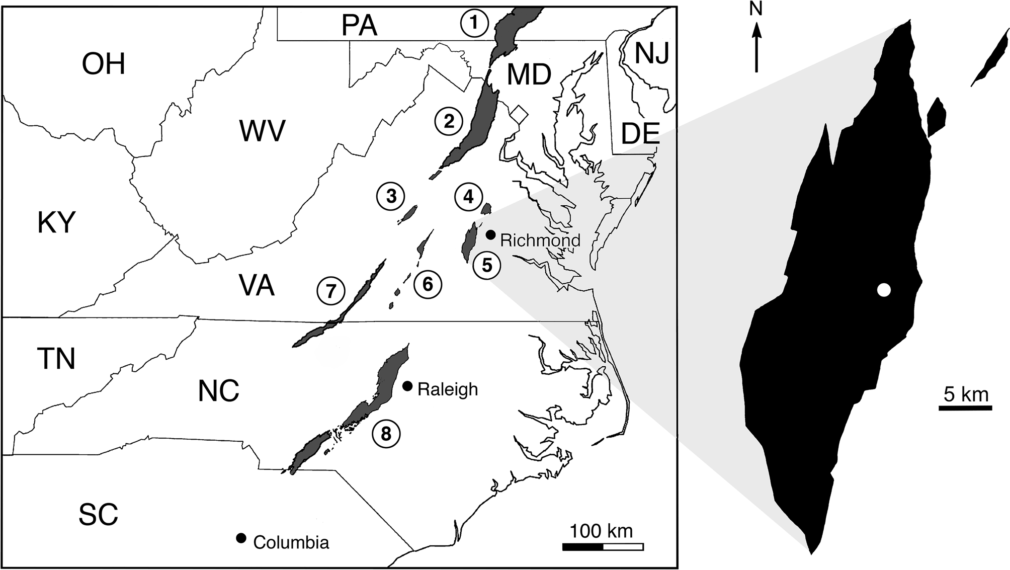
The locality exposed strata of the Tomahawk Creek Member of the Vinita Formation in the Richmond basin of the Newark Supergroup (Fig. 1). Vertebrate remains were most common in a 15–20 cm thick layer of massive mudstone. They include partial skulls and other associated skeletal elements but, for the most part, comprise dissociated but well-preserved bones, bone fragments, and teeth representing a considerable diversity of tetrapods and fishes. The tetrapods include a traversodontid and a prozostrodontian cynodont, a pseudosuchian of uncertain affinities with distinctive dorsal armor, phytosaurs, various lepidosauromorph reptiles, and possible parareptiles. The vertebrate fossils were associated with poorly preserved root traces and small carbonate nodules (with diameters of up to 1 cm). The depositional environment was probably a marginal lacustrine setting (Olsen, personal communication).
Distribution of the exposed rift basins of the Newark Supergroup in the southeastern United States with enlarged outline of the Richmond basin (in black) on which the position of the locality is marked by a white dot. Modified from an illustration courtesy of P.E. Olsen. Circled numbers denote individual basins: 1 = Gettysburg basin; 2 = Culpeper basin; 3 = Scottsville basin; 4 = Taylorsville basin; 5 = Richmond basin; 6 = Farmville and associated basins; 7 = Danville basin; and 8 = Deep River basin. Individual states are labeled with their customary abbreviations.

The Vinita Formation is late Carnian (Tuvalian) in age (Huber et al., Reference Huber, Lucas and Hunt1993; Olsen et al., Reference Olsen, Kent and Whiteside2011; Kent et al., Reference Kent, Olsen and Muttoni2017). This date is based on the astrochronology and geomagnetic polarity scale for the Newark Supergroup (Newark-APTS) developed by P.E. Olsen (Columbia University) and D.V. Kent (Rutgers University) and their associates (Kent et al., Reference Kent, Olsen and Muttoni2017). The Vinita Formation is the stratigraphic equivalent of the Falling Creek Formation in the neighboring Taylorsville basin of the Newark Supergroup, where it lies below the Newfound and Port Royal formations (LeTourneau, Reference LeTourneau, LeTourneau and Olsen2003). The Newfound Formation correlates with the upper part of the Stockton Formation in the Newark basin and the Port Royal Formation with the Lockatong Formation in the latter basin. The Lockatong Formation and the upper part of the Stockton Formation are now considered earliest Norian in age based on astrochronological and magnetostratigraphic data (Kent et al., Reference Kent, Olsen and Muttoni2017).
Materials and methods
Repository and institutional abbreviation
The holotype and a referred specimen of the new sphenodontian taxon are housed in the collections of the Department of Paleobiology at the National Museum of Natural History, Smithsonian Institution, Washington, DC (abbreviated as USNM).
Systematic paleontology
Lepidosauromorpha Gauthier et al., Reference Gauthier, Estes, de Queiroz, Estes and Pregill1988
Lepidosauria Haeckel, Reference Haeckel1866
Rhynchocephalia Günther, Reference Günther1867 sensu Gauthier et al., Reference Gauthier, Estes, de Queiroz, Estes and Pregill1988
Sphenodontia Williston, Reference Williston1925
Micromenodon new genus
Type species
Micromenodon pitti n. gen. n. sp. (by monotypy)
Diagnosis
As for the type and only known species, given below.
Occurrence
Upper Triassic (upper Carnian, Tuvalian), Richmond basin, Newark Supergroup; east-central Virginia, United States of America.
Etymology
Derived from Greek mikros, small, menos, strong, and odous (odon), tooth.
Remarks
Based on the acrodont tooth attachment and the regionalization of the maxillary dentition with enlarged additional teeth, Micromenodon pitti n. gen. n. sp. is referable to Sphenodontia. The low number of maxillary teeth and the fully acrodont tooth implantation indicate a position among Rhynchocephalia crownward of the early-diverging Gephyrosaurus spp. and Diphydontosaurus avonis Whiteside, Reference Whiteside1986 (Evans, Reference Evans1980; Whiteside, Reference Whiteside1986; Whiteside and Duffin, Reference Whiteside and Duffin2017).
Micromenodon pitti new species
Figures 2, 3
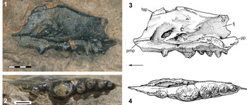
Right maxilla of Micromenodon pitti n. gen. n. sp. (USNM PAL 770096, holotype), photographs and drawings in (1, 3) lingual (=medial) view and (2, 4) occlusal view. Scale bars = (1) 2 mm and (2) 1 mm. Abbreviations: fap = facial (=dorsal) process of maxilla; fec = facet for ectopterygoid; fj = facet for jugal; fpl = facet for palatine; pf = palatine foramen; pmp = premaxillary process; pp = posterior process of maxilla. Arrow points in anterior direction.

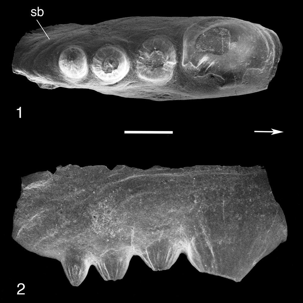
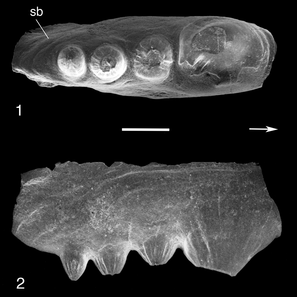
Fragment of right maxilla of Micromenodon pitti n. gen. n. sp. (USNM PAL 770098) in (1) occlusal view and (2) labial (=lateral) view. Scale bar = 400 μm. Abbreviation: sb = secondary bone. Arrow points in anterior direction.
Holotype
USNM PAL 770096, a nearly complete right maxilla with six teeth (Fig. 2).
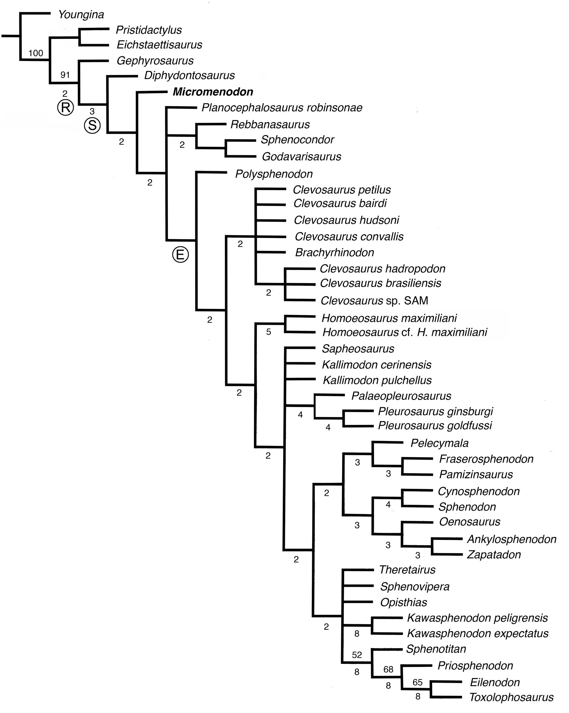
Strict consensus tree of Rhynchocephalia for 10 most-parsimonious trees generated by parsimony analysis with TNT to illustrate the phylogenetic position of Micromenodon n. gen. (highlighted in bold-face type). Character-taxon matrix modified from Hsiou et al. (Reference Hsiou, Nydam, Simões, Pretto, Onary, Martinelli, Liparini, de Vivar Martínez, Soares, Schultz and Caldwell2019) (see Supplemental Data). Numbers above branches indicate bootstrap frequencies greater than 50% and numbers below branches are Bremer Support values greater than one. Circled letters denote major nodes: E = Eusphenodontia; R = Rhynchocephalia; S = Sphenodontia.

Diagnosis
Maxilla with dorsoventrally deep facial process that extends for most of the anteroposterior length of bone*. Dentition fully acrodont. Presence of series of broad-based, obtusely conical additional teeth with pronounced radial ridging and lacking flanges*. Anterior tooth of additional series elliptical in outline, distinctly larger than more rounded teeth posterior to it. (* marks autapomorphy)
Occurrence
Upper Triassic (upper Carnian, Tuvalian); east-central Virginia, United States of America.
Description
The well-preserved holotypic maxilla is exposed in medial (lingual) view. It only lacks a few small pieces of bone, mostly along the anterior margin. The element is 9.4 mm long. It has an anteroposteriorly wide and dorsoventrally tall, nearly vertical facial (=nasal) process, which is much wider relative to the total length of the maxilla than in most other known early Mesozoic rhynchocephalians (compare Jones, Reference Jones2008, fig. 2). In the latter and in Sphenodon punctatus, the facial process arises more anteriorly, lending the maxilla an ‘L’ shape in side view. The posterior edge of the facial process is concave dorsoventrally. Just ventral to this concave edge there is a second, much smaller concavity. The maxilla has a distinct, mediolaterally slightly flattened anterior process that contacted the premaxilla; there is no facet for a long posterior process of the premaxilla as in Clevosaurus spp. (Fraser, Reference Fraser1988). The anterior edge of the holotypic maxilla is damaged but, based on the phylogenetic position of M. pitti n. gen. n. sp. (see below), it probably formed the posterior margin of the external narial fenestra. Posteriorly, the maxilla forms a robust process that extends beyond the posterior end of the tooth row and, based on comparison with Sphenodon punctatus (Jones et al., Reference Jones, Curtis, Fagan, O'Higgins and Evans2011, fig. 20), contacted the ectopterygoid and jugal. The process is not laterally bowed as in various other sphenodontians. The areas for sutural contact with the nasal and prefrontal on the medial surface of the facial process are not clearly delineated. The tooth-bearing portion of the maxilla forms a robust medial shelf. Dorsal to this shelf, there are a well-defined area served for contact with the palatine (as in Sphenodon punctatus; Jones et al., Reference Jones, Curtis, Fagan, O'Higgins and Evans2011, fig. 20) and a large foramen for passage of the superior alveolar nerve and associated vessels (=palatine foramen of authors).
The teeth are indistinguishably fused to the apical crest of the maxilla. This acrodont mode of tooth attachment was confirmed by micro-CT scans. Most successional and hatchling teeth presumably had been already obliterated by wear. An anterior conical tooth represents a remnant of the successional teeth and is separated by a short gap from a posterior series of five additional teeth (terminology following Robinson, Reference Robinson, Bellairs and Cox1976) with broad-based, obtusely conical crowns that bear pronounced, radially arranged apicobasal ridges labially and lingually. Of the latter, the anterior tooth is at least twice as large as each of the four teeth behind it. It is longer mesiodistally than wide labiolingually in occlusal view, whereas the two dimensions are more equal on the more posterior teeth. The latter decrease in size and become slightly more lingually displaced toward the posterior end of the tooth row in the holotype. The apices of all additional teeth are damaged, but none of the teeth has any traces of flanges, unlike in most other rhynchocephalians, nor apical troughs (as in Deltadectes elveticus Whiteside, Duffin, and Furrer, Reference Whiteside, Duffin and Furrer2017). Radial ridging of the tooth crowns is also variously developed mainly on additional teeth in other rhynchocephalians (e.g., Planocephalosaurus robinsonae Fraser, Reference Fraser1982; P. lucasi Heckert, Reference Heckert2004; Deltadectes elveticus) although it appears to be particularly pronounced in Micromenodon pitti n. gen. n. sp.
USNM PAL 770098 is the posterior portion of a right maxilla with four additional teeth (Fig. 3). The first of these teeth is much larger than the three teeth behind it and is longer mesiodistally than wide labiolingually. The three posterior teeth, especially the posteriormost, are more conical than those in USNM PAL 770096. The maxillary fragment has a slight band of secondary bone labial to the teeth. It represents a slightly smaller individual than the holotype with four rather than five additional teeth.
Etymology
The species name recognizes Kenneth A. Pitt for his indefatigable assistance in the field.
Hypodigm
Holotype and USNM PAL 770098, posterior portion of a right maxilla with four teeth (mounted and coated for examination under a scanning electron microscope; Sues et al., Reference Sues, Olsen, Kroehler, Fraser and Sues1994, fig. 7A,B; Fig. 3).
Remarks
As for the genus.
Phylogenetic position and discussion
In order to assess the phylogenetic position of Micromenodon n. gen. among Rhynchocephalia, the features of the available material for this taxon (see Appendix) were scored in the character-taxon matrix by Hsiou et al. (Reference Hsiou, Nydam, Simões, Pretto, Onary, Martinelli, Liparini, de Vivar Martínez, Soares, Schultz and Caldwell2019), which itself is based on the matrix compiled by Apesteguía et al. (Reference Apesteguía, Gómez and Rougier2014). Two characters (54 and 69) proved phylogenetically uninformative. First, the modified matrix (comprising 44 taxa and 73 characters) was analyzed using TNT 1.1. A heuristic search of 1,000 replications of Wagner trees (random addition sequence) was followed by the application of the tree bisection and reconnection (TBR) branch-swapping algorithm (holding 10 trees per replicate). The analysis yielded 10 most-parsimonious trees under the Traditional Search option (TL = 258 steps, CI = 0.4147; RI = 0.691). All consensus trees found Micromenodon n. gen. crownward of Diphydontosaurus and as the sister-taxon to all other known sphenodontians.
The maxillary dentition of Micromenodon pitti n. gen. n. sp. resembles that of the extant scincid Cyclodomorphus gerrardii (Gray, Reference Gray1845) from Australia, which also has a single greatly enlarged maxillary tooth followed by a few smaller teeth posteriorly. Although the diet of this lizard is varied and includes diverse invertebrates as well as fruit, it frequently employs its massive teeth to crush the shells of snails (Estes and Williams, Reference Estes and Williams1984). Thus, it is plausible that the enlarged maxillary teeth of M. pitti n. gen. n. sp. were similarly used for crushing hard objects such as arthropod exoskeletons and snail shells. The pronounced apicobasal ridges on the tooth crowns likely strengthened the teeth during powerful biting.
The Late Triassic to Early Jurassic basal rhynchocephalian Gephyrosaurus spp. (Evans, Reference Evans1980; Whiteside and Duffin, Reference Whiteside and Duffin2017) and the Late Triassic sphenodontians Diphydontosaurus avonis Whiteside, Reference Whiteside1986 and Planocephalosaurus spp. (Fraser, Reference Fraser1982; Heckert, Reference Heckert2004) have conical tooth crowns suitable for piercing (Jones, Reference Jones2009). More derived sphenodontian taxa have tooth crowns that are functionally suitable either for cutting and slicing (e.g., Clevosauridae, Sphenodon punctatus) or for shredding and grinding (e.g., Opisthodontia) (Jones, Reference Jones2008, Reference Jones2009; Martínez et al., Reference Martínez, Apaldetti, Colombi, Praderio, Fernandez, Malnis, Correa, Abelin and Alcober2013). The maxillary teeth of Micromenodon pitti n. gen. n. sp. resemble the conical teeth in basal sphenodontians more closely in overall structure than those of the more derived taxa. Among known sphenodontians, they are comparable to those of Planocephalosaurus robinsonae, which has a large additional tooth in the maxilla followed by three small teeth posteriorly (Fraser, Reference Fraser1982; personal observation). However, the maxillary teeth of Micromenodon pitti n. gen. n. sp. differ from those of P. robinsonae in the absence of posterolingual flanges, and the teeth of the latter and of P. lucasi typically have slightly recurved, finely ridged crowns. Furthermore, the anterior teeth of the more derived P. robinsonae retain pleurodont rather than acrodont implantation (Fraser and Shelton, Reference Fraser and Shelton1988; Jenkins et al., Reference Jenkins, Jones, Zikmund, Boyde and Daza2017), suggesting that the presence of a fully acrodont dentition in M. pitti n. gen. n. sp. is convergent.
Micromenodon pitti n. gen. n. sp. is the oldest known sphenodontian from the Triassic of eastern North America to date and represents a previously unknown dental morphotype among early Mesozoic sphenodontians.
Acknowledgments
Many thanks to E.B. Sues, P.A. Kroehler, and K.A. Pitt for their invaluable assistance in the field. J. Morrison prepared the drawings for Figure 2.3 and 2.4. S. Whittaker prepared the SEM photographs and S. Jabo and P. Tardelli Canedo photographed the holotype. A.S. Hsiou provided the NEXUS file for her 2019 paper. D.I. Whiteside shared images of Triassic sphenodontian jaws for comparisons. N.C. Fraser and an anonymous reviewer provided helpful comments on a draft of this paper. The software TNT is freely available courtesy of the Willi Hennig Society. Support for the fieldwork was provided by grants from the National Science Foundation and National Geographic Society to H.-D.S. and P.E. Olsen.
Appendix
Character-states for Micromenodon pitti n. gen. n. sp. for the character-taxon matrix by Hsiou et al. (Reference Hsiou, Nydam, Simões, Pretto, Onary, Martinelli, Liparini, de Vivar Martínez, Soares, Schultz and Caldwell2019).
?????0?0??????????????????????0??????????21??00??00????????????????????12