Introduction
Elevated ambient temperatures result in heat stress, a major concern for agriculture production in tropical and subtropical countries worldwide due to its negative effect on reproduction (Ealy et al., Reference Ealy, Howell, Monterroso, Arechiga and Hansen1995). Egypt boasts a subtropical climate. Buffaloes, the principal dairy animal of Egypt, contribute over 60% of the milk production in this country (Abou-Bakr, Reference Abou-Bakr2008) and 45% of meat production (Ibrahim, Reference Ibrahim2012). Although buffaloes are well adapted to hot and humid climatic conditions, they are more susceptible to heat stress than cattle (Manjari et al., Reference Manjari, Yadav, Ramesh, Uniyal, Rastogi, Sejian and Hyder2015) due to scarcely distributed sweat glands, dark body colour and sparse hair on their body surface (Das et al., Reference Das, Upadhyay and Madan1999). Anestrus, longer intercalving periods, silent heat, poor conception rate, and high incidence of early embryonic death are major constraints on reproductive performance and productivity in buffaloes under heat stress conditions. Studies in cattle suggest that oocytes are directly susceptible to heat stress in addition to changes in the follicular gonadotropic environment (Ashraf et al., Reference Ashraf, Shah, Saini, Dhanda, Singh and Chauhan2014). Heat stress may indirectly affect the follicular surge of gonadotropins (Badinga et al., Reference Badinga, Thatcher, Diaz, Drost and Wolfenson1993) or may directly affect oocyte quality (Nandi et al., Reference Nandi, Chauhan and Palta2001). Studies in buffaloes from different parts of the world have examined the effects of seasonal fluctuation, with conflicting results (Nandi et al., Reference Nandi, Chauhan and Palta2001; Khairy et al., Reference Khairy, Zoheir, Mahrous, Amer, Zaher, Li-Guo and El-Nahass2007).
In buffaloes, reduced fertility during the summer months is mostly due to hyperthermia. There are several possible mechanisms by which heat stress may affect buffalo reproduction. One mechanism that causes infertility is reduction in oocyte developmental competence. During the early stages of folliculogenesis, oocyte damage from heat stress has been indicated as ovarian recovery from summer thermal stress requires two to three oestrous cycles before competent oocytes are present (Roth et al., Reference Roth, Arav, Bor, Zeron, Braw-Tal and Wolfenson2001). Oocytes recovered during the summer have exhibited lower quality (Rutledge et al., Reference Rutledge, Monson, Northey and Leibfried-Rutledge1999; Al-Katanani et al., Reference Al-Katanani, Paula-Lopes and Hansen2002), and reduced developmental potential (Rutledge et al., Reference Rutledge, Monson, Northey and Leibfried-Rutledge1999; Al-Katanani et al., Reference Al-Katanani, Paula-Lopes and Hansen2002; Paula-Lopes et al., Reference Paula-Lopes, Milazzotto, Assumpcao and Visintin2008) and pre-implantation embryonic development (Paula-Lopes & Hansen, Reference Paula-Lopes and Hansen2002). In buffaloes, elevated temperatures in the summer reduced the yield of good quality oocytes, maturation and cleavage rates and yield of embryos that developed to the blastocyst stage (Sadeesh et al., Reference Sadeesh, Sikka, Balhara and Balhara2016). In addition, Abdoon et al. (Reference Abdoon, Gabler, Holder, Kandil and Einspanier2014) found that higher temperatures during spring and summer had a detrimental effect on oocyte maturation and increased the percentage of degenerated oocytes compared with oocytes in autumn and winter; the percentage of oocytes arrested at the 2-cell stage was also high. Heat stress during the summer induced a decrease in oocyte function due to a sequence of cellular changes that altered nuclear and cytoplasmic compartments of the oocyte, and in turn destroyed intracellular events associated with both nuclear and cytoplasmic maturation (Payton et al., Reference Payton, Lawrence, Saxton, Dunlap and Edwards2003; Maya-Soriano et al., Reference Maya-Soriano, López-Gatius, Andreu-Vázquez and López-Béjar2013). Furthermore, there is evidence that oocyte cytoplasm is more susceptible to the adverse effects of increased temperature than the nucleus (Shen et al., Reference Shen, Lee, Cheng, Su, Lee and Liu2010).
Various studies have focused on the effect of elevated temperatures on oocyte competency and embryo development through in vivo experiments following exposure of heifers to heat stress between the onset of estrous and insemination. Scientists have started to study the effect of heat stress on oocyte competency and embryo development in vitro. In bovine, various experiments examined the effects of heat stress on in vitro development of oocytes (Roth & Hansen, Reference Roth and Hansen2004) and different developmental stages (Paula-Lopes & Hansen, Reference Paula-Lopes and Hansen2002; Jousan & Hansen, Reference Jousan and Hansen2004; Sakatani et al., Reference Sakatani, Kobayashi and Takahashi2004; De Castro e Paula & Hansen, Reference De Castro e Paula and Hansen2008; Sakatani et al., Reference Sakatani, Yamanaka, Kobayashi and Takahashi2008). The molecular and cellular mechanisms of heat-induced reduced buffalo oocyte competence and their effect on embryo viability in vitro are poorly understood (Ashraf et al., Reference Ashraf, Shah, Saini, Dhanda, Singh and Chauhan2014) and few data regarding this issue have been published. The first report was released by Yadav et al. (Reference Yadav, Singh, Singh, Saini, Palta, RS Manik, SK Singla, Upadhyayand and Chauhan2013) in which they exposed buffalo oocytes and embryos to elevated temperatures of 39.5 and 40.5°C. They found a compromised effect of heat stress on embryo development and altered relative mRNA abundance of the stress-related genes for heat shock protein (HSP) 70.1 and HSP 70.2, pro-apoptotic genes CASPASE-3, BID and BAX and anti-apoptotic genes BCL-XL and MCL-1. Another study by Ashraf et al. (Reference Ashraf, Shah, Saini, Dhanda, Singh and Chauhan2014) examined the direct effect of physiologically relevant high temperatures (40.5 and 41.5°C) for two time periods (12 and 24 h) on bubaline oocytes during in vitro maturation. They found that the cleavage rates, blastocyst yield and mean cell number decreased markedly. Overall these were currently the only published data on the effect of heat stress on buffalo embryos in vitro at the developmental and molecular level.
In vitro conditions such as heat stress usually increase the production of reactive oxygen species in bubaline oocytes and have been implicated as a major cause of reduced developmental competence. Ashraf et al. (Reference Ashraf, Raies-ul-Haq, Dhanda, Kumar, Sridhar, Chauhan and Upadhyay2016) studied the effects of elevated temperature and heat stress during meiotic maturation on bubaline cumulus–oocyte complexes, denuded oocytes, and cumulus cell mass in terms of their oxidative status. Their results indicated that the production of reactive oxygen species, lipid peroxides, and nitric oxide was significantly higher in oocytes subjected to heat stress (40.5 and 41.5°C). Furthermore, the antioxidant enzymatic activities of superoxide dismutase, catalase, glutathione peroxidase, and glutathione reductase were significantly increased in all treatment groups compared with the control group.
Studies were carried out to identify the causes of heat-induced reduced developmental competence of bubaline oocytes, none of these studied the relationship between elevated temperature on oocytes and their cumulus cells at the molecular level. The aims of the current study were to examine the effect of heat shock on the developmental competence of oocytes and to describe its effects on the quantitative expression of some genes related to heat shock protein (HSP-70, HSP-90 and HSF-1), pro-apoptotic proteins (BAX and p53), oxidative stress proteins (SOD1 and COX1) and mediator of extracellular signals (MAPK14) in denuded oocytes and their isolated cumulus cells using quantitative real-time polymerase chain reaction (qRT-PCR).
Materials and Methods
Chemicals and reagents
All reagents and medium used in the current study were obtained from Sigma-Aldrich (St. Louis, MO, USA) unless otherwise mentioned.
Experimental design
This study was carried out in two experiments. The first experiment studied bubaline oocyte competency and embryo development up to the blastocyst stage under different levels of in vitro heat stress. In this experiment, the groups (average 250 oocyte/group) were immature oocytes exposed to 38.5°C and considered as the control (C), plus oocytes at 39.5°C called treatment 1 (T1) or at 40.5°C for treatment 2 (T2) for the first 6 h of in vitro maturation (IVM), followed by 38.5°C over the next 18 h/IVM, and early embryonic development up to the blastocyst stage. In the second experiment, the mRNA expression of genes related to heat shock protein (HSF-1, HSF-2, HSP-70 and HSP-90), pro-apoptotic proteins (BAX and p53), oxidative stress proteins (SOD1 and COX1) and mediator of extracellular signals (MAPK14) in denuded oocytes and their isolated cumulus cells were investigated by quantitative real-time PCR (qRT-PCR).
Collection of ovaries and recovery of oocytes
Buffalo ovaries were collected from nearby abattoirs and transported to the laboratory within 4 h in warmed (30°C) physiological saline (0.9% NaCl) fortified with (100 µg/ml streptomycin sulfate and 100 IU/ml penicillin). Ovaries were washed once in 70% ethanol and three times in phosphate-buffered saline (PBS). Cumulus–oocyte complexes (COCs) were collected by aspiration of antral follicles (2–8 mm in diameter) with an 18-gauge needle attached to a 10-ml syringe containing the aspiration (HEPES-buffered) medium 199 (medium 199; 22340; Gibco, UK) containing 25 mM 4-(2-hydroxyethyl)-1-piperazine ethanesulfonic acid (HEPES), Earle's salts, l-glutamine and non-essential amino acids supplemented with 100 µg/ml streptomycin sulfate and 100 IU/ml penicillin. The COCs were then washed three times with washing medium (HEPES-buffered) medium 199 (medium 199; 22340; Gibco, UK) supplemented with 0.73 µg/ml sodium bicarbonate, 0.05 µg/ml gentamicin, 0.23 µg/ml sodium pyruvate, 1.27 µg/ml HEPES, 0.55 µg/ml calcium lactate and 0.3 cysteine + 10% inactivated fetal bovine serum (FBS).
Oocytes with uniform cytoplasm and multi-layered cumulus cells were selected for further processing.
In vitro maturation
Oocytes were washed three times in the maturation medium consisting of medium 199 (31150; Gibco, UK) containing Earle's salts, l-glutamine, supplemented with 10% inactivated (56°C/20 min) FBS, 0.02 IU/ml FSH, 1 µg/ml estradiol 17 β (E2), 22 µg/ml Na-pyruvate, 0.3 mM cystine and 50 µg/ml gentamycin. Groups of COCs (25–30/well) were cultured in 400 µl of maturation medium overlaid with sterile 400 µl mineral oil in 4-well plates under a 5% CO2 in air atmosphere with high humidity. COCs matured at 38.5°C for 24 h was designated as the control group. Heat treatment was given to two groups of COCs at 39.5 and 40.5°C for 6 h of maturation and subsequently matured at 38.5°C for the remaining 18–24 h.
In vitro fertilization and in vitro culture
Developmental competence of mature oocytes in the control, and T1 and T2 groups were evaluated after maturation by fertilization and early embryonic development up to the blastocyst stage. The expanded COCs were recorded for maturation data, afterwards washed three times in Fert-TALP medium, placed into the sperm suspension (25–30 oocytes/well) then kept at 38.5°C in a Multi-gas incubator (5% CO2 and 5% O2) for 18 h. The spermatozoa were processed for IVF as described earlier (Ghanem & El-Sayed, Reference Ghanem and El-Sayed2012) with some modifications. Briefly, one straw of frozen semen from Egyptian buffalo bulls tested for IVF earlier was thawed at 37°C for 30 s. Afterwards, sperm were washed twice in sperm Tyrode's Albumin Lactate Pyruvate medium (Sperm-TALP) containing 2.2 mg/ml sodium pyruvate, 10 μg/ml heparin and bovine serum albumin (BSA) F-V (6 µg/ml) + 50 μg/ml gentamycin using a centrifuge adjusted to 500 g for 10 min. After washing, the sperm pellets were suspended in 0.5 ml of fresh Fert-TALP medium supplemented with 6 µg/ml BSA (fatty acid free) + 10 μg/ml heparin + 3 μl penicillamine–hypotaurine–epinephrine (PHE) and 50 μg/ml gentamycin. Sperm concentration was adjusted to 2 × 10⁶ spermatozoa/ml. In total, 400 μl of motile sperm suspension was placed in a 4-well culture plate and overlaid with 400 μl sterile mineral oil. The presumptive zygotes obtained after IVF were denuded by gentle repetitive pipetting, washed 3–4 times with in vitro culture (IVC) medium [modified synthetic oviductal fluid (mSOFaa)] supplemented with 5 mg/ml BSA + 5 ng/ml insulin and 50 μg/ml gentamycin and subsequently cultured in the same medium for 9 days post-insemination at 38.5°C in a humidified Multigas CO2 incubator. The medium was replaced with 50% of fresh IVC medium every 48 h. The cleavage rate was checked on day 2 post-insemination, and the percentage of oocytes that developed to the 8–16-cell stage and blastocyst stage was recorded on days 4 and 7 post-insemination.
RNA isolation and cDNA synthesis
After 24 h of maturation, COCs were denuded from cumulus cells by gentle pipetting in maturation medium supplemented with 1 mg/ml hyaluronidase. Oocyte maturation, as determined by the presence of the first polar body extruded was recorded. The denuded oocytes and the cumulus cell mass from control and T1 groups [three replicates of denuded oocytes (60 oocytes/pool) and three replicates from its counterparts cumulus cells] were washed twice in PBS and snap frozen separately in cryo-tubes containing 20 µl of lysis buffer [0.8% IGEPAL, 40 U/µl RNasin (Promega Madison WI, USA), 5 mM dithiothreitol (DTT) (Promega Madison WI, USA)]. Finally, samples were stored in liquid nitrogen at −196°C until RNA isolation.
Total RNA was isolated using the PicoPure™ RNA isolation kit (MDS Analytical Technologies GmbH, Ismaning, Germany) according to manufacturer's instructions. For each sample, cDNA synthesis was performed using an oligo(dT) 23 primer, random primer and superscript reverse transcriptase II (Invitrogen, Karlsruhe, Germany). One microlitre of oligo(dT) 23 primer and 1 μl random primer were added to the 10 μ1 RNA sample and the mixture was incubated for 3 min at 70°C and then immediately chilled on ice. Eight microlitres of the master mix containing 4 μl of 5× first strand buffer, 2 μl of 0.1 M DTT, 1 μl of dNTP (10 pmol/μl) and 0.3 μl of RNase inhibitor and 0.7 μl of Super Script II™ reverse transcriptase (200 unit/μl) were added to the mixture and incubated for 90 min at 42°C followed by heat inactivation for 15 min at 70°C. The synthesized cDNA was stored at −20°C for further use.
Quantitative real-time PCR
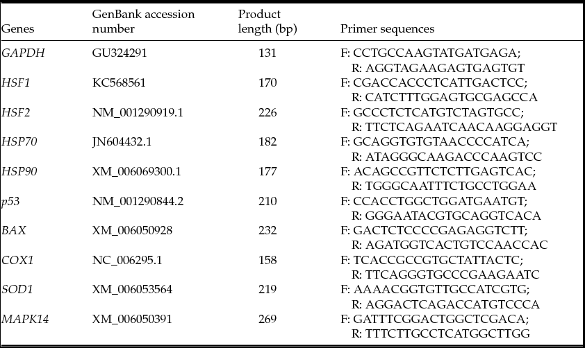
Gene-specific primers were designed with a target to amplify fragments of approximately 150–220 bp. The primers were designed using PRIMER 3 Software (http://www-genome.wi.mit.edu/cgi-bin/prime/primer3-www.cgi). Real-time PCR primers were designed based on the cDNA sequences of the nine transcripts available in GenBank (Table 1). Quantitative analyses of cDNA samples from independent oocytes and cumulus cells were performed in comparison with the GAPDH gene (endogenous control), and were run in separate wells using Step One Plus real-time PCR system (Applied Biosystems). Independent qRT-PCR (three replicates for each group) was performed in a 20-μl reaction volume containing iTaq SYBR Green master mix with ROX (Bio-Rad Laboratories, Munich, Germany), the cDNA samples and the specific forward and reverse primers. The thermal cycling parameters were set at 95°C for 10 min, 40 cycles of 95°C for 15 s and 60°C for 1 min. After the end of the last cycle, a dissociation curve was generated by starting fluorescence acquisition at 60°C and taking measurements every 7-s interval until the temperature reached 95°C. The comparative cycle threshold (Ct) method was used to quantify expression levels as previously described (Yadav et al., Reference Yadav, Singh, Singh, Saini, Palta, RS Manik, SK Singla, Upadhyayand and Chauhan2013). Briefly, during data analysis, the Ct value of the housekeeping gene was subtracted from the Ct value of the target gene to obtain change in Ct (ΔCt). The ΔCt value of the target gene sample was subtracted from the calibrator (control) to get ΔΔCt values. Difference in the transcript abundance for the target genes was calculated using the equation 2−ΔΔCt.
Details of primers used for quantitative real-time PCR

Statistical analysis
Statistical analysis for maturation rate and embryonic development was performed using one-way analysis of variance (ANOVA) and SPSS 19.0 software. Data are presented as means and standard errors (mean ± SE), significance was determined at P-value < 0.01.
Gene expressions transcripts were analysed using real-time PCR, fold changes were calculated using the ∆∆Ct method and significance was tested by T-test at P < 0.05.
Results
First experiment
Effect of heat shock on maturation rate of oocytes
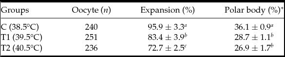
Our results indicated that culture at 39.5 (T1) and 40.5°C (T2) reduced the number of expanded COCs and the numbers of oocytes that had a discernible polar body compared with the 38.5°C (control). The percentage of mature oocytes decreased with increase in maturation temperature. It was found that maturation rate based on cumulus cells expansion decreased in T1 (83.4 ± 3.9%) and T2 (72.7 ± 2.5%) compared with the control group (95.9 ± 3.3%). In addition, the effect of heat shock persisted for nuclear maturation rate, which decreased in T1 (28.7 ± 1.1%) and T2 (26.9 ± 1.7%) groups compared with 36.1 ± 0.9% in the control group. Therefore, a decrease in the percentage of oocytes that progressed to metaphase II after 24 h of maturation was observed at higher temperatures (39.5 or 40.5°C) compared with oocytes matured at optimal temperature (38.5°C), as shown in Table 2.
Effect of heat shock on buffalo oocyte maturation rate [mean ± standard error of the mean (SEM)]

Superscripts a and b within the same column differ significantly at P < 0.01.
*Evaluated after 24 h.
Effect of heat shock on the developmental competence of oocytes
The cleavage rate on day 2, and the percentage of embryos that developed to the 8–16-cell stage on day 4 and to blastocyst stage on day 7 post-insemination (Table 3) was significantly lower (P < 0.01) for oocytes exposed to 39.5°C or 40.5 for 6 h of IVM compared with those for the control. These results showed that the percentage of cleaved embryos arrested at the 2- and 4-cell stages was higher (P < 0.01) in matured oocytes under elevated temperatures (39.5 or 40.5°C) compared with the control 38.5°C. Not a single oocyte exposed to 40.5°C developed to the blastocyst stage.
Effect of heat shock on cleavage rate and the development of in vitro produced buffalo embryos (mean ± SE)

Superscripts a and b within the same column differ significantly at P < 0.01.
*Based on cumulus cells expansion.
**Calculated as % of mature oocytes.
***Calculated as % of cleavage embryos.
Second experiment
Analysis of mRNA expression
Heat shock-related genes
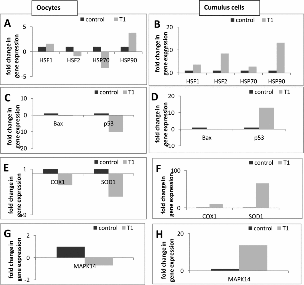
The effect of elevated temperature (39.5°C) during maturation on mRNA expression of heat shock-related genes (HSF-1, HSF-2, HSP-70 and HSP-90) of mature oocytes and their counterpart cumulus cells is depicted in Fig. 1A, B.
Relative abundance of heat shock protein genes (HSF-1, HSF-2, HSP-70, and HSP-90) in bubaline denuded oocytes and their counterpart cumulus cells (A, B respectively) in the control and heat-treated groups (T1). The relative abundance of pro-apoptotic (BAX and p53), oxidative stress (SOD1 and COX1) and mediator of extracellular signals (MAPK14) in denuded oocytes (C, E, G respectively) and its isolated cumulus cells (D, F, H respectively) as affected by heat shock (T1) compared with control. Control group represents the optimal maturation conditions (38.5°C for 24 h) and T1 represents COCs matured at 39.5°C for 6 h followed by 38.5°C through the next 18 h/IVM.

Results showed an increase (P < 0.05) in mRNA expression of HSF-1 and HSP-90 genes and a decrease (P < 0.05) in expression of HSF-2 and HSP-70 genes in the treated group compared with the control, as shown in Fig. 1A. Conversely, it was found that mRNA expression of heat shock-related genes (HSF-1, HSF-2, HSP-70 and HSP-90) increased significantly (P < 0.05) in cumulus cells isolated from treated COCs (T1) compared with the control as shown in Fig. 1B.
Pro-apoptotic and oxidative stress-related genes
In the current study, we observed a significantly lower (P < 0.05) mRNA abundance of a pro-apoptosis related gene (p53), however downregulation of BAX gene was not significant for oocytes matured at higher temperatures (39.5°C) compared with control, as shown in Fig. 1C. In the corresponding cumulus cells, the BAX gene followed the same trend but in contrast p53 showed an opposite trend as levels were greatly increased (P < 0.05) in the treated group compared with the control (Fig. 1D). The same pattern (P < 0.05) was observed for oxidative stress (SOD1 and COX1) genes in treated oocytes (Fig. 1E). For cumulus cells, both SOD1 and COX1 were upregulated (P < 0.05) in cumulus cells isolated from treated COCs (T1) compared with the control, as shown in Fig. 1F. The results showed high transcript abundance of SOD1 (65.9-fold change) in the cumulus cells of the treated group (Fig. 1F).
Discussion
Fertility is a multi-factorial condition and its retrogradation is related to genetic and environmental factors. High environmental temperatures during the summer are a major constraint for the production and fertility of agricultural animals worldwide. To maintain the ‘internal milieu’ under stressful conditions, numbers of physiological and behavioural changes occur that vary in duration and intensity of environmental stress (Das et al., Reference Das, Upadhyay and Madan1999), particularly for fertility. When exposed to heat stress of sufficient magnitude, mammalian females experience infertility, demonstrated for large domestic animals like cattle and buffalo (Yadav et al., Reference Yadav, Singh, Singh, Saini, Palta, RS Manik, SK Singla, Upadhyayand and Chauhan2013; Ashraf et al., Reference Ashraf, Shah, Saini, Dhanda, Singh and Chauhan2014). Buffaloes are more susceptible to heat stress than cattle as they are poorly thermoregulated due to their morphological and anatomical characteristics (Das et al., Reference Das, Upadhyay and Madan1999). Heat stress affects their biological functions including reproduction (Marai & Haeeb, Reference Marai and Haeeb2010). Ovaries showing inactivity represent 93% of cases of summer infertility in buffaloes raised in the Middle Egypt, as reported by Ali et al. (Reference Ali, Abdel-Razek, Derar, Abdel-Rheem and Shehata2009). The core body temperature of buffaloes ranges from 38.5 to 39°C and may reach up to 41.5°C on exposure to solar radiation during the summer (Sethi et al., Reference Sethi, Bharadwaj and Chopra1994; Aggarwal & Upadhyay, Reference Aggarwal and Upadhyay1998). For local breeds such as Egyptian buffaloes, internal temperature as indicated by rectal assessments ranged from 38 to 41°C with most around 39°C during the summer in hot regions in (Upper) Egypt (Taqi Reference Taqi2012), therefore it was appropriate to study the effect of heat shock at 39°C at the molecular level. Effects of elevated temperatures manifested as heat stress are particularly prominent during oestrus (day 1) when the oocyte is undergoing meiotic maturation in preparation for fertilization (Gwazdauskas et al., 1973, Cavestany et al., 1985). Reduced quality of buffalo oocytes was reported in summer and autumn compared with winter and spring (Zoheir et al., Reference Zoheir, Abdoon, Mahrous, Amer, Zaher and Li Guo2007). Furthermore, heat stress during the summer induced a decrease in oocyte function as a result of a series of cellular alterations affecting nuclear and cytoplasmic compartments of the oocyte (Paula-Lopes et al., Reference Paula-Lopes, Lima, Risolia, Ispada, Assumpção and Visintin2012). Therefore many studies have been carried out on bovine oocytes related to heat stress to find the cause behind these catastrophic effects on oocyte competency (Payton et al., Reference Payton, Romar, Coy, Saxton, Lawrence and Edwards2004; Ju et al., Reference Ju, Jiang, Tseng, Parks and Yang2005; Roth & Hansen, Reference Roth and Hansen2005; Paula-Lopes et al., Reference Paula-Lopes, Milazzotto, Assumpcao and Visintin2008; Nabenishi et al., Reference Nabenishi, Takagi, Kamata, Nishimoto, Morita, Ashizawa and Tsuzuki2012; Maya-Soriano et al., Reference Maya-Soriano, López-Gatius, Andreu-Vázquez and López-Béjar2013, Pavani et al., Reference Pavani, Baron, Correia, Lourenço and Bettencourt2016). To date, the availability of published data on the effect of heat stress on bubaline oocytes and embryos has been very limited (Khairy et al., Reference Khairy, Zoheir, Mahrous, Amer, Zaher, Li-Guo and El-Nahass2007; Yadav et al., Reference Yadav, Singh, Singh, Saini, Palta, RS Manik, SK Singla, Upadhyayand and Chauhan2013, Abdoon et al., Reference Abdoon, Gabler, Holder, Kandil and Einspanier2014; Ashraf et al., Reference Ashraf, Shah, Saini, Dhanda, Singh and Chauhan2014, Reference Ashraf, Raies-ul-Haq, Dhanda, Kumar, Sridhar, Chauhan and Upadhyay2016; Sadeesh et al., Reference Sadeesh, Sikka, Balhara and Balhara2016) and there is no literature available investigating the mechanisms by which heat shock can cause such catastrophic effects on buffalo COCs. Therefore, it appears necessary to define the molecular mechanisms that identify potential local effects of elevated heat temperature on buffalo cumulus–oocyte complexes (COCs) and oocyte developmental competence. A better understanding of the molecular mechanisms underlying how heat stress could impair oocytes’ developmental competence may lead to the development of additional approaches to alleviate these effects and subsequently increase the fertility during hot seasons. Therefore, we report for the first time the developmental changes and molecular alterations in bubaline oocytes and their cumulus cell counterparts with physiologically relevant elevated temperatures that manifest in the form of heat stress.
In the present study, it was found that exposure of buffalo COCs to higher temperatures (39.5 and 40.5°C) during in vitro maturation (first 6 h only) resulted in a significant decrease (P < 0.01) in oocyte maturation rate based on cumulus cells expansion and first polar body extrusion.
Heat stress during maturation may induce a decrease in oocyte function due to a sequence of cellular changes that affect nuclear and cytoplasmic compartments of the oocyte and which in turn affect intracellular events associated with both nuclear and cytoplasmic maturation. Our results are in agreement with previous studies in bovine (Payton et al., Reference Payton, Lawrence, Saxton, Dunlap and Edwards2003, Paula-Lopes et al., Reference Paula-Lopes, Lima, Risolia, Ispada, Assumpção and Visintin2012; Maya-Soriano et al., Reference Maya-Soriano, López-Gatius, Andreu-Vázquez and López-Béjar2013) and buffaloes (Khairy et al., Reference Khairy, Zoheir, Mahrous, Amer, Zaher, Li-Guo and El-Nahass2007; Abdoon et al., Reference Abdoon, Gabler, Holder, Kandil and Einspanier2014; Ashraf et al., Reference Ashraf, Raies-ul-Haq, Dhanda, Kumar, Sridhar, Chauhan and Upadhyay2016; Sadeesh et al., Reference Sadeesh, Sikka, Balhara and Balhara2016). Heat stress impairs some intracellular events associated with maturation, such as alteration phospholipids composition of oocytes (Zeron et al., Reference Zeron, Ocheretny, Kedar, Borochov and Skla Arav2001), translocation of cortical granules to the oolemma (Payton et al., Reference Payton, Lawrence, Saxton, Dunlap and Edwards2003), cytoskeletal rearrangement (Roth & Hansen, Reference Roth and Hansen2005), and spindle formation (Ju et al., Reference Ju, Jiang, Tseng, Parks and Yang2005; Roth & Hansen, Reference Roth and Hansen2005). Edwards et al. (Reference Edwards, Saxton, Lawrence, Payton and Dunlap2005) found the opposite effect when oocytes underwent maturation under heat stress conditions. Their study reported that culture of oocytes at 41.0°C did not compromise their ability to undergo nuclear (i.e., progression to MII) or cytoplasmic maturation (i.e. cortical granule translocation to the oolemma). Heat stress did reduce the time required for these critical processes (i.e., heat-stressed oocytes matured faster than controls) and may be inducing premature ageing of the maturing oocyte which likewise reduces developmental competence (Fissore et al., Reference Fissore, Kurokawa, Knott, Zhang and Smyth2002). Cumulus cells surrounding the oocyte probably have a significant role as they are intimately connected with the oocyte, projecting through the zona pellucida and oolemma to establish an intercellular bidirectional form of communication. Therefore some effects of heat stress to reduce oocyte development may be mediated through the cumulus cells. Lenz et al. (Reference Lenz, Ball, Leibfried, Ax and First1983) showed that prolonged exposure to a physiologically relevant elevated temperature altered cumulus cell function because culture at 41.0°C for 24 h reduced hyaluronic acid production. Therefore crosstalk between cumulus cells and oocyte at molecular level was considered in our current study.
On developmental competence of the oocyte, results obtained from the present study revealed the reduced ability of oocytes to become blastocysts after fertilization compared with the control group. Exposure of buffalo oocytes to higher temperatures (39.5 and 40.5°C) during in vitro maturation (first 6 h) resulted in a significant decrease (P < 0.01) in the cleavage rate and therefore blastocyst production. The percentage of cleaved embryos arrested at the 2- and 4-cell stages was higher (P < 0.01) in matured oocytes under elevated heat temperature (39.5 or 40.5°C) compared with the control 38.5°C. Not a single oocyte exposed to 40.5°C was able to develop to the blastocyst stage.
Our results are in agreement with Ashraf et al. (Reference Ashraf, Shah, Saini, Dhanda, Singh and Chauhan2014) who found that exposure of buffalo oocytes to higher temperatures (40.5 and 41.5°C) during in vitro maturation (first 12 h only or entire 24 h) resulted in a significant decrease in the cleavage rate and in blastocyst production. Similarly, Yadav et al. (Reference Yadav, Singh, Singh, Saini, Palta, RS Manik, SK Singla, Upadhyayand and Chauhan2013) found that physiologically relevant heat shock (40.5°C) to buffalo oocytes or embryos for 2 h once every day throughout IVM, IVF and IVC resulted in a decreased percentage of buffalo oocytes that developed to the 8–16-cell or blastocyst stage. Our results are further in accordance with Tseng et al. (Reference Tseng, Chen, Chou, Yeh and Ju2004) who found decreased developmental competence of porcine embryos at 2 and 4 h at 41.5°C compared with the control group (39°C). Moreover, studies carried out in bovine have also shown similar results, for example, it has been reported that exposure of bovine oocytes to increased temperature (40–41°C for 12 h) reduced the numbers of embryos developing to the blastocyst stage, 4–8-cell embryos and the percentage of cleaved embryos (Edwards & Hansen, Reference Edwards and Hansen1997; Roth & Hansen, Reference Roth and Hansen2004; Jousan & Hansen, 2007; Rispoli et al., Reference Rispoli, Lawrence, Payton, Saxton, Schrock and Schrick2011). Collectively, in vitro studies that evaluated the developmental competence of oocytes exposed to heat stress during maturation support our present data in which a similar pattern, i.e., marked decreased (P < 0.01) in the percentages of buffalo oocytes that developed to 2-cell, 8–16-cell or blastocyst stages was observed. Likewise Mishra et al. (Reference Mishra, Misra and Sharma2007), Abdoon et al. (Reference Abdoon, Gabler, Holder, Kandil and Einspanier2014), and Sadeesh et al. (Reference Sadeesh, Sikka, Balhara and Balhara2016) illustrated that IVM and IVF of buffalo oocytes during the hot season had significantly (P < 0.01) showed decreased cleavage rate and embryo development up to the blastocyst stage.
The present findings, therefore, provide clear evidence that oocyte damage caused by heat shock in vitro is likely to be relevant to the understanding of early embryonic loss in buffalo following heat stress conditions. Therefore high ambient temperature may affect the maturation process and subsequent embryo development at various cell stages and may ultimately cause a negative effect on buffalo reproduction.
Cellular homeostasis in stress conditions is maintained by balancing net growth and death rate of cells (Lockshin & Zakeri, Reference Lockshin and Zakeri2007). Cells tackle adverse conditions by upregulating certain sets of genes and downregulating others.
In the present investigation, we have studied the effects of heat shock on the quantitative expression of some genes related to heat shock protein (HSP-70, HSP-90, HSF-1 and HSF-2), pro-apoptotic proteins (BAX and p53), oxidative stress proteins (SOD1 and COX1) and mediator of extracellular signals (MAPK14) in denuded oocytes and its isolated cumulus cells. Our results showed an increase in mRNA expression of heat shock-related genes (HSP-70, HSP-90, HSF-1 and HSF-2) in both oocytes and cumulus cells except for HSF-2 and HSP-70 genes, with decreased mRNA expression in the oocyte treated (39.5°C) group compared with the control (38.5°C).
Results for HSP-70 in oocytes and cumulus cells are completely in accordance with those obtained by Abdoon et al. (Reference Abdoon, Gabler, Holder, Kandil and Einspanier2014) who stated that the transcript abundance of HSP70 mRNA was significantly lower in mature oocytes collected during the hot season than those collected during the cold season and was affected by elevated temperatures; the opposite trend was observed in COCs. HSP70 transcription may be increased by stressful conditions such as heat shock (Beckham et al., Reference Beckham, Mackanos, Crooke, Takahashi, O'Connell Rodwell and Contag2004; Mishra & Palai, Reference Mishra and Palai2014; Archana et al., Reference Archana, Aleena, Pragana, Vidya, Abdul Niyas, Bagath, Krishnan, Manimaran, Beena, Kurien, Sejian and Bahtta2017). Its expression has been used as an indicator of heat stress in bovine and buffalo embryos (Payton et al., Reference Payton, Rispoli, Saxton and Edwards2011; Yadav et al., Reference Yadav, Singh, Singh, Saini, Palta, RS Manik, SK Singla, Upadhyayand and Chauhan2013, Abdoon et al., Reference Abdoon, Gabler, Holder, Kandil and Einspanier2014; Ashraf et al., Reference Ashraf, Shah, Saini, Dhanda, Singh and Chauhan2014; Manjari et al., Reference Manjari, Yadav, Ramesh, Uniyal, Rastogi, Sejian and Hyder2015; Sadeesh et al., Reference Sadeesh, Sikka, Balhara and Balhara2016). This gene has a pro-apoptotic function and its upregulation indicated signs of degeneration in buffalo COCs recovered during the hot season (Abdoon et al., Reference Abdoon, Gabler, Holder, Kandil and Einspanier2014). Exposure of COCs to elevated temperatures increased HSP70 mRNA levels in cumulus cells, and some of the negative effects of heat stress on oocytes were mediated through cumulus cells (Payton et al., Reference Payton, Rispoli, Saxton and Edwards2011).
In addition to the internal characteristics of the oocyte, cumulus cells surrounding the bovine oocyte have also been reported to increase HSP70 and HSP71 expression during heat shock maturation (Edwards & Hansen, Reference Edwards and Hansen1997); these authors suggested a supporting role for cumulus cells involving HSP in a protective role. In our study, treated oocytes may have insufficient HSP70 for protection against cellular stress and apoptosis, reflected by the reduction in maturation rate and further development to 8–16-cell embryos and blastocysts. This hypothesis is confirmed by data from Sharma et al. (Reference Sharma, Nath, Prasad, Singhal, Singh, Gade, Dubey and Saikumar2012) who found differing HSP70 expression between competent and non-competent buffalo embryos, with an increase in competent embryos. The above-mentioned reports strongly support our results regarding HSP70 expression. Results for HSP-90 showed an opposite trend to those for HSP70, which was upregulated in oocytes and highly upregulated in cumulus cells in the treated group compared with the control. This result is in accordance with the previous reports in farm animals in which heat stress was shown to increase HSP-90 levels in sheep (Salces-Ortiz et al., Reference Salces-Ortiz, Gonzalez, Moreno-Sanchez, Calvo, Perez-Guzman and Serrano2013), buffalo (Kapila et al., Reference Kapila, Kishore, Sodhi, Sharma, Mohanty, Kumar and Mukesh2013, Kishore et al., Reference Kishore, Sodhi, Kumari, Mohanty, Sadana, Kapila, Khate, Shandilya, Kataria and Mukesh2014), cattle (Deb et al., Reference Deb, Sajjanar, Singh, Kumar, Singh, Sengar and Sharma2014; Kishore et al., Reference Kishore, Sodhi, Kumari, Mohanty, Sadana, Kapila, Khate, Shandilya, Kataria and Mukesh2014; Li et al., Reference Li, Wu, Luo, Sun and Wang2016) and goats (Dangi et al., Reference Dangi, Gupta, Nagar, Yadav, Dangi, Shankar, Chouhan, Kumar, Singh and Sarkar2014).
HSP90 protein is anti-apoptotic in mammalian cells (Garrido et al., Reference Garrido, Gurbuxani, Ravagnan and Kroemer2001). Studies have indicated that stressors, such as heat shock in cells, may halt activities such as pre-mRNA splicing (Biamonti & Caceres, Reference Biamonti and Caceres2009). However, for HSPs, appropriate splicing for HSP90a and HSP90b has been shown to occur in human cells under heat shock, indicating that they may escape this shutdown (Jolly et al., 1999; Biamonti & Caceres, Reference Biamonti and Caceres2009). Thus, the multiple isoforms of HSP may be critical in aiding the cell during times of stress and should be characterized further to understand their presence and role during early development.
HSP are families that can work independently or jointly to address protein aggregation and misfolding (Mathew & Morimoto, Reference Mathew and Morimoto1998). In addition, specific roles maybe assigned to individual families, as both members of HSP70 and HSP90 have been implicated in protein degradation pathways (Mathew & Morimoto, Reference Mathew and Morimoto1998). Thermo-tolerance development is mainly correlated with HSP70 and HSP90 in livestock species (Belhadj Slimen et al., Reference Belhadj Slimen, Najar, Ghram and Abdrrabba2016). HSP70 and HSP90 are the predominant HSPs, having a protective role during heat stress in farm animals. Among the HSPs studied, HSP70 was identified to be the ideal biological marker for quantifying heat stress in animals (Archana et al., Reference Archana, Aleena, Pragana, Vidya, Abdul Niyas, Bagath, Krishnan, Manimaran, Beena, Kurien, Sejian and Bahtta2017). Therefore, HSP can display both time- and stimuli-dependent expression to ensure survival of an organism.
Components of the heat shock response include heat shock factors (HSFs), heat shock element (HSE) and HSP. Heat shock proteins are regulated through HSF (Akerfelt et al., Reference Akerfelt, Morimoto and Sistonen2010). Different isoforms of HSF are present in different livestock species. HSF1 to HSF4 have been reported to date in large eukaryotes; HSF1 has been the main focus of study and may have a direct correlation with thermo-tolerance in livestock (Archana et al., Reference Archana, Aleena, Pragana, Vidya, Abdul Niyas, Bagath, Krishnan, Manimaran, Beena, Kurien, Sejian and Bahtta2017). In the current study, we found that HSF1 expression followed the same trend as HSP90, and was upregulated in both oocytes and cumulus cells from the treated group (39.5°C) compared with the control (38.5°C). Our results are in agreement with those obtained by Ashraf et al. (Reference Ashraf, Shah, Saini, Dhanda, Singh and Chauhan2014) who found that the relative mRNA expression of HSF1 was significantly higher (P < 0.05) in bubaline mature oocytes exposed to heat shock (40.5 and 41.5°C) during in vitro maturation. Moreover, Bulman & Nelson (Reference Bulman and Nelson2005) and Li et al. (Reference Li, Ju, Huang, Li, Li and Hou2011) found that after detection of heat stress, HSF1 acquired a high level of transcriptional activity. HSF1 is the main heat stress transcription factor; it binds to the promoter region of the HSP genes, resulting in rapid induction of HSP expression in cells that are submitted to a stressor (Xiao et al., Reference Xiao, Zuo, Davis, McMillan, Curry and Richardson1999). Considering the similar expression in both HSP90 and HSF1 in our study, it was reported that HSP90 has a role in regulating transcriptional competence of HSF1 (Nadeau et al., Reference Nadeau, Das and Walsh1993, Ali et al., Reference Ali, Bharadwaj, O'Carroll and Ovsenek1998). Further studies in embryos developed under heat stress are needed to find the exact molecular basis for HSP90–HSF1 interactions. Collectively, our results on expression levels of HSP70, HSF1 and HSP90 in treated oocytes are confirmed by a recent report by Tang et al. (Reference Tang, Chen, Cheng, Nasir, Kemper and Bao2016) who found that HSP90 and HSF1 expression increased in the cells exposed to heat stress, whereas HSP70 followed an opposite trend. HSF2 expression was downregulated in oocytes exposed to heat stress (39.5°C) during maturation and the opposite trend was found in cumulus cells. The similar trend between HSP70 and HSF2 in oocytes and cumulus cells points to an interaction between these two genes. Our findings concur with an earlier report by Tang et al. (Reference Tang, Chen, Cheng, Nasir, Kemper and Bao2016). Furthermore, Le Masson & Christians (Reference Le Masson and Christians2011) reported that paternal HSF2 modifies endogenous HSP70 expression in mice embryos. In addition, Ostling et al. (Reference Ostling, Bjork, Roos-Mattjus, Mezger and Sistonen2007) demonstrated that both HSF1 and HSF2 bind to an upstream promoter region of the HSP70 gene. Interestingly, the binding affinity of HSF2 was dependent on the presence of HSF1, indicating a complex interplay between them (Ostling et al., Reference Ostling, Bjork, Roos-Mattjus, Mezger and Sistonen2007). Therefore, it is conceivable that interactions between HSP70 and HSF2 and HSP90 and HSF1 may play a vital role in the survival and development of cells exposed to heat stress. However, the specific mechanisms through which HSF interacts with HSP in oocytes and cumulus cells remain to be determined.
One mechanism through which heat shock adversely affects embryonic development may be mediated through apoptosis. In the present investigation, decreased mRNA expression of pro-apoptotic genes (BAX and p53) was observed in buffalo oocytes matured in vitro at 39.5°C. This result is in agreement with the data obtained by Sadeesh et al. (Reference Sadeesh, Sikka, Balhara and Balhara2016) who found a slightly decrease in BAX expression in mature bubaline oocytes exposed to heat stress during maturation. Moreover our results are in agreement with Li et al. (Reference Li, Zhang, He, Zhu, Xu, Ma and Liu2015) who found downregulation of p53 in porcine oocytes exposed to heat stress. These results differ from previously reported data (Yadav et al., Reference Yadav, Singh, Singh, Saini, Palta, RS Manik, SK Singla, Upadhyayand and Chauhan2013, Ashraf et al., Reference Ashraf, Shah, Saini, Dhanda, Singh and Chauhan2014) describing a positive relationship between heat stress and BAX expression levels in bubaline oocytes.
Cumulus cells showed slight downregulation of the BAX gene under heat stress, whereas p53 was highly upregulated in cumulus cells isolated from treated COCs (T1) compared with the control. These data are in agreement with the study by Fu et al. (Reference Fu, He, Ji, Zhuo, Tian, Wang and Liu2014) who found upregulation of p53 in sheep granulosa cells under thermal stress. Furthermore, Hu et al. (Reference Hu, Wang, Gao, Li, Zhang and Zheng2015) reported downregulation of BAX and upregulation of p53 in bovine mammary epithelial cells during heat stress. Therefore cumulus cells and p53 may act in a protective manner. The conflicting results of BAX as a pro-apoptotic gene may be explained by Stankiewicz et al. (Reference Stankiewicz, Lachapelle, Foo, Radicioni and Mosser2005) who suggested that interaction of BAX expression and HSP70 may occur under stress conditions, with suppression of BAX activation in cells with high HSP70 levels, as in the current study regarding cumulus cells. Other conflicting results for BAX as a pro-apoptotic gene were reported by Cánepa et al. (Reference Cánepa, Ortega, Monteleone, Mucci, Kaiser, Brocco and Mutto2014), Therefore, BAX expression needs further study to determine heat shock mechanisms, especially during oocyte maturation.
Antioxidant-related genes (SOD1 and COX1) had significantly lower mRNA abundance in oocytes matured at higher temperatures (39.5°C) compared with the control. Downregulation of SOD1 in oocytes may explain the high percentage of arrested embryos at the 2- and 4-cell stages and agrees with results by Kimura et al. (Reference Kimura, Tsunoda, Iuchi, Abe, Totsukawa and Fujii2010) and Tsunoda et al. (Reference Tsunoda, Kimura and Fujii2014) who found that development of all embryos from SOD1-deficient oocytes was arrested at the 2-cell stage under oxidative stress conditions.
SOD1 is involved in the breakdown of reactive oxygen species (ROS) during stress conditions, and its downregulation leads to loss of protecting cells through antioxidant enzymes; ROS from heat stress can induce development block and retardation. ROS induce lipid peroxidations with related effects on cell division, metabolite transport and mitochondrial dysfunction. The 2-cell block observed in mouse embryos is associated with a rise in lipid peroxides (Nasr-Esfahani et al., Reference Nasr-Esfahani, Aitken and Johnson1990; Noda et al., Reference Noda, Matsumot and Umaoka1991). The mechanisms for acquisition of thermo-tolerance involve changes in ROS production in response to heat shock, as well as accumulation of antioxidants in the embryo (Hansen, Reference Hansen2013). Hansen also reported that the oocyte is susceptible to disruption by heat shock early in folliculogenesis that continued throughout oocyte maturation. Exposure of maturating oocytes to heat stress increased ROS production (Nabenishi et al., Reference Nabenishi, Takagi, Kamata, Nishimoto, Morita, Ashizawa and Tsuzuki2012). In the current study, the high abundance of SOD1 in cumulus cells acted as a first defence of antioxidant enzymes to scavenge ROS and consequently protect the oocytes. Li et al. (Reference Li, Zhang, He, Zhu, Xu, Ma and Liu2015) reported a similar trend of high SOD1 expression level in COCs affected by heat stress condition during maturation. They reported that HSP70 may trigger SOD1 expression under such conditions, in agreement with our data for genes upregulated in cumulus cells in the heated COC group.
Previous data in hamster and pig (Zuelke & Perreault, Reference Zuelke and Perreault1994; Funahashi & Day, Reference Funahashi and Day1995) support our results; they reported that cumulus cells synthesized large amounts of glutathione, which appears to be the main non-enzymatic defence system against ROS in embryos (Gardiner et al., Reference Gardiner, Salmen and Brandt1998).
Cytochrome c oxidase subunit 1 (COX1) is the main subunit of the cytochrome c oxidase complex, which is a component of the mitochondrial electron transfer chain and plays an important role as an antioxidant during stress.
In many studies in which oxidative stress is induced by extracellularly administered ROS or pro-oxidants, the abnormal development of embryos is attributed to energy depletion by mitochondrial malfunction (Kimura et al., Reference Kimura, Tsunoda, Iuchi, Abe, Totsukawa and Fujii2010). Similar to our SOD1 results, we found upregulation of COX1 in cumulus cells isolated from heat-shocked COCs, therefore this protein may act as an antioxidant. This result is in agreement with that for COX1 as a scavenger and activated in a ROS-dependent fashion (Atlante et al., Reference Atlante, Calissano and Bobba2000; Wang et al., Reference Wang, Li, Zhao and Xu2003). Due to limited published data on this gene in buffalo embryos, further study is needed to ascertain the exact role of COX1 in oocytes matured under heat stress conditions.
The p38MAPK superfamily is comprised of four members, p38MAPKα (MAPK14), p38MAPKβ (MAPK11), p38MAPKγ (MAPK12), and p38MAPKδ (MAPK13). In most mammalian tissues, mitogen-activated protein kinase (MAPK14) is the most abundant isoform (Cowan & Storey, Reference Cowan and Storey2003). MAPK14 is part of the key signalling pathway in heat-induced germ cell apoptosis (Rahman et al., Reference Rahman, Vandaele, Rijsselaere, Maes, Shamsuddin and Van Soom2012). This gene was reported to be not involved in meiotic induction and is not affected by heat treatment of either oocytes or cumulus cells (LaRosa & Downs, Reference LaRosa and Downs2006; Liu et al., Reference Liu, Fan, Wang and Richards2010). Interestingly, we found that MAPK14 is downregulated in heat-shocked oocytes and upregulated in isolated cumulus cells compared with control cells. It is downregulated in buffalo oocytes affected by cryopreservation (El-Sayed et al., Reference El-Sayed, Ashour, Khalifa and Gad2015), which reduces the antioxidant capability of these cells (Bilodeau et al., Reference Bilodeau, Chatterjee, Sirard and Gagnon1999).
MAPK14 was reported to play an important role in a paracrine- and autocrine-positive feedback loop between mouse granulosa and cumulus cells, in which prostaglandin E2 (PGE2), through the PGE2 receptor and MAPK14, enhanced the expression of EGF-like factors that stimulate expression of expansion-related genes, including PTGS2 (Prochazka et al., Reference Prochazka, Blaha and Nemcova2012). Therefore, through this mechanism, MAPK14 is involved in the regulation of mouse cumulus expansion. Heat stress and ROS trigger MAPK14 activation through caspase 2, which constitutes a critical component of the apoptotic signalling of male germ cells (Nolan et al., Reference Nolan, Verker, Lynch and Lynch2003, Jia et al., Reference Jia, Sinha Hikim, Swerdloff, Lue, Vera, Zhang, Hu, Li, Liu and Wang2007; Johnson et al., Reference Johnson, Jia, Wang, Lue, Swerdloff, Zhang, Hu, Li, Liu and Sinha Hikim2008). However, further research is needed to elucidate other signalling pathways possibly involved in heat-induced oocyte damage.
Conclusion
The results of the present study demonstrated that exposure of buffalo COCs to elevated temperatures (39.5°C and 40.5°C) for 6 h during in vitro maturation decreased nuclear and cytoplasmic maturation, compromised oocyte developmental competence and increased embryo arrest during the early stages. The present data show for the first time in buffaloes the molecular crosstalk of oocytes and their cumulus cells as represented by the mRNA expression of genes related to heat shock protein (HSF-1, HSF-2, HSP-70 and HSP-90), pro-apoptotic proteins (BAX and p53), oxidative stress proteins (SOD1 and COX1) and mediator of extracellular signals (MAPK14). We suggest another example of a mechanism in which the supporting nature of cumulus cells involves a group of genes in a protective manner. High ambient temperature may affect the processes of maturation and subsequent embryo development at various cell stages that may ultimately cause negative effects on buffalo reproduction. Our findings indicated that seasonal change should be taken into account in the systematic breeding programmes for buffalo.
Conflict of interest
The authors declare that there is no conflict of interest that could be perceived as prejudicing the impartiality of the research reported.
Financial support
The authors greatly appreciated the financial support from Cairo University, Egypt through the project ‘Improving productivity of the Egyptian buffalo (Bubalus bubalis) through cloning by nuclear transfer (NT) from elite aborigine national breeds’.
Acknowledgements
The authors also greatly appreciate the support by the Animal Production Research Institute, Egypt.