





Introduction
Nasopharyngeal carcinoma (NPC), predominantly represented by undifferentiated carcinoma of the nasopharyngeal type (UNPC), shows marked geographic and ethnic variation in incidence. Tunisia is considered an intermediate endemic area, with an estimated incidence ranging from 3 to 9 cases per 100,000 population. Reference Brennan1–Reference Cammoun, Hoerner and Mourali3 The standard treatment for NPC is external beam radiotherapy, often combined with chemotherapy. Surgical resection is rarely feasible due to the deep location of the nasopharynx and its proximity to critical skull base structures, making radiotherapy planning particularly complex. An optimal therapeutic dose must be delivered to the tumour volume while sparing surrounding organs at risk. Reference Jardel, Thariat and Blanchard4 Advances in imaging techniques—including computed tomography (CT), magnetic resonance imaging (MRI) and positron emission tomography (PET)—have significantly improved disease staging and target volume delineation. Reference Law, Peters and Dutu5,Reference Liao, Mao and Liu6 Intensity-modulated radiotherapy (IMRT) has become the preferred technique for NPC, offering superior target conformity, improved local control and increased long-term survival rates. Reference Zhang, Li and Shen7,Reference Sun, Su and Chen8 Despite these improvements, late toxicities remain frequent and can impair quality of life. Reference McDowell, Rock and Xu9 Among them, radiation-induced hypothyroidism is one of the most common complications following neck irradiation, with a reported incidence of 40–50% in NPC patients. Reference Vogelius, Bentzen and Maraldo10–Reference Jereczek-Fossa, Alterio and Jassem12 Although a dose–response relationship between thyroid irradiation and hypothyroidism has been proposed, reported threshold values vary between 40 and 50 Gy. Reference Vogelius, Bentzen and Maraldo10 Given these uncertainties, a more specific analysis of dosimetric and clinical risk factors in NPC is essential. The aim of this study was to evaluate the correlation between thyroid dosimetric parameters, epidemiological factors and the occurrence of primary hypothyroidism in patients with NPC treated at the Salah Azaiez Institute.
Materials and Methods
Study design
This was a retrospective, descriptive, single-centre study including 50 histologically confirmed cases of nasopharyngeal carcinoma. Data were collected from 110 clinical and technical records archived at the Salah Azaiez Institute, covering patients treated between 1 January 2016 and 1 January 2019. Dosimetric information was retrieved from a digital registry used in our department since the implementation of IMRT for patient follow-up.
The sample size was determined by the number of eligible patients with complete clinical, dosimetric and thyroid function data available during the study period.
Patient selection
Included patients had histologically confirmed undifferentiated nasopharyngeal carcinoma (UNPC) without distant metastases (stages I–IVB), treated with IMRT at the Salah Azaiez Institute. All patients had complete clinical and dosimetric records, underwent regular follow-up at the same center and had baseline thyroid function tests (TSH and FT4) before treatment.
Patients were excluded if they had a non-UNPC histology, pre-existing thyroid dysfunction, prior thyroid surgery, were treated with 2D or 3D radiotherapy, had recurrent disease or were classified as stage IVC. Further exclusion criteria included central hypothyroidism, voluntary treatment discontinuation, disease progression at evaluation, local or distant recurrence during follow-up, missing or incomplete medical records, loss to follow-up after treatment, absence of post-radiotherapy thyroid function tests or death before assessment.
Data collection
Epidemiological, clinical, outcome and dosimetric data were collected from three sources: patients’ medical records, radiotherapy technical sheets from the Salah Azaiez Institute and the Eclipse software database used by medical staff. All data were recorded on standardized data collection forms and entered into a digital spreadsheet for statistical analysis using SPSS software.
The data supporting the findings of this study are openly available. Reference Kouti, Mousli and Ben Zid13
Statistical analysis
Data analysis was performed using SPSS software version 23·0, and graphs and tables were generated with Microsoft Excel. Both descriptive and analytical approaches were applied. Qualitative variables were described using frequencies and percentages. For quantitative variables, normality was assessed using skewness, kurtosis and standard normality tests. Variables with normal distribution were reported as means and standard deviations; otherwise, medians and interquartile ranges were used. Associations between qualitative variables were analyzed using Pearson’s chi-square test or Fisher’s exact test when appropriate. Comparisons between qualitative and quantitative variables were performed using Student’s t-test.
To evaluate the predictive performance of dosimetric parameters for primary hypothyroidism, receiver operating characteristic (ROC) curves were used, with calculation of area under the curve (AUC), sensitivity, specificity, positive predictive value (PPV) and negative predictive value (NPV). A p-value ≤ 0·05 was considered statistically significant, and effect estimates were reported with 95% confidence intervals.
Ethics statement
This study was conducted in accordance with the principles of the Declaration of Helsinki. Ethical approval was obtained from the ETHICS COMMITTEE OF THE SALAH AZAIEZ INSTITUTE OF TUNIS, reference number ISA/2025/27.
Informed consent
Verbal informed consent was obtained from all participants, as approved by the Ethics Committee of the Salah Azaiez Institute of Tunis. Written consent was not required since the study was retrospective and based on routine outpatient follow-up of previously treated patients, with anonymized data collected from existing clinical records.
Results
The mean age of the patients was 46 years (range: 16–64), and a male predominance was observed with a sex ratio of 2 (33 men and 17 women). Although one patient was 16 years old, treatment was delivered according to the adult radiation protocol used at our institution for NPC including identical radiotherapy technique and dose prescription. All patients received IMRT with conventional fractionation (2 Gy/day, 5 days/week). The prescribed dose to the high-risk planning target volume (PTV70) was 70 Gy. Intermediate-risk PTVs received 59·4 Gy (1·7 Gy/fraction), and low-risk PTVs were treated with 56 Gy (1·6 Gy/fraction). The mean overall treatment duration was 54 days (range: 49–70 days), with 86% of patients completing radiotherapy within 7 weeks. A minority of patients experienced prolonged treatment due to hematologic toxicity. A total of 33 patients (66%) developed biochemical hypothyroidism, defined by elevated thyroid-stimulating hormone (TSH) levels (>4·2 mIU/L) and low free thyroxine (FT4 < 10 pmol/L). The median time to onset of hypothyroidism was 26 months (range: 6–42 months).
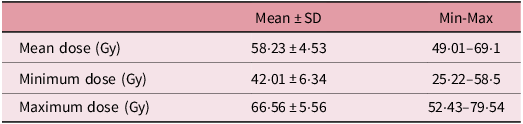
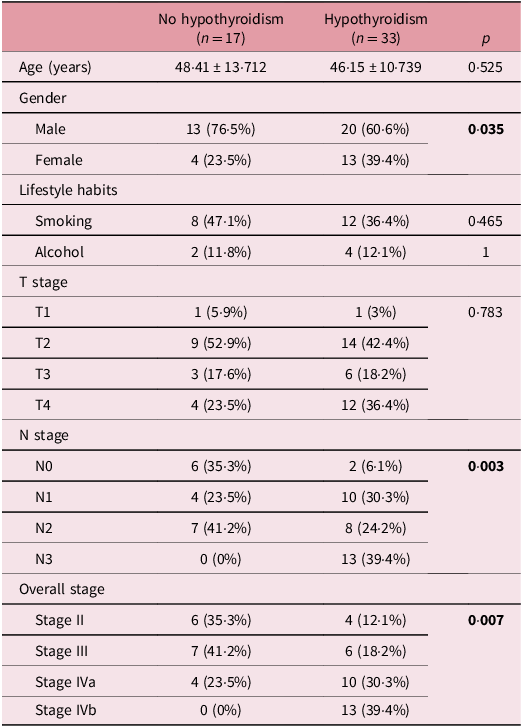
The mean volume of the thyroid gland was 16·24 ± 6·74 cc, with a range of 7 to 38·2 cc. The mean dose received by the thyroid gland was 58·23 ± 4·53 Gy (range: 49·01–69·1 Gy). The minimum dose to the thyroid was 42·01 ± 6·34 Gy (range: 25·22–58·5 Gy) (Tables 1 and 2). The development of hypothyroidism was significantly associated with smaller thyroid volumes (14·73 ± 6·14 cc vs. 19·17 ± 7·07 cc; p = 0·026) and higher mean thyroid doses (59·8 ± 4·27 Gy vs. 55·19 ± 3·41 Gy; p < 0·001). In addition, several clinical parameters were found to be predictive of HPPT. Female sex was significantly associated with increased risk (p = 0·035). A higher T stage (p = 0·007) and greater nodal involvement (≥N2) (p = 0·003) were also identified as predictive factors (Table 3). Use of 5FU–CDDP regimen as a neoadjuvant chemotherapy regimen was associated with a significantly higher incidence of HPPT (42·4% vs. 5·9%; p = 0·008). Concurrent chemotherapy consisted of cisplatin administered weekly at a dose of 40 mh/m2. No significant association was observed between concurrent chemotherapy administration and the occurrence of hypothyroidism.
Doses received by the thyroid gland during treatment

Table 1. Long description
A table with three rows and three columns summarizing the doses received by the thyroid gland during treatment. The columns are labeled Mean ± SD, Min-Max, and the rows are labeled Mean dose (Gy), Minimum dose (Gy), and Maximum dose (Gy). Row 1: Mean dose (Gy), Mean ± SD, 58-23 ± 4-53, Min-Max, 49-01–69-1. Row 2: Minimum dose (Gy), Mean ± SD, 42-01 ± 6-34, Min-Max, 25-22–58-5. Row 3: Maximum dose (Gy), Mean ± SD, 66-56 ± 5-56, Min-Max, 52-43–79-54.
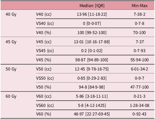
Doses received according to the irradiated volume of the thyroid gland

Table 2. Long description
A table with four rows and three columns. The columns are labeled ‘40 Gy’, ‘45 Gy’, ‘50 Gy’, and ‘60 Gy’. Each row represents different dose metrics: V40 (cc), VS40 (cc), V40 (%), V45 (cc), VS45 (cc), V45 (%), V50 (cc), VS50 (cc), V50 (%), V60 (cc), VS60 (cc), and V60 (%). Each cell contains values for Median [IQR] and Min-Max. For example, Row 1: 40 Gy, V40 (cc) Median [IQR] 1396 [111822], Min-Max 7382. Row 2: 40 Gy, VS40 (cc) Median [IQR] 0 [0007], Min-Max 078. Row 3: 40 Gy, V40 (%) Median [IQR] 100 [9952100], Min-Max 70100. Row 4: 45 Gy, V45 (cc) Median [IQR] 1301 [10161788], Min-Max 737. Row 5: 45 Gy, VS45 (cc) Median [IQR] 02 [0102], Min-Max 0793. Row 6: 45 Gy, V45 (%) Median [IQR] 9887 [9489100], Min-Max 5594100. Row 7: 50 Gy, V50 (cc) Median [IQR] 1245 [9781675], Min-Max 601342. Row 8: 50 Gy, VS50 (cc) Median [IQR] 085 [029283], Min-Max 097. Row 9: 50 Gy, V50 (%) Median [IQR] 948 [84998], Min-Max 4777100. Row 10: 60 Gy, V60 (cc) Median [IQR] 586 [3181111], Min-Max 0213. Row 11: 60 Gy, VS60 (cc) Median [IQR] 58 [4121425], Min-Max 1283408. Row 12: 60 Gy, V60 (%) Median [IQR] 4897 [22276945], Min-Max 09243.
V (cc): volume of the thyroid gland receiving the dose; VS (cc): volume spared from receiving the dose; V (%): percentage of the volume receiving the dose relative to the total volume of the gland.
Univariate analysis of predictor factors of post-therapeutic primary hypothyroidism

Table 3. Long description
The table compares various factors between patients with and without hypothyroidism. It has 12 rows and 6 columns. The columns are labeled as No hypothyroidism (n = 17), Hypothyroidism (n = 33), and p. The rows are labeled as Age (years), Gender (Male, Female), Lifestyle habits (Smoking, Alcohol), T stage (T1, T2, T3, T4), N stage (N0, N1, N2, N3), and Overall stage (Stage II, Stage III, Stage IVa, Stage IVb). Row 1: Age (years), No hypothyroidism 48.41 ± 13.712, Hypothyroidism 46.15 ± 10.739, p 0.525. Row 2: Gender Male, No hypothyroidism 13 (76.5 percent), Hypothyroidism 20 (60.6 percent), p 0.035. Row 3: Gender Female, No hypothyroidism 4 (23.5 percent), Hypothyroidism 13 (39.4 percent), p 0.035. Row 4: Lifestyle habits Smoking, No hypothyroidism 8 (47.1 percent), Hypothyroidism 12 (36.4 percent), p 0.465. Row 5: Lifestyle habits Alcohol, No hypothyroidism 2 (11.8 percent), Hypothyroidism 4 (12.1 percent), p 1. Row 6: T stage T1, No hypothyroidism 1 (5.9 percent), Hypothyroidism 1 (3 percent), p 0.783. Row 7: T stage T2, No hypothyroidism 9 (52.9 percent), Hypothyroidism 14 (42.4 percent), p 0.783. Row 8: T stage T3, No hypothyroidism 3 (17.6 percent), Hypothyroidism 6 (18.2 percent), p 0.783. Row 9: T stage T4, No hypothyroidism 4 (23.5 percent), Hypothyroidism 12 (36.4 percent), p 0.783. Row 10: N stage N0, No hypothyroidism 6 (35.3 percent), Hypothyroidism 2 (6.1 percent), p 0.003. Row 11: N stage N1, No hypothyroidism 4 (23.5 percent), Hypothyroidism 10 (30.3 percent), p 0.003. Row 12: N stage N2, No hypothyroidism 7 (41.2 percent), Hypothyroidism 8 (24.2 percent), p 0.003. Row 13: N stage N3, No hypothyroidism 0 (0 percent), Hypothyroidism 13 (39.4 percent), p 0.003. Row 14: Overall stage Stage II, No hypothyroidism 6 (35.3 percent), Hypothyroidism 4 (12.1 percent), p 0.007. Row 15: Overall stage Stage III, No hypothyroidism 7 (41.2 percent), Hypothyroidism 6 (18.2 percent), p 0.007. Row 16: Overall stage Stage IVa, No hypothyroidism 4 (23.5 percent), Hypothyroidism 10 (30.3 percent), p 0.007. Row 17: Overall stage Stage IVb, No hypothyroidism 0 (0 percent), Hypothyroidism 13 (39.4 percent), p 0.007.
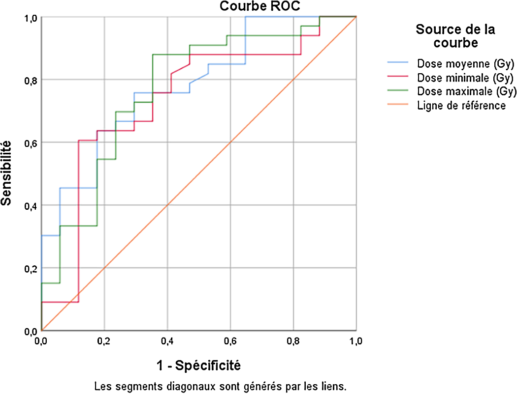
Receiver operating characteristic (ROC) analysis demonstrated that a mean thyroid dose ≥ 56·67 Gy was significantly associated with an increased risk of HPPT (AUC = 0·782, 95% CI: 0·651–0·912), with a sensitivity of 75·8% and a specificity of 64·7% (Figure 1).
Performance of dose parameters in predicting the occurrence of post-therapeutic hypothyroidism.

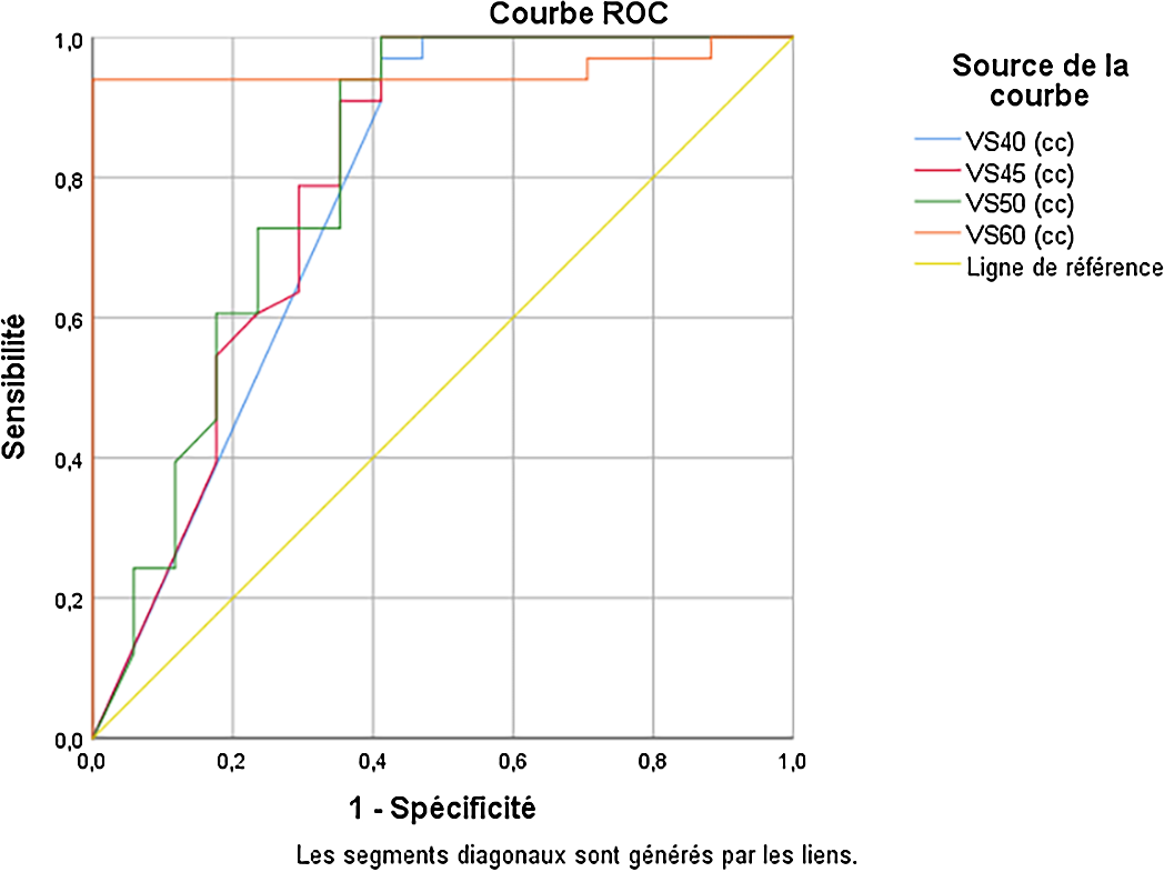
The most robust dosimetric predictor of hypothyroidism was the proportion of thyroid volume spared from doses ≥ 60 Gy (Figure 2). A spared thyroid volume < 9·9 cc at 60 Gy was highly predictive of HPPT, with an AUC of 0·952 (95% CI: 0·886–1), sensitivity of 93·9% and specificity of 100%. This suggests that maintaining at least 9·9 cc of the thyroid below the 60 Gy threshold could represent an optimal planning constraint to minimize the risk of hypothyroidism.
Performance of spared irradiated volume in predicting the occurrence of post-therapeutic hypothyroidism.

Discussion
Nasopharyngeal carcinomas, particularly the undifferentiated type, which is the most common histological subtype, exhibit epidemiological, clinical and management characteristics that vary by region and population. Our study of 50 patients provides insights into these features in our Mediterranean context, showing an incidence consistent with intermediate-frequency regions and a demographic profile similar to published data. The male predominance with a sex ratio of 2:1 (66% men) aligns with literature reporting marked male predominance. Reference Chang, Ye and Zeng14 The mean age at diagnosis was 46 years, within the typical range described, which includes two incidence peaks: an early peak between 15 and 25 years and a later peak around 50 years. Reference Chang, Ye and Zeng14,Reference Busson, Ooka and Corbex15 In our cohort, age was not significantly associated with the development of hypothyroidism (p = 0·515). Cervical lymphadenopathy was the most common presenting symptom (80%), consistent with prior reports, while rhinological (40%) and otological (36%) symptoms often reflected locoregional tumour extension. Diagnosis was frequently delayed due to the deep tumour location. Reference El Korbi, Ben Tkhayat and Bouatay16,Reference Saleh-Ebrahimi, Zwicker and Muenter17 Most patients presented with advanced TNM stages (III-IVB: 80%), in agreement with the literature. Reference EAR, Irekeola and Yean18
Regarding primary hypothyroidism after radiotherapy, it was observed in 66% of patients over a 60-month follow-up, defined by elevated TSH (>4·2 mUI/L) and low FT4 (<10 pmol/L). This incidence is higher than that reported in some studies, where rates generally range from 40% to 50%. Reference Chyan, Chen and Shugard19,Reference Diaz, Jaboin and Morales-Paliza20 The cumulative incidence of hypothyroidism post-radiotherapy in our cohort was 6·1% at 12 months, 42·4% at 24 months and 90·9% at 36 months. These findings partly concur with literature data. Yoden et al. reported a 40% incidence at 24 months but less marked long-term increase, whereas Alterio et al. described a slower progression with 30% at 12 months and 50% at 36 months. Reference Jereczek-Fossa, Alterio and Jassem12,Reference Prayongrat and Lertbutsayanukul21 The median time to hypothyroidism onset was 26 months (IQR 18–30), ranging from 6 to 42 months, comparable to Tell et al.’s median of 24 months, though some studies report peak incidence between 12 and 24 months post-irradiation. Reference Mercado, Adelstein and Saxton22–Reference Murthy, Narang and Ghosh-Laskar24 This relatively long latency reflects the progressive radiation-induced damage mechanisms, including vascular and cellular injury leading to delayed thyroid dysfunction. Reference Rai, Kumar and Mahobiya25
The average thyroid gland volume in our study was 16·24 ± 6·74 cm3 (range 7–38·2 cm3), consistent with adult reference values between 15 and 18 cm3. Reference Chaudhary and Bano26 Thyroid volume is an important factor, as smaller glands may be more susceptible to radiation damage at equivalent doses. Reference Zhou, Chen and Shen27,Reference Nowicka, Tomasik and Papis-Ubych28 Analysis of predictive factors for post-therapeutic hypothyroidism showed female sex to be significant (p = 0·035), indicating women are more prone to develop primary hypothyroidism after treatment, consistent with Zhai et al.’s findings of female sex as an independent risk factor. Reference Zhai, Lyu and Ni29 This increased susceptibility may relate to biological, hormonal and immunological sex differences, including a typically smaller thyroid volume in women, resulting in higher radiation dose per tissue unit, and direct effects of female sex hormones such as estrogens on thyrocyte function and oxidative stress response. Reference Chaudhary and Bano26,Reference Lamichhane, Pant and Lamichhane30 Hypothyroidism occurrence was also significantly associated with advanced tumour stage (p = 0·007) and extensive nodal involvement (p = 0·003). The nodal extension (N stage) showed good predictive performance (p = 0·006; AUC = 0·74; 95% CI = 0·602–0·878), with sensitivity and specificity of 63·6% and 58·8%, respectively, for threshold ≥ N2. Overall, the TNM stage was a statistically significant predictor (p = 0·001) with an AUC of 0·784 (95% CI = 0·66–0·909), identifying stage ≥ IVa as the optimal cut-off (sensitivity 69·7%, specificity 76·5%). These results align with literature where higher nodal stages requiring larger irradiation volumes increase hypothyroidism risk due to greater thyroid radiation exposure. Reference Prayongrat and Lertbutsayanukul21,Reference Zhai, Lyu and Ni29 The relationship between thyroid volume and post-radiotherapy hypothyroidism is well documented. Smaller thyroid volumes (<12·82 cm3) are linked to higher hypothyroidism rates (75% versus 37% for larger volumes). Reference Zhou, Chen and Shen27,Reference Nowicka, Tomasik and Papis-Ubych28 Dosimetric parameters also correlate strongly with hypothyroidism risk. Vogelius et al. reported a mean thyroid dose of 45 Gy as a threshold associated with a 50% hypothyroidism risk, increasing beyond this dose. Reference Vogelius, Bentzen and Maraldo10 Similarly, Bhandare et al. observed a 16% rise in incidence when mean dose exceeded 45 Gy. Reference Bhandare, Kennedy and Malyapa31 In our cohort, patients developing hypothyroidism received significantly higher mean thyroid doses (59·8 ± 4·27 Gy) compared to those without hypothyroidism (55·19 ± 3·41 Gy; p < 0·001), reinforcing a dose-effect relationship. The mean thyroid dose predicted hypothyroidism well (p = 0·001) with an AUC of 0·782 (95% CI = 0·651–0·912), and an optimal cut-off at 56·67 Gy yielded 75·8% sensitivity and 64·7% specificity, consistent with thresholds reported in similar studies. Reference Chyan, Chen and Shugard19
Dose–volume parameters further highlighted the importance of volume irradiated at higher doses. The percentage of thyroid volume receiving 60 Gy (V60) showed excellent predictive performance (p < 0·001; AUC = 0·934; 95% CI = 0·87–0·999). A V60 threshold of ≥34·87% had 90·9% sensitivity and 82·4% specificity for hypothyroidism risk. This supports that larger thyroid volumes exposed to high doses substantially increase hypothyroidism risk, emphasising dose–volume constraints in treatment planning. Reference Nowicka, Tomasik and Papis-Ubych28,Reference Bhandare, Kennedy and Malyapa31 Other volumes spared (VS40, VS45) had acceptable but lower predictive value (AUCs 0·774 and 0·793, respectively), with VS40 specificity at only 58·8%, limiting clinical utility. Notably, the thyroid volume spared from 60 Gy (VS60) was an excellent protective factor, with a threshold of 9·9 cm3 yielding 93·9% sensitivity and 100% specificity. This aligns with literature underscoring VS60 as a key parameter in preserving thyroid function post-radiotherapy and outperforming lower-dose spared volumes. Reference Zhai, Lyu and Ni29,Reference Zhou, Chen and Tao32 Although radiotherapy remains the main risk factor for hypothyroidism, certain systemic treatments may exacerbate thyroid damage. Chemotherapy agents can increase radiation-induced inflammation or fibrosis of the thyroid, impairing hormone production. Reference Yeung, Chiu and Vassilopoulou-Sellin33 In our study, neoadjuvant chemotherapy with 5FU-CDDP was significantly associated with higher hypothyroidism prevalence (42·4% vs. 5·9%; p = 0·008), supporting a contributory role of chemotherapy in thyroid dysfunction among nasopharyngeal carcinoma patients treated with radiotherapy. Reference Zhou, Chen and Tao32,Reference Yeung, Chiu and Vassilopoulou-Sellin33
Conclusion
External radiotherapy, often combined with chemotherapy, is the standard treatment for nasopharyngeal cancer. This study on 50 patients treated with IMRT showed that primary hypothyroidism is a common side effect linked to high radiation doses to the thyroid, smaller thyroid volumes, advanced tumour stages and chemotherapy. A mean thyroid dose above 45 Gy significantly increases the risk, with a critical threshold around 56·67 Gy. Sparing sufficient thyroid volume during treatment reduces this risk. These findings highlight the need to balance effective tumour control with thyroid protection to minimize hypothyroidism and improve patient quality of life. Future research should focus on refining dosimetric constraints, exploring protective agents for thyroid function and developing personalized treatment plans to further reduce thyroid toxicity without compromising cancer control.
Data availability statement
Underlying data: The statistical analysis outputs supporting the findings of this study are openly available in Figshare at https://doi.org/10.6084/m9.figshare.30355081 under a Creative Commons Attribution 4.0 International (CC BY 4.0) license (https://creativecommons.org/licenses/by/4.0/).
The data supporting the findings of this study are openly available. Reference Kouti, Mousli and Ben Zid13
Author contributions
-
• Kouti A: Study conception, data collection, manuscript drafting, and writing—original draft
-
• Mousli A: Statistical analysis, data interpretation, and critical revision of the manuscript
-
• Fatma Missaoui: critical revision of the manuscript and submission
-
• Ben Zid K: Patient follow-up and contribution to the discussion
-
• Ghorbel A: Radiotherapy supervision, protocol validation, and methodological input
-
• Abidi R: Methodological support and scientific and clinical manuscript revision
-
• Nasr C: Study supervision, overall oversight and final manuscript approval
Financial support
This research received no specific grant from any funding agency in the public, commercial or not-for-profit sectors.
Competing interests
The authors declare no competing interests.
Informed consent
Verbal informed consent was obtained from all participants prior to inclusion in the study. Verbal consent was preferred over written consent because the study was retrospective and based on routine outpatient follow-up of previously treated patients. During these follow-up visits, patients were informed that their anonymized thyroid function test results could be included in the study. No personal identifying data were collected, and the study involved only retrospective analysis of existing clinical records. The procedure for obtaining and documenting verbal consent was approved by the institutional ethics committee.

 Open access

