Introduction
Allergic rhinitis, a chronic inflammatory disease of the upper respiratory tract, is a global health problem associated with severe consequences.Reference Bousquet, Schünemann, Samolinski, Demoly, Baena-Cagnani and Bachert1, Reference Dykewicz and Hamilos2 According to the International Study of Asthma and Allergies in Childhood, the prevalence of allergic rhinitis among children varies widely, ranging from less than 1 per cent to almost 40 per cent,Reference Asher, Montefort, Björkstén, Lai, Strachan and Weiland3, Reference Baena-Cagnani, Passalacqua, Gómez, Zernotti and Canonica4 reaching 50 per cent in some populations.Reference Katelaris, Lee, Potter, Maspero, Cingi and Lopatin5 The underlying pathogenic mechanism remains to be elucidated, although previous studies have suggested that genetic, environmental and socioeconomic factors are responsible for the occurrence and development of allergic rhinitis.Reference Hatakka, Piirainen, Pohjavuori, Poussa, Savilahti and Korpela6, Reference Larsson, Hägerhed-Engman, Sigsgaard, Janson, Sundell and Bornehag7
Earlier research on allergic rhinitis has demonstrated that an imbalance of type 1 helper to type 2 helper cells influences the development of allergic rhinitis by affecting the production of related cytokines.Reference Varga, Jacobson, Till, Masuyama, O'Brien and Rak8, Reference Ozkara, Keles, Ilhan, Gungor, Kaygusuz and Alpay9 Recently, CD4+CD25+ regulatory T cells have received general attention in the medical scientific field. These cells are characterised by their ability to suppress the development of type 1 helper and type 2 helper cell-mediated immune responses.Reference Mamessier, Milhe, Guillot, Birnbaum, Dupuy and Lorec10, Reference Sogut, Yilmaz, Kirmaz, Ozbilgin, Onur and Celik11 CD4+CD25+ regulatory T cells can also regulate immune function by inhibiting cell-to-cell contact, and by secreting interleukin 10 and transforming growth factor β.Reference Dasgupta and Saxena12, Reference Taylor, Verhagen, Akdis and Akdis13 O'Hehir et al. found that transforming growth factor β1 and regulatory T cells have suppressive functions in sublingual immunotherapy.Reference O'Hehir, Gardner, de Leon, Hales, Biondo and Douglass14 Current available data suggest that CD4+CD25+ T cells play an important role in allergy or autoimmune diseases.
Recent studies have focused on an imbalance between CD4+CD25+ regulatory T cells and type 2 helper cells in allergic diseases.Reference Akdis, Verhagen, Taylor, Karamloo, Karagiannidis and Crameri15 The specific transcription factor for regulatory T cells is the forkhead-winged helix transcription factor (FOXP3) and that for type 2 helper cells is GA-TA binding protein 3 (GATA3), which can differentiate naive CD4+ cells into regulatory T or type 2 helper cells.Reference Paik, Dahl, Fang and Calhoun16, Reference Hori, Nomura and Sakaguchi17 Mo et al. reported that higher numbers of type 2 helper cells and a deficiency of regulatory T cells are the hallmarks of asthma disorders.Reference Mo, Chung and Kim18 Therefore, we hypothesised that an imbalance of type 2 helper/regulatory T cells may contribute to the development of allergic rhinitis.
The present study aimed to investigate whether a type 2 helper/regulatory T cell-mediated immunity imbalance exists, and whether such an imbalance is related to allergic rhinitis disease severity. This may provide a new insight into the pathogenesis of allergic rhinitis and act as a basis for potential therapeutic approaches.
Materials and methods
Subjects and symptom scoring
The study included 46 patients (24 males and 22 females) with allergic rhinitis. Mean age was 6.1 years (range, 4.4–8.8 years). We also enrolled 42 healthy control subjects without allergy (22 males and 20 females). Mean age was 6.4 years (range, 4.2–9.4 years).
All selected patients were of Han nationality, and were diagnosed using the Allergic Rhinitis and its Impact on Asthma initiative criteria.Reference Bousquet, Khaltaev, Cruz, Denburg, Fokkens and Togias19 Characteristics of patients included: a history of paroxysmal sneezing, watery rhinorrhea and nasal obstruction, positive specific immunoglobulin E (IgE) results, and positive skin prick test results. All patients underwent a nasal cavity examination. Prior to blood sample collection, all medications, including systematic glucocorticoids, were prohibited for at least four weeks. Patients with infectious, occupational or drug-induced rhinitis, and those with any complications, were excluded. Subjects in the control group had no history of allergic diseases, no detectable, high levels of specific IgE antibodies and serum total IgE levels below the average standard of the general population.
This study was approved by the ethical committee of Chongqing Medical University. Informed consent was obtained from all subjects' parents or guardians.
Allergic rhinitis symptoms were assessed using the Total 5 Symptom Score (‘T5SS’), which assessed watery rhinorrhea, paroxysmal sneezing, nasal congestion, and nasal and ocular pruritus. Each sign or symptom was scored on a scale from 0 to 3 (whereby 0 = none and 3 = severe). Patients were enrolled if their Total 5 Symptom Score was more than 6, with symptoms persisting for at least 4 days every week during each episode of allergic rhinitis.
Enzyme-linked immunosorbent assay
For the interleukin (IL)-4, IL-5 and transforming growth factor β1 assay, anticoagulated blood samples were collected from patients on their first visit to the out-patient clinic in the First Affiliated Hospital of Chongqing Medical University and/or when they were hospitalised for further treatment. These samples were obtained using vacuum tubes containing heparin, centrifuged at 200 × g for 5 minutes. The supernatant (i.e. plasma) was collected and stored at −80 °C until cytokine measurement. Plasma cytokine levels were detected (according to the manufacturer's instructions) using: a Human IL-4 High Sensitivity ELISA Kit (eBioscience, San Diego, California, USA), a Human IL-5 Platinum ELISA Kit (eBioscience) and a Human TGF-β1 Platinum ELISA Kit (eBioscience). The detection limits for IL-4, IL-5 and transforming growth factor β1, respectively, were 0.1, 10 and 8.6 pg/ml.
RNA extraction, complementary DNA synthesis and polymerase chain reaction
Total RNA was extracted using a Total RNA Extraction Kit (BioTeke, Beijing, China), and was reverse transcribed to complementary DNA using a Prime Script RT Reagent Kit (TaKaRa, Dalian, China), according to the manufacturer's instructions. The primer sequences used (Sangon Biotech, Shanghai, China) were as follows: FOXP3 forward, 5′-AGGGACCAAGAAGTGAGGTTTC-3′, and reverse, 5′-TGGGGTTTGTGTTGAGTGAG-3′; GATA-3 forward, 5′-AGACCACCACAACCACACTCT-3′, and reverse, 5′-GATGCCTTCCTTCTTCATAGTCA-3′; and β-actin forward, 5′-AGCGAGCATCCCCCAAAGTT-3′, and reverse, 5′-GGGCACGAAGGCTCATCATT-3′. Reaction mixtures were heated to 95 °C for 4 minutes. This was followed by: 30 cycles of denaturation at 95 °C for 45 seconds; annealing at 55 °C (FOXP3), 58 °C (GATA3) or 59 °C (β-actin) for 5 seconds; and extension at 72 °C for 10 minutes. All polymerase chain reactions were performed in duplicate.
To confirm the specificity of the polymerase chain reaction, polymerase chain reaction products were analysed by 3 per cent agarose gel electrophoresis and ethidium bromide staining, followed by visualisation using an ultraviolet transilluminator (Bio-Rad, Hercules, California, USA).
Peripheral blood mononuclear cell preparation
Peripheral blood mononuclear cells were prepared by standard Ficoll–Hypaque density gradient centrifugation for 30 minutes at 800 × g at 4 °C. The layer of peripheral blood mononuclear cells was collected, then washed twice with phosphate-buffered saline, and cells were re-suspended at a density of 1 × 10⁶ cells/ml in Roswell Park Memorial Institute 1640 medium. Cell viability was assessed with a trypan blue exclusion assay under an optical microscope, and was found to be greater than 95 per cent.
FOXP3 and GATA3 Western blotting analysis
Proteins were extracted from the peripheral blood mononuclear cells of allergic rhinitis and control patients. The cells were lysed with radio-immunoprecipitation assay lysis buffer (containing 1 per cent phenylmethylsulfonyl fluoride; Beyotime, Shanghai, China) according to the manufacturer's instructions, and kept on ice for 30 minutes at 4 °C. Then, the lysates were centrifuged at 15 000 rpm at 4 °C for 15 minutes. The supernatants were collected and stored at −20 °C until detection. Protein concentrations were measured using an enhanced bicinchoninic acid protein assay kit (Beyotime). Samples were diluted with loading buffer, boiled for 8 minutes and loaded onto sodium dodecyl sulphate-polyacrylamide gels. Samples were electrophoresed at 65–75 V for 2–3 hours; the separated proteins were transferred onto polyvinylidene difluoride membranes (Bio-Rad). The protein bands were measured using a Bio-Rad densitometer and analysed using Quantity One software, version 4.52. Anti-FOXP3 and anti-GATA-3 antibodies were purchased from Sigma-Aldrich (St Louis, Missouri, USA).
Statistical analysis
All parametric results are expressed as mean ± standard deviation. Results were analysed using the Student's t-test (between-group comparisons), non-parametric Mann–Whitney test and Pearson correlation. Differences were considered statistically significant when the p value was less than 0.05. All analyses were performed using IBM SPSS Statistics® version 20.0.
Results
Patient characteristics
The subjects in the two groups were matched for age and sex (Table I). Total IgE levels significantly differed between the control group and the allergic rhinitis group (6.6 ± 1.9 kU/l vs 122.2 ± 16.0 kU/l, p < 0.05). Total 5 Symptom Score in the allergic rhinitis group was 9.75 ± 2.35, which was significantly different to that of the control group (score of 0) (p < 0.001). Similarly, the eosinophil ratio was significantly greater in the allergic rhinitis group than in the control group.
Demographic and clinical characteristics of participants

NS = non-significant; SD = standard deviation; M = males; F = females; IgE = immunoglobulin E; T5SS = Total 5 Symptom Score
Interleukins 4 and 5, and transforming growth factor β1
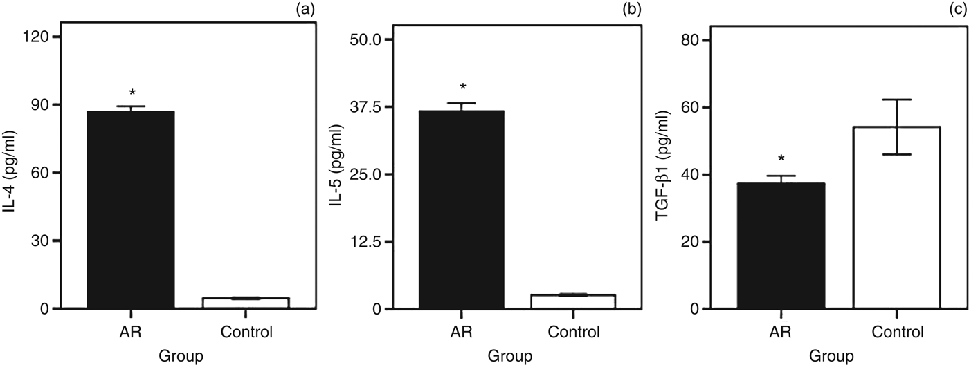
The plasma levels of interleukin (IL)-4 and IL-5 were significantly higher in patients with allergic rhinitis (86.9 ± 2.36 pg/ml and 36.7 ± 1.49 pg/ml) than in healthy controls (4.6 ± 0.3 pg/ml and 2.6 ± 0.2 pg/ml) (Figure 1a and b). However, patients with allergic rhinitis had a significantly lower level of transforming growth factor β1 than did the control subjects (37.5 ± 2.61 pg/ml vs 54.8 ± 3.76 pg/ml, p < 0.05; Figure 1c).
Enzyme-linked immunosorbent assay of type 2 helper/regulatory T type cytokines ((a) interleukin (IL)-4, (b) IL-5 and (c) transforming growth factor (TGF)-β1)) in plasma samples of allergic rhinitis (AR) patients and healthy controls. Interleukin-4 and IL-5 levels were significantly higher in allergic rhinitis patients than in controls; transforming growth factor β1 level was significantly lower in allergic rhinitis patients than in controls. Significance level was set at 0.05.*p < 0.05
FOXP3 and GATA3 messenger RNA expression
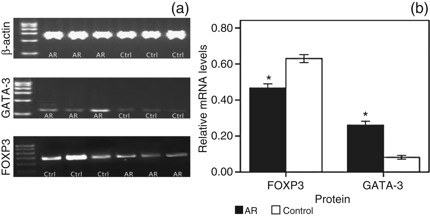
FOXP3 messenger RNA (mRNA) expression (measured in arbitrary units) was higher in control subjects (0.63 ± 0.02) than in allergic rhinitis patients (0.47 ± 0.02, p < 0.001; Figure 2), whereas GATA3 mRNA expression (measured in arbitrary units) was higher in allergic rhinitis patients than in controls (0.26 ± 0.02 vs 0.08 ± 0.01, p < 0.001; Figure 2).
Reverse transcription polymerase chain reaction quantification analysis of GATA3 and FOXP3 messenger RNA (mRNA) in peripheral blood mononuclear cells of allergic rhinitis (AR) patients and controls (ctrl). (a) Reverse transcription polymerase chain reaction analysis of regulatory T transcription factor FOXP3 mRNA expression, type 2 helper transcription factor, GATA3 mRNA expression and β-actin expression. (b) Quantification of GATA-3 and FOXP3 mRNA expression (measured in arbitrary units). Significance level was set at 0.05. *p < 0.001
FOXP3 and GATA3 protein analysis
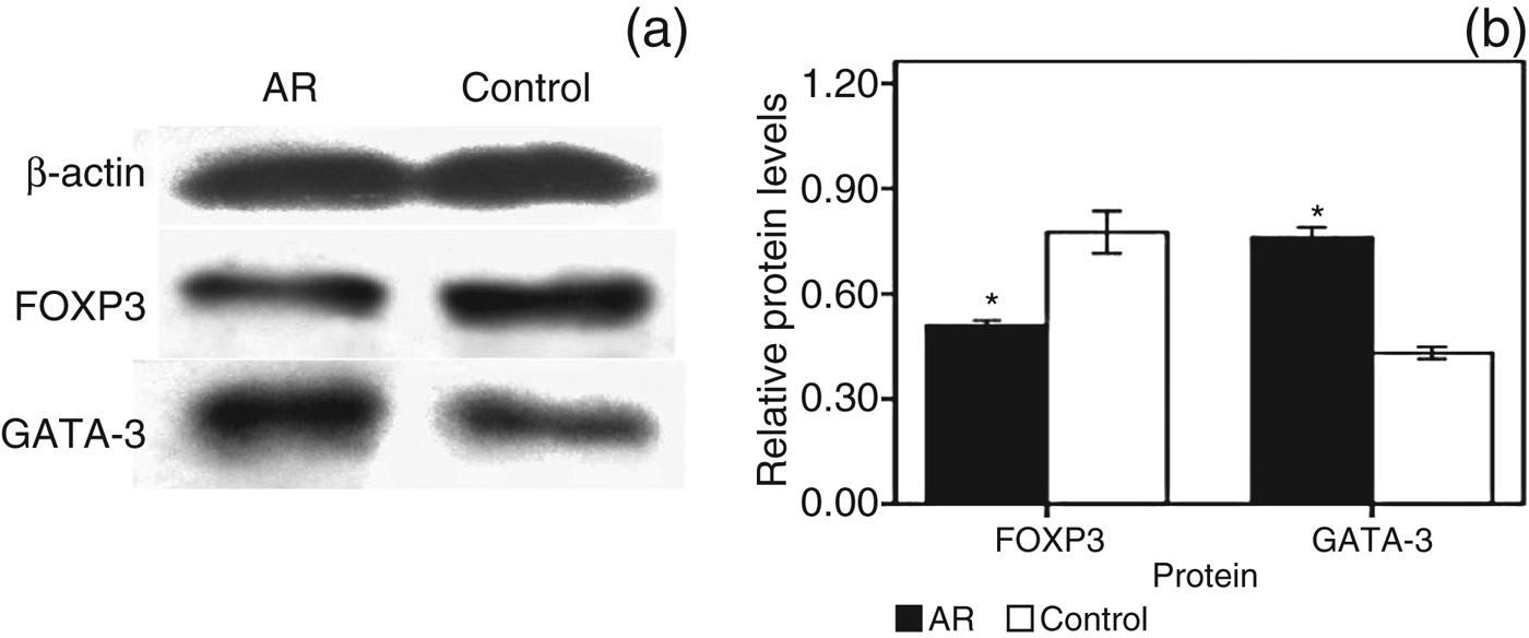
GATA3 protein expression level (measured in arbitrary units) was considerably higher in peripheral blood mononuclear cells from allergic rhinitis patients (0.76 ± 0.01) than in those from controls (0.43 ± 0.01, p < 0.05; Figure 3). However, FOXP3 protein expression level (measured in arbitrary units) was significantly lower in the allergic rhinitis group than in the control group (0.51 ± 0.01 vs 0.77 ± 0.03, p < 0.05; Figure 3). These results were consistent with the reverse transcription polymerase chain reaction results.
Upregulation of GATA3 and downregulation of FOXP3 protein expression in peripheral blood mononuclear cells from patients with allergic rhinitis (AR) compared with control subjects. (a) Western blot analysis for GATA3 and FOXP3 expression in peripheral blood mononuclear cells from allergic rhinitis patients and healthy control subjects. (b) Quantification of GATA3 and FOXP3 protein expression (measured in arbitrary units). Significance level was set at 0.05.*p < 0.05
Correlational analysis
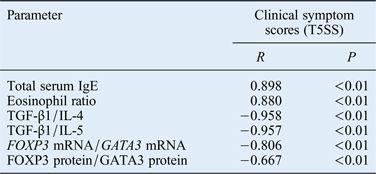
To understand the role of regulatory T/type 2 helper cell-mediated immune imbalance in allergic rhinitis, we examined the relation between the regulatory T/type 2 helper cell ratio and clinical symptom scores. The ratio of regulatory T/type 2 helper cells was assessed by determining the ratios of: transforming growth factor β1/IL-4, transforming growth factor β1/IL-5, FOXP3 mRNA/GATA3 mRNA, and FOXP3 protein/GATA3 protein. The results showed significant negative correlations between clinical symptom scores (Total 5 Symptom Scores) and the regulatory T/type 2 helper cell ratio (Table II).
Correlations between total serum immunoglobulin e, eosinophil ratio, regulatory t/type 2 helper cell ratio and nasal symptom score

T5SS = Total 5 Symptom Score; IgE = immunoglobulin E; TGF = transforming growth factor; IL = interleukin; mRNA = messenger RNA
Discussion
We investigated the expression and immune imbalance of regulatory T/type 2 helper cells in paediatric allergic rhinitis patients by measuring the levels of cytokines (interleukin (IL)-4, IL-5 and transforming growth factor β1), and mRNA and intracellular proteins of transcription factors (FOXP3 and GATA3). Levels of IL-4 and IL-5 (type 2 helper cytokines) were higher in patients with allergic rhinitis than in controls, and mRNA and intracellular protein levels of GATA3, a type 2 helper cell-specific transcription factor, were higher in the allergic rhinitis group than in the control group. However, mRNA and intracellular protein levels of FOXP3, a regulatory T cell-specific transcription factor, were lower in allergic rhinitis patients than in control subjects. These findings are consistent with the results of Mo et al. and Xu et al.Reference Mo, Chung and Kim18, Reference Xu, Mou, Jiang, Cheng, Shi and Xu20
Allergic rhinitis, defined by immediate hypersensitivity in the upper respiratory tract, is perhaps the most familiar disease in rhinology. However, the underlying pathogenesis is not fully clear. Traditionally, allergic rhinitis is considered a type 2 helper dominant response mediated by IgE. In recent years, owing to their unique function to suppress the immune response, regulatory T cells have returned as a focus of research. The ‘suppressor T cells’ were one of the dominant themes of immunology in the 1970s. However, because of the limitations of research techniques at that time, initial attempts to define populations and their mechanisms of action were not successful.Reference Abul, Andrew and Shiv21
The role of regulatory T cells in the pathogenesis of allergic disease was not defined until recently. Regulatory T cells may block the transition from the early activation stage to the differentiated type 2 helper state, limit allergic inflammation of the airways and act to prevent inappropriate type 2 helper responses to environmental allergens.Reference Lewkowich, Herman, Schleifer, Dance, Chen and Dienger22, Reference Kearley, Barker, Robinson and Lloyd23 Gambineri et al. reported that patients who lack CD4+CD25+ regulatory T cells develop severe eczema, elevated IgE levels, eosinophilia and food allergy.Reference Gambineri, Torgerson and Ochs24 However, the reported frequency of regulatory T cells in peripheral blood from patients with allergy is inconsistent.Reference Ling, Smith, Nguyen, Pridgeon, Dallman and Arbery25–Reference Zheng, Wu, Huang, Chen, Yang and Li28 The difference in these observed frequencies might be related to the patients' age, disease severity and disease status. With regard to allergic rhinitis, several studies have reported lower FOXP3 mRNA expression in peripheral blood mononuclear cells, nasal secretions and nasal tissue,Reference Xu, Mou, Jiang, Cheng, Shi and Xu20, Reference Lee, Gao, Dahl, Calhoun and Fang29 implying a critical role of regulatory T cells in dampening the allergic response. However, data concerning the role of regulatory T cells in the pathogenesis of paediatric allergic disease are rare.
To our knowledge, only Lee et al. have reported that paediatric patients with airway allergy have significantly lower numbers of CD4+CD25+ T cells.Reference Lee, Yu, Wang, Yang, Lin and Chiang30 This finding is consistent with our results. However, the relationship between the number of regulatory T cells and type 2 helper cells was not emphasised in their study. In the present study, we found that downregulated expression of regulatory T cells may worsen the aberrant distribution of regulatory T/type 2 helper cells and exacerbate the clinical severity of allergic rhinitis.
The total IgE level in serum showed a positive correlation with patients' clinical symptom scores; however, Total 5 Symptom Score showed a negative correlation with the ratios of FOXP3 mRNA/GATA3 mRNA and FOXP3 protein/GATA3 protein in peripheral blood mononuclear cells. These findings are consistent with a previous report which indicated that adaptive and functional regulatory T cells might be generated in response to aggravated atopy and disease severity, with increased expression of FOXP3 and IL-10 in regulatory T cells from patients with relatively severe allergic disease.Reference Lee, Yu, Wang, Yang, Lin and Chiang30 Zheng et al. also reported a negative correlation between FOXP3, VgammaIII and visual analogue scale scores.Reference Zheng, Wu, Huang, Chen, Yang and Li28 The above experimental results imply that regulatory T/type 2 helper imbalance is correlated with disease severity, from which we conclude that an imbalance of regulatory T/type 2 helper cells may be an important mechanism in the pathogenesis of allergic rhinitis.
This study has certain limitations. First, the numbers of subjects were limited. If possible, a more comprehensive study will be carried out to draw a more detailed conclusion. Second, because we did not acquire local tissue specimens through nasal operation, the local changes of T lymphocyte subgroups in mucosa could not be investigated. If these studies were performed, we could show a more synthesised and compelling conclusion. Despite these limitations, the present study presents novel information about the differentiation of T cell subsets, and variations of transcription factors and cytokines, from a systematic view in allergic rhinitis, and helps us understand the pathogenesis of allergic rhinitis in terms of T cell subsets.
• Cytokine levels (interleukins 4 and 5, and transforming growth factor β1) differ between children with allergic rhinitis and healthy control subjects
• The regulatory T cell/T-helper 2 cell ratio is inversely correlated with clinical symptom scores for allergic rhinitis
• The study findings provide new insight into the pathogenesis of allergic rhinitis and suggest possible therapeutic approaches
Taken together, the data showed that paediatric patients with allergic rhinitis had fewer regulatory T cells and more type 2 helper cells on average. These findings suggest that lower numbers of regulatory T cells might represent a deficiency in the regulatory T cell population in children with allergic airway disease. The reduction in the suppression function of regulatory T cells may contribute to sustaining and exaggerating type 2 helper cell response, resulting in the development of allergic inflammation. The immune network imbalance helps us to comprehend a possible mechanism of allergic rhinitis, and seek a better strategy through ameliorating the imbalance between effector and regulatory T cells.