INTRODUCTION
The microbiota of non-ruminant animals influences host health and performance. Although commensal bacteria could also become a burden for the animal, the gut microbiota has many positive effects including providing nutrients for the host and the development of intestinal host defences. Moreover, microbiota aids in protecting the host from colonization by pathogens by competitive exclusion, thereby leading to decreased morbidity and mortality (Metzler et al. Reference Metzler, Bauer and Mosenthin2005; Richards et al. Reference Richards, Gong and De Lange2005). As a result of its beneficial influence on animal well-being, investigating the intestinal microbiota of pigs is of considerable importance. It is important to provide a suitable methodological strategy and useful tools to characterize such complex communities as present in the gastrointestinal tract. Several methods are available to study the composition and complexity of the intestinal microflora. Previously, classical culture-based methods were most frequently employed and provided a way to enumerate bacterial communities as well as to investigate the viability and metabolism of these organisms. However, due to medial selectivity and the presence of viable but non-culturable bacteria in the intestine, cultivation methods do not give sufficient information on the real microbial ecology in the gastrointestinal tract. To overcome these limitations, a wide range of modern molecular analysis techniques such as polymerase chain reaction (PCR)-based methods or genetic fingerprinting approaches are currently available (McCartney Reference McCartney2002; Namsolleck et al. Reference Namsolleck, Thiel, Lawson, Holmstrøm, Rajilic, Vaughan, Rigottier-Gois, Collins, De Vos and Blaut2004). Hence, a combination of different methods would be the best strategy to obtain a comprehensive view on the gut microbiota.
In the current study, fluorescence in situ hybridization (FISH) and real-time PCR were applied and results of bacterial counts were compared to those obtained with classical culture-based methods. FISH allows the visualization, identification, enumeration and localization of micro-organisms within complex samples and microbial communities simultaneously. Real-time PCR analysis allows the identification as well as the quantification of bacteria with increased specificity, sensitivity and accuracy compared to conventional PCR methods. With the use of rRNA-targeted oligonucleotide probes (FISH) and specific primers (real-time PCR) unculturable prokaryotic organisms can also be identified, such as highly anaerobic micro-organisms. However, these modern molecular methods also have some drawbacks, such as possible lack of specificity of probes and primers or insufficient efficiency of the DNA extraction. Moreover, the presence of multiple copies of 16S rDNA requires a calibration on plate countings when performing real-time PCR analyses (Moter & Göbel Reference Moter and Göbel2000; Wagner et al. Reference Wagner, Horn and Daims2003; Castillo et al. Reference Castillo, Martín-Orúe, Manzanilla, Badiola, Martín and Gasa2006; Holzapfel & Wickert Reference Holzapfel and Wickert2007).
The current study initially focused on the enumeration of different parameters of pig gut microbiota using three different methods (cultivation method, FISH and real-time PCR) with the aim of comparing and evaluating the methods. Subsequently these techniques were used to investigate the influence of different dietary treatments on bacterial counts in the gastrointestinal tract of pigs. For this purpose, 48 newly weaned piglets were offered four different diets with the addition of insoluble dietary fibre differing in lignin content. Wheat bran (low lignin) and pollen from Chinese Masson pine (Pinus massoniana) (rich in lignin) were added to the basal diet. Dietary fibre, which is defined as those components of plants or analogous carbohydrates that are resistant to digestion by endogenous enzymes, is claimed to promote beneficial physiological effects and contribute to animal health (AACC Report 2001). Examples of beneficial influences of dietary fibre on pig gut microbiota have been reported previously, as they affect digestion, absorption, metabolism and intestinal morphology. Furthermore, a high fibre content of digesta shows an increase of peristaltic action and a reduction of transit time, which prevents constipation (Wenk Reference Wenk2001). Pollen from the Chinese Masson pine (P. massoniana) represents a dietary fibre complex, which is especially rich in undigestible carbohydrates and may also contribute to a healthy gut microbiota (Zhao et al. Reference Zhao, Windisch, Roth, Eder and Ettle2007; Schedle et al. Reference Schedle, Plitzner, Ettle, Zhao, Domig and Windisch2008a, Reference Schedle, Pfaffl, Plitzner, Meyer and Windischb).
MATERIALS AND METHODS
Sampling
Forty-eight newly weaned piglets (crossbred of German Large White×Pietrain) were fed four diets enriched with varying amounts of different sources of insoluble fibre (wheat bran or pollen from the Chinese Masson pine; P. massoniana) for 37 days. The piglets (8·5 kg mean body weight) were fed ad libitum. The control group received a basal diet with no further addition of fibre. The other three dietary treatment groups included an addition of 30 g wheat bran/kg (Herzig Muehle, Lichtenwoerth, Austria) (added total dietary fibre (TDF): 16·0 g/kg dry matter (DM)), 12·7 g/kg (added TDF: 8·0 g/kg DM, pollen low group) and 25·5 g/kg (added TDF: 16·0 g/kg DM, pollen high group) pine pollen (GUOZHEN, Yantai, P. R. China) to the basal diet. Animals were housed in eight conventional pens equipped with slatted floors, air conditioning, heated lying area, cup drinker and feeder. Following slaughter at days 36 and 37 with mean body weight of 23±3·7 kg, the terminal ileum (approximately 150 mm proximal to the ileo-caecal valve) and the colon (at the flexura centralis) of 45 animals were opened and chyme samples were collected in sterile 60 ml twist-seal sample bags (VWR, Vienna, Austria) and stored under anaerobic conditions (GENbag anaer, Biomérieux, Marcy l'Etoile, France) at −80°C. The analysed compositions of the diets have been reported previously (Schedle et al. Reference Schedle, Plitzner, Ettle, Zhao, Domig and Windisch2008a).
Cultivation method
Microbial enumeration analyses of chyme samples of the following microbial parameters were performed as described previously: total anaerobic colony count, Gram-negative anaerobic bacteria, lactobacilli, bifidobacteria, clostridia, total microbial colony count, Escherichia coli and Enterobacteriaceae. Growth conditions of parameters which are relevant for the evaluation of results given in the current study were: total anaerobic colony count (37°C, 72 h, anaerobic, Wilkins–Chalgren agar, Oxoid, Basingstoke, UK), lactobacilli (37°C, 72 h, anaerobic, MRS-agar, Merck, Germany) and bifidobacteria (37°C, 72 h, anaerobic, Wilkins–Chalgren agar, Oxoid Ltd, Basingstoke, UK plus mupirocin 100 mg/l, LGC Promochem, Germany). Anaerobic incubation was performed in an anaerobic workstation (Don Whitley Scientific, West Yorkshire, UK) at anaerobic conditions (0·85 N2, 0·10 CO2 and 0·05 H2). Colony forming units (cfu) were counted and expressed as log10 of viable bacteria/g of fresh matter (Schedle et al. Reference Schedle, Plitzner, Ettle, Zhao, Domig and Windisch2008a).
Fluorescence in-situ hybridization
Colonic samples were analysed with FISH using commercial FISH-Kits (Ribotechnologies/Microscreen, Groningen, The Netherlands) according to the manufacturer's instructions. With these ready-to-use kits, bacterial counts were determined for all bacteria (total viable count), lactobacilli and bifidobacteria. Enumeration was performed using a reflected-light fluorescence microscope (Olympus BX-51, Hamburg, Germany) at 550 nm and a Cy3-filter-set. Eleven pictures were taken per sample and bacteria that appeared red were calculated. Subsequently the average values of bacterial counts were calculated according to instructions in the protocol and expressed as log10/g fresh sample.
Bacterial strains, growth conditions and quantification of real-time PCR products
To quantify lactobacilli, bifidobacteria, Bacterioides vulgatus and the total cell count on the basis of domain V of the 23S rRNA in ileum and colon samples, standard curves were prepared. For this purpose, strains of Lactobacillus rhamnosus (DSM 20021), Bifidobacterium thermophilum (DSM 20210) and B. vulgatus (LMG 17767) were applied as positive controls and used to establish a standard curve for real-time PCR analyses. Starting from glycerine stock cultures, L. rhamnosus was grown in De Man Rogosa Sharpe media (Merck, Darmstadt, Germany), while B. thermophilum and B. vulgatus were cultured in Brain Heart Infusion media (Oxoid Ltd, Basingstoke, UK). All three strains were incubated at 37°C under anaerobic conditions. Subsequently, colonies were suspended in NaCl solution (8·5 g/l) and inocula were adjusted to McFarland standard 1 using photometric measurements at a wavelength of 625 nm. Serial decimal dilutions in NaCl solution (8·5 g/l) of the bacterial strains were used as standards to quantify real-time PCR products. To confirm the accuracy of the dilutions, 100 μl aliquots were plated on agar media and colonies were counted, except for B. vulgatus, which could not grow on solid agar media.
DNA isolation
DNA of the investigated samples was isolated using the QIAamp® DNA Stool Mini Kit (Qiagen, Hilden, Germany) according to the provided instructions for pathogen detection. This ready-to-use kit contains InhibitEX tablets that remove substances that could degrade DNA. It has previously been shown to provide a fast and appropriate isolation method for faecal samples (Li et al. Reference Li, Gong, Cottrill, Yu, De Lange, Burton and Topp2003). A 180–220 mg sample was taken for DNA isolation which should result in a DNA yield of 15–60 μg and a DNA concentration of 75–300 ng/μl. DNA of bacterial strains used as positive controls and dilutions of B. vulgatus were isolated using the Puregene DNA isolation kit (Qiagen). DNA of serial dilutions of positive controls L. rhamnosus and B. thermophilum were directly applied to PCR analyses after cell lysis at 99°C for 15 min.
Real-time PCR amplification
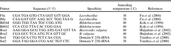
The real-time PCR experiments were performed using the Rotorgene 3000 cycler (Corbett Research, Sydney, Australia) with SybrGreen as the fluorescent marker. Primers used are listed in Table 1. The reaction mixture with a total amount of 20 μl consisted of 0·5 μl DNA template, 0·5 μl of each primer (10 pmol/μl, Table 1), 10 μl QuantiMix EASY KIT (Biotools, Madrid, Spain) and 8·5 μl distilled sterile water. Real-time PCR conditions were as follows: initial denaturation at 95°C for 3 min, followed by 35 cycles with denaturation at 95°C for 20 s, annealing temperature (Table 1) for 20 s and elongation at 72°C for 20 s. After the PCR run, a melting point analysis (72–95°C, 5 s per 0·5°C) and an agarose gel electrophoresis were performed to determine and confirm the specificity of the amplification products.
Primers used in this study

Recovery experiment
To check the reliability of the QIAamp® DNA Stool Mini Kit (Qiagen) and to determine the detection limit of the real-time PCR method, a recovery experiment was performed. Ileal samples, in which B. vulgatus could not be detected, were dosed with aliquots of different dilutions of B. vulgatus (LMG 17767; 107−0 cfu/ml). DNA isolation was undertaken and real-time PCR analyses of the samples were performed to determine the amount of detected B. vulgatus. Additionally, electrophoresis on agarose gel was performed to confirm the results.
Statistical analysis
Statistical analysis was performed using SPSS version 17 (SPSS Inc., Chicago, USA). The analysis of variance (ANOVA) was used to determine the influence of the dietary treatment on bacterial counts. Using the Student–Newman–Keuls test, means of each dietary treatment were compared. Means of the measured variables from different methods were compared with an ANOVA Tukey test.
RESULTS
Real-time PCR results showed specific peaks in melting point analysis, indicating that the primers and PCR conditions were appropriate for the identification of lactobacilli, bifidobacteria, B. vulgatus and the total cell count. Melting point temperatures reached rounded values of 88°C for lactobacilli, 92°C for bifidobacteria, 85°C for B. vulgatus and 89°C for total bacterial counts (primer Sot1/Sot2).
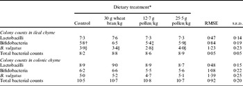
Minimum detection limits for positive controls were 104 cfu/ml of diluted B. thermophilum and B. vulgatus solutions and 105 cfu/ml of diluted L. rhamnosus solution. The detection limit was not low enough to determine bacterial cell counts in 13 ileal samples investigating bifidobacteria and 24 ileal samples analysing B. vulgatus. Mean values of microbial colony counts per feeding group obtained with real-time PCR are shown in Table 2. Statistical analysis of average values showed no significant differences between the four different dietary treatments for all investigated bacteria groups. Average colonic microbial counts were higher compared to ileal counts, except for ileal bifidobacteria counts in the high pollen treatment that showed a higher value than colonic counts. Total microbial counts received with the primer pair Sot1/Sot2 showed the highest results in all feeding groups, followed by lactobacilli, bifidobacteria and finally B. vulgatus. A recovery analysis was performed to check the reliability of the DNA isolation kit and the real-time PCR method and to find the detection limit of the method. For this purpose, ileum samples in which B. vulgatus could not be detected were dosed with different concentrations of B. vulgatus. After DNA isolation and real-time PCR, B. vulgatus could be found in the samples up to 25 cfu/PCR-approach which relates to 104 cfu/ml.
Colony counts received with real-time PCR analyses – results are expressed as log10 of bacteria/g of fresh matter. (n=11 (control), 12 (wheat bran), 12 (12·7 g pollen/kg) and 10 (25 g pollen/kg); RMSE, root mean square error; s.e.d., standard error of difference; total bacterial counts received with primer pair Sot1/Sot2)

* Differences between means were not significant (ANOVA and Student–Newman–Keuls test).
† Three,
‡ four and
§ six values were outside the detection limit and therefore omitted from statistical analysis.
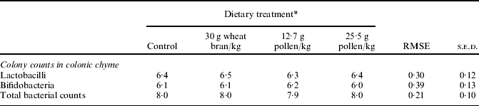
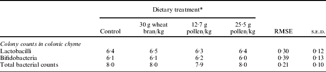
Average values of microbial colony counts received with FISH are shown in Table 3. Total bacterial counts showed the highest colony counts in colonic samples followed by lactobacilli and bifidobacteria. No significant differences could be detected between the four diet groups.
Colony counts received with FISH – results are expressed as log10 of bacteria/g of fresh matter. (n=11 (control), 12 (wheat bran), 12 (12·7 g pollen/kg) and 10 (25·5 g pollen/kg); RMSE, root mean square error; s.e.d., standard error of difference)
* Differences between means are not significant.
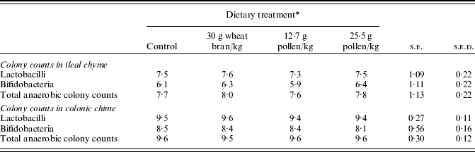
Results obtained with the cultivation method (Table 4) have been reported previously and were included in Table 5 to evaluate present results (Schedle et al. Reference Schedle, Plitzner, Ettle, Zhao, Domig and Windisch2008a).
Colony counts received with the cultivation method – results are expressed as log10 of bacteria/g of fresh matter. (n=11 (control), 12 (wheat bran), 12 (12·7 g pollen/kg) and 10 (25·5 g pollen/kg); s.e., pooled standard error; s.e.d., standard error of difference (Schedle et al. Reference Schedle, Plitzner, Ettle, Zhao, Domig and Windisch2008a))

* Differences between means are not significant.
Statistical differences between different enumeration analyses for colonic chyme (Tukey test)

Culture, cultivation method; PCR, real-time PCR; FISH, fluorescence in situ hybridization.
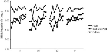
Comparing all three methods, the ANOVA showed significant differences for all investigated parameters. Considering bifidobacteria, the highest bacterial counts were received with the cultivation method. No significant differences were observed between real-time PCR and FISH results, while the cultivation method compared to FISH and real-time PCR showed significant differences (Fig. 1 and Table 5). Comparing the results of all three investigated methods, significant differences could be detected between methods for lactobacilli and for the total bacterial counts (Table 5). In both analyses, FISH showed the lowest values while real-time PCR results and plate counts were about the same. The cultivation method generally presented slightly higher results in lactobacilli and real-time PCR generally showed higher results in total bacterial counts.
Comparison of bifidobacteria counts received with real-time PCR, FISH and the cultivation method (c, control group; p1, low pollen group; p2, high pollen group; w, wheat bran group).

DISCUSSION
Real-time PCR and FISH are appropriate tools to identify and quantify bacterial populations and were used successfully to study human or animal gut microbiota (Gueimonde et al. Reference Gueimonde, Tölkkö, Korpimäki and Salminen2004; Rintillä et al. Reference Rintillä, Kassinen, Malinen, Krogius and Palva2004; Tzortzis et al. Reference Tzortzis, Goulas, Gee and Gibson2005; Child et al. Reference Child, Kennedy, Walker, Bahrami, MacFarlane and MacFarlane2006; Carey et al. Reference Carey, Kirk, Ojha and Kostrzynska2007; Pieper et al. Reference Pieper, Janczyk, Zeyner, Smidt, Guiard and Souffrant2008; Solano-Aguilar et al. Reference Solano-Aguilar, Dawson, Restrepo, Andrews, Vinyard and Urban2008).
The aim of the current study was to evaluate a culture-independent, fast and reliable method to monitor effects of different diets on the gut microbiota of pigs during feeding trials. Therefore real-time PCR and FISH were applied and compared to conventional cultivation based methods. Since real-time PCR and FISH results are compared with those obtained with the cultivation method, all results are expressed as cfu/g fresh sample.
Recovery analysis and minimum detection limits proved the reliability of the DNA isolation kit, and the real-time PCR method and showed that the sensitivity of the PCR method is sufficient for the monitoring of intestinal bacterial populations. The detection limit of the real-time PCR method is comparable with those reported by other authors (Lahtinen et al. Reference Lahtinen, Gueimonde, Ouwehand, Reinikainen and Salminen2006). Total microbial counts received with the Sot1/Sot2 primer pair were highest in all feeding groups, followed by counts for lactobacilli, bifidobacteria and finally B. vulgatus. This can be explained by the specificity of the primers. The applied primer pair Sot1/Sot2 targets the highly conserved region of domain V of 23S RNA and was therefore used as a universal primer to detect bacteria. Furthermore, genus-specific primer pairs were used to detect lactobacilli and bifidobacteria and finally a species-specific primer to analyse B. vulgatus. B. vulgatus was investigated as a member of the genus Bacteroides, which is an important representative of the Gram-negative part of pig gut microbiota (Leser et al. Reference Leser, Lindecrona, Jensen, Jensen and Møller2000). The reported increase in bacterial cell counts from the proximal to the distal part of the gut could be confirmed in the current study as average counts of colonic samples were higher than ileal values (Richards et al. Reference Richards, Gong and De Lange2005). Only ileal average bifidobacteria counts in the high pollen group were higher than in the colon. This may be because six samples did not reach the detection limit; since only four values were obtained, this result is not representative.
To monitor a possible prebiotic effect of the investigated insoluble fibre sources, lactobacilli and bifidobacteria were enumerated. No significant differences could be found between the dietary treatments and therefore no prebiotic effect could be observed. Similar results have been reported in previous studies (Van Nevel et al. Reference Van Nevel, Decuypere, Dierick and Molly2005; Schedle et al. Reference Schedle, Plitzner, Ettle, Zhao, Domig and Windisch2008a). Generally, it is difficult to compare the cell counts found in the current work with other studies because of differences in the methods used or the use of other analytical tools.
Only colonic samples were analysed with FISH because cell counts of the ileal samples were slightly lower than the described detection limit of 105–106 cells/g intestinal sample (Harmsen et al. Reference Harmsen, Gibson, Elfferich, Raangs, Wildeboer-Veloo, Argaiz, Roberfroid and Welling2000; Hogardt et al. Reference Hogardt, Trebesius, Geiger, Hornef, Rosenecker and Heesemann2000). No significant differences could be found between the dietary treatments for all three investigated parameters.
Considering bifidobacteria, the highest bacterial counts were received with the cultivation method, while no significant differences were observed between real-time PCR and FISH results. However, the cultivation method showed significant differences compared to FISH and real-time PCR. Although real-time PCR, in contrast to cultivation methods, also detects dead cells, higher bifidobacterial counts were obtained in the current study with the classical culture technique. This fact may be explained by possible losses due to DNA isolation or the presence of PCR inhibitors in complex chyme samples. Furthermore, the homogeneity of the analysed aliquot needs to be considered. Another reason could be insufficient specificity of the growth media. Rada & Petr (Reference Rada and Petr2002) stated that Tryptone (casein)-Peptone (soy)-Yeast extract (TPY) medium with the addition of mupirocin (100 mg/l), glacial acetic acid (1 ml/l) and colistin (25 mg/l) is appropriate to detect bifidobacteria in pig faeces. However, using Wilkins–Chalgren agar with addition of mupirocin (100 mg/l) and the anaerobic incubation provided by an anaerobic workstation could provide excellent growing conditions and therefore explain higher culture counts.
When examining lactobacilli and the total bacterial counts, all three investigated methods showed significant differences, while in both analyses FISH showed the lowest values. Lower FISH results could be explained by higher specificity of the probe and imperfect hybridization rates. Furthermore, the probe permeation in Gram-positive samples was described as being problematic and may result in low signal intensity. Moreover, due to three-dimensional structure of the rRNA, probe design is difficult and detection problems may occur investigating cells with low rRNA content (Moter & Göbel Reference Moter and Göbel2000; Matsuki et al. Reference Matsuki, Watanabe, Fujimoto, Takada and Tanaka2004). Generally, cultivation results showed slightly higher results in lactobacilli and real-time PCR presented higher results in total bacterial counts. Higher cultivation results might be caused by the application of MRS agar, which is not specific for the enumeration of Lactobacillus spp. but used for counting total lactic acid bacteria. Higher real-time PCR results may be explained due to multiple 16S rRNA operons in chromosomes and the additional detection of dead and viable but not culturable cells (Huijsdens et al. Reference Huijsdens, Linskens, Mak, Meuwissen, Vandenbroucke-Grauls and Savelkoul2002; Bartosch et al. Reference Bartosch, Woodmansey, Paterson, McMurdo and MacFarlane2005; Fu et al. Reference Fu, Carter, Li, Porter and Kerley2006). The presence of PCR inhibitors and low DNA extraction efficiency may be the reason for higher cultivation method results.
In conclusion, it was shown that real-time PCR can be regarded as a good alternative to conventional techniques in practical applications, also compared to FISH, which shows a high limit of quantification. However, real-time PCR also has its drawbacks regarding the choice of primers and interfering factors in complex samples. Therefore, currently the classic cultivation method of plating still remains an important tool when studying effects of feed materials and feed additives on the gut microbiota. Each described method with its benefits is justifiable for certain analysis conditions and targets. Furthermore, the methods provide relevant information on complex communities like the gut microbiota of pigs, both investigating the foregut (performance studies) and the hindgut flora (study of diseases). Dietary additions of wheat bran and pine pollen did not influence total bacterial counts, lactobacilli or bifidobacteria of weanling pigs using real-time PCR as well as FISH. Real-time PCR, FISH and the cultivation method generally gave different results, while consistency of the results depended on the investigated bacteria.
The H. Wilhelm Schaumann Stiftung (Hamburg, Germany) is gratefully acknowledged for granting a scholarship to Agnes Petersson and supporting this work. Many thanks to Elisabeth Moser and Silvia Pfalz for contributing to this project.