INTRODUCTION
Malaria remains a major health problem in the world. The latest survey published by the World Health Organization estimates that 3·2 billion people live in malaria-endemic areas, which accounts for 214 million cases each year and 438 000 deaths (WHO, 2015). Malaria is caused by Plasmodium parasites injected into the mammalian host through the bite of an infected female Anopheles mosquito. The life cycle of Plasmodium parasites oscillates between a mammalian host and an invertebrate vector. In the mammalian host, sporozoites injected by an infected mosquito home to the liver, where they undergo an asymptomatic, yet obligatory, phase of development inside hepatocytes (Prudêncio et al. Reference Prudêncio, Rodriguez and Mota2006). This results in the formation of thousands of erythrocyte-infectious merozoites that invade red blood cells and cause the symptoms of malaria. The transmission of the human parasite to the mosquito is due to the gametocyte sexual form, which, when present in the bloodstream of the human host, can eventually be ingested by the mosquito vector during feeding (Nilsson et al. Reference Nilsson, Childs, Buckee and Marti2015). While P. falciparum is the deadliest human-infective malaria parasite, there are an additional four species of Plasmodium, Plasmodium vivax, Plasmodium ovale, Plasmodium malariae and Plasmodium knowlesi, capable of causing malaria in humans. Moreover, P. vivax and P. ovale are capable forming hypnozoites, dormant parasite forms that may lead to disease relapses long after the initial symptomatic infection has been treated.
Chemotherapy has long been a mainstay in the combat against malaria, but increasing emergence of drug resistance is limiting malarial control (Petersen et al. Reference Petersen, Eastman and Lanzer2011; Price et al. Reference Price, von Seidlein, Valecha, Nosten, Baird and White2014). Another problem regarding current therapies is that most available drugs have a narrow spectrum of action. The front-line antimalarial quinolines chloroquine (CQ), mefloquine and amodiaquine present long half-lives, affordable cost and safety profile, but lack strong activity against sexual and liver parasite forms, which are important stages for interrupting transmission and avoiding relapse, respectively (Prudêncio et al. Reference Prudêncio, Bringmann, Frischknecht and Mueller2015; Stone et al. Reference Stone, Gonçalves, Bousema and Drakeley2015). Therefore, it is necessary to develop new antimalarial drugs with a multistage spectrum of action.
Compounds containing transition metals (i.e. metal complexes) are considered promising antimalarial agents (Scovill et al. Reference Scovill, Klayman and Franchino1982; Gabbiani et al. Reference Gabbiani, Messori, Cinellu, Casini, Mura, Sannella, Severini, Majori, Bilia and Vincieri2009; Khanye et al. Reference Khanye, Smith, Lategan, Smith, Gut, Rosenthal and Chibale2010; Glans et al. Reference Glans, Taylor, de Kock, Smith, Haukka, Moss and Nordlander2011, Reference Glans, Ehnbom, de Kock, Martínez, Estrada, Smith, Haukka, Sánchez-Delgado and Nordlander2012a , Reference Glans, Hu, Jöst, de Kock, Smith, Haukka, Bruhn, Schatzschneider and Nordlander b ; Adams et al. Reference Adams, Li, Khot, De Kock, Smith, Land, Chibale and Smith2013; Barbosa et al. Reference Barbosa, Corrêa, de Oliveira, Rodrigues, Ellena, Nascimento, Rocha, Nonato, Macedo, Barbosa-Filho, Soares and Batista2014; Chellan et al. Reference Chellan, Land, Shokar, Au, An, Taylor, Smith, Riedel, Dyson, Chibale and Smith2014; Adams et al. Reference Adams, de Kock, Smith, Land, Liu, Hopper, Hsiao, Burgoyne, Stringer, Meyer, Wiesner, Chibale and Smith2015). A substantial number of metal complexes containing CQ as a ligand have been investigated as antimalarial agents, employing various metals, a variety of ligands and different numbers of chloroquine molecules. An improvement in the potency against intraerythrocytic stages of P. falciparum was observed in most cases, in comparison with metal-free CQ. An interesting finding is that the antiparasitic potency is not superior to complexes containing two CQ molecules in the structure, which indicates that metal complexes are not mere drug delivery systems that act by releasing CQ and that, instead, the entire chemical structure is involved in the antiparasitic activity (Sánchez-Delgado et al. Reference Sánchez-Delgado, Navarro, Pérez and Urbina1996; Goldberg et al. Reference Goldberg, Sharma, Oksman, Gluzman, Wellems and Piwnica-Worms1997; Lewis et al. Reference Lewis, Behrends, Sá, Cunha, Mendes, Lasitschka, Sattler, Heiss, Kooij, Navarro, Pérez and Sánchez-Delgado1997; Navarro et al. Reference Lewis, Behrends, Sá, Cunha, Mendes, Lasitschka, Sattler, Heiss, Kooij, Navarro, Pérez and Sánchez-Delgado1997, Reference Navarro, Vásquez, Sánchez-Delgado, Pérez, Sinou and Schrével2004, Reference Navarro, Castro, Higuera-Padilla, Sierraalta, Abad, Taylor and Sánchez-Delgado2011a , Reference Navarro, Castro, Martínez and Sánchez Delgado b , Reference Navarro, Castro, Madamet, Amalvict, Benoit and Pradines2014; Rajapakse et al. Reference Rajapakse, Martínez, Naoulou, Jarzecki, Suárez, Deregnaucourt, Sinou, Schrével, Musi, Ambrosini, Schwartz and Sánchez-Delgado2009). Given this promising outlook, CQ analogs have been employed in the metal complex composition in the last years and given rise to many successful outcomes (Dubar et al. Reference Dubar, Egan, Pradines, Kuter, Ncokazi, Forge, Paul, Pierrot, Kalamou, Khalife, Buisine, Rogier, Vezin, Forfar, Slomianny, Trivelli, Kapishnikov, Leiserowitz, Dive and Biot2011, Reference Dubar, Slomianny, Khalife, Dive, Kalamou, Guérardel, Grellier and Biot2013; Glans et al. Reference Glans, Ehnbom, de Kock, Martínez, Estrada, Smith, Haukka, Sánchez-Delgado and Nordlander2012a , Reference Glans, Hu, Jöst, de Kock, Smith, Haukka, Bruhn, Schatzschneider and Nordlander b ; Salas et al. Reference Salas, Herrmann, Cawthray, Nimphius, Kenkel, Chen, de Kock, Smith, Patrick, Adam and Orvig2013; Ekengard et al. Reference Ekengard, Glans, Cassells, Fogeron, Govender, Stringer, Chellan, Lisensky, Hersh, Doverbratt, Lidin, de Kock, Smith, Smith and Nordlander2015). Investigations of the mechanism of action demonstrated that CQ-metal complexes display different interactions with β-hematin, optimal oil-aqueous partition permeability and fast accumulation in the parasitic vacuoles (Martínez et al. Reference Martínez, Rajapakse, Jalloh, Dautriche and Sánchez-Delgado2009, Reference Martínez, Suárez, Shand, Magliozzo and Sánchez-Delgado2011; Navarro et al. Reference Navarro, Castro, Higuera-Padilla, Sierraalta, Abad, Taylor and Sánchez-Delgado2011a , Reference Navarro, Castro, Martínez and Sánchez Delgado b ). Furthermore, it has been shown that the antimalarial activity of ferroquine, a CQ-derived iron organometallic compound, is superior to that of CQ, due to its ability to produce reactive oxygen species (ROS), which induce oxidative stress (Dubar et al. Reference Dubar, Egan, Pradines, Kuter, Ncokazi, Forge, Paul, Pierrot, Kalamou, Khalife, Buisine, Rogier, Vezin, Forfar, Slomianny, Trivelli, Kapishnikov, Leiserowitz, Dive and Biot2011, Reference Dubar, Slomianny, Khalife, Dive, Kalamou, Guérardel, Grellier and Biot2013). However, CQ-metal complexes other than ferroquine have not been fully examined against P. falciparum, especially when regarding their multistage spectrum of action and their in vivo efficacy.
From a pharmaceutical point of view, ruthenium is considered the most promising transition metal for drug development because of its safe and drugable profile (Meier et al. Reference Meier, Novak, Kandioller, Jakupec, Arion, Metzler-Nolte, Keppler and Hartinger2013; Maschke et al. Reference Maschke, Alborzinia, Lieb, Wölfl and Metzler-Nolte2014; Clavel et al. Reference Clavel, Păunescu, Nowak-Sliwinska, Griffioen, Scopelliti and Dyson2015). Regarding the use of CQ during composition of metal complexes, it presents a variety of advantages for drug development, especially pleiotropic effects and the possibility of tuning the spectrum of action by structural modification (Pérez et al. Reference Pérez, Teixeira, Albuquerque, Gut, Rosenthal, Gomes, Prudêncio and Gomes2013; Lin et al. Reference Lin, Spaccapelo, Schwarzer, Sajid, Annoura, Deroost, Ravelli, Aime, Capuccini, Mommaas-Kienhuis, O'Toole, Prins, Franke-Fayard, Ramesar, Chevalley-Maurel, Kroeze, Koster, Tanke, Crisanti, Langhorne, Arese, Van den Steen, Janse and Khan2015; Huta et al. Reference Huta, Mehlenbacher, Nie, Lai, Zubieta, Bou-Abdallah and Doyle2016). Bearing this in mind, we investigated here the activity of CQ-containing organoruthenium complexes. We found that, in addition to the classical activity of CQ against blood stages of P. falciparum, organoruthenium complexes presented a fast-action profile, promising activity against P. berghei liver stages and strong activity against gametocytes. Moreover, compounds presented in vivo efficacy and a mechanism of action involving toxic free heme accumulation in the parasite, which consequently induces oxidative stress.
MATERIALS AND METHODS
Drugs and dilutions
CQ-containing organoruthenium complexes (BCQ, MCQ, FCQ and FFCQ) and organoruthenium complex lacking CQ (FCL) were prepared as described in the literature (Colina-Vegas et al. Reference Colina-Vegas, Villarreal, Navarro, de Oliveira, Graminha, Maia, Deflon, Ferreira, Cominetti and Batista2015). All manipulations were carried out under argon using common Schlenk techniques. Solvents were purified by standard procedures immediately prior to use. Chloroquine, Mefloquine and Artesunate were supplied by FarManguinhos (Rio de Janeiro, Brazil). Primaquine was purchased from Sigma-Aldrich (St. Louis, MO). All drugs were dissolved in DMSO (PanReac, Barcelona, Spain) prior to use, and then diluted in culture medium. The final concentration of DMSO was less than 0·5% in all in vitro experiments.
Drug stability
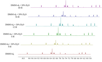
The stability of the complexes in solution was monitored using the 1H NMR technique. Approximately 15 mg of each complex was dissolved in DMSO-d 6 solution containing 20% of D2O (CIL, Tewksbury, MA) and incubated up to 60 h. Aliquots were collected in the indicated time and analyzed on a 9·4 T Bruker Advance III (Billerica, MA) spectrometer with a 5 mm internal diameter indirect probe with Automatic Tuning Matching, holding the temperature stable at 300 K.
Animals
Male Swiss Webster mice (4–6 weeks), housed at Centro de Pesquisas Gonçalo Moniz (Fundação Oswaldo Cruz, Bahia, Brazil), were maintained in sterilized cages under a controlled environment, receiving a rodent balanced diet and water ad libitum. All experiments were carried out in accordance with the recommendations of Ethical Issues Guidelines and were approved by the local Animal Ethics Committee (protocol number 016/2013).
Cell culture
CQ-sensitive 3D7 and CQ-resistant W2 strains of P. falciparum, NK65 strain of P. berghei as well as transgenic P. berghei expressing green fluorescent protein (GFP) and firefly luciferase (Luc), (PbGFP-Luccon, parasite line 676m1cl1) and 3D7 strain P. falciparum 3D7elo1-pfs-16-CBG99 expressing the Pyrophorus plagiophthalamus CBG99 luciferase under the gametocyte specific promoter pfs16 (Cevenini et al. Reference Cevenini, Camarda, Michelini, Siciliano, Calabretta, Bona, Kumar, Cara, Branchini, Fidock, Roda and Alano2014) were used here. P. falciparum was cultivated in human O+ erythrocytes (donated by HEMOBA, Salvador, Brazil) at 5% hematocrit with daily maintenance in Roswell Park Memorial Institute medium (RPMI-1640, Sigma-Aldrich) supplemented with 10% v/v human plasma (donated by HEMOBA, Salvador, Brazil), 25 mm HEPES (ChemCruz, Dallas, TX), 300 µ m hypoxanthine (MP Biomedicals, Santa Ana, CA), 11 mm glucose (Sigma-Aldrich) and 20 µg mL−1 of gentamicin (Life, Carlsbad, CA). Five days prior to use, P. falciparum was cultivated without hypoxanthine and synchronized to rings by 5% D-sorbitol (USB, Santa Clara, CA). Gametocytes were obtained from cultures of asexual parasites as described elsewhere (D'Alessandro et al. Reference D'Alessandro, Silvestrini, Dechering, Corbett, Parapini, Timmerman, Galastri, Basilico, Sauerwein, Alano and Taramelli2013). J774 macrophages were cultured in Dulbecco′s modified Eagle′s medium (DMEM) (Sigma-Aldrich) supplemented with 10% heat-inactivated fetal bovine serum (FBS, Gibco, Gaithersburg, MD) and 50 µg mL−1 of gentamicin (Life). Hepatocellular carcinoma cells (HepG2) were cultivated in RPMI supplemented with 10% heat-inactivated FBS (Gibco) and 50 µg mL−1 of gentamicin (Life).
Mammalian cell toxicity
HepG2 or J774 cells were seeded in 100 µL of RPMI and DMEM, respectively, at 1 × 10⁴ cells mL−1 in 96-well plates. Drugs were added 24 h later in a volume of 100 µL suspended in medium and the plates were incubated for 72 h at 37 °C and 5% CO2. Drugs were tested in eight concentrations (150–0·78 µ m), each one in triplicate. Gentian violet (Synth) was used as positive control, while untreated cells were employed as negative controls. Then, 25 µL of 10% AlamarBlue (Life) were added and incubated for 24 h. Colorimetric readings were performed at 570 and 600 nm using SpectraMAx 190 instrument (Molecular Devices, Sunnyvale, CA). CC50 values were calculated using data-points gathered from three independent experiments.
Drug-induced hemolysis
Fresh and uninfected human O+ erythrocytes were washed three-times with sterile phosphate-buffered saline (PBS), adjusted for 1% hematocrit and 100 µL dispensed in a 96-well round bottom plate. Then, 100 µL of drugs previously in DMSO and suspended in PBS were dispensed in the respective wells. Each drug was tested in seven concentrations (100–0·1 µ m) assayed in triplicate. Untreated cells received 100 µL of PBS containing 0·5% DMSO (negative control), while positive controls received saponin (Sigma-Aldrich) at 1% v/v. Plates were incubated for 1 h at 37 °C under 5% CO2. Plates were centrifuged at 1500 rpm for 10 min and 100 µL of supernatant were transferred to another plate, in which absorbance at 540 nm was measured using a SpectraMax 190 instrument. The percentage of hemolysis was calculated in comparison with positive and negative controls, and plotted against drug concentration generated using GraphPad Prism 5·01. Three independent experiments were performed.
Cytostatic activity against intraerythrocytic P. falciparum
One hundred μL of rings at 1% parasitaemia and 2·5% hematocrit in RPMI were dispensed in a 96-well round bottom plate. Then, 100 µL of drugs (4·0–0·003 µ m) previously suspended in RPMI were dispensed in the respective wells. Each drug was tested in triplicate, in seven different concentrations. Untreated parasite samples received 100 µL of medium containing 0·5% DMSO. Chloroquine and mefloquine were used as positive controls. Plates were incubated for 24 h at 37 °C under 3% O2, 5% CO2 and 91% N2 atmosphere. Then, 25 µL of tritiated hypoxanthine (0·5 µCi well−1, PerkinElmer, Shelton, CT) in RPMI was added to each well and incubated for 24 h. Plates were frozen at −20 °C and subsequently thawed and the contents transferred to UniFilter-96 GF/B PEI coated plates (PerkinElmer) using a cell harvester. After drying, 50 µL of scintillation cocktail (MaxiLight, Hidex, Turku, Finland) was added in each well, sealed and plate read in a liquid scintillation microplate counter (Chameleon, Turku, Finland). The per cent of inhibition was determined in comparison to untreated and inhibitory concentration for 50% (IC50) values were determined by using non-linear regression with Logistic equation available at OriginPro 8·5. Three independent experiments were performed.
Cytocidal activity against intraerythrocytic P. falciparum
One hundred μL of trophozoites at 2% parasitaemia and 3·0% hematocrit in RPMI were dispensed in a 96-well round bottom plate. Then, 100 µL of drugs (10–0·07 µ m) previously suspended in RPMI were added to the respective wells. Each drug was tested in seven concentrations, each one in triplicate. Untreated parasites received 100 µL of medium containing 0·5% DMSO, artesunate was used as positive control. Plates were incubated for 18 h at 37 °C under 3% O2, 5% CO2 and 91% N2 atmosphere. The plate was centrifuged three times with 200 µL of drug-free medium at 1500 rpm for 5 min., then 200 µL of media containing tritiated hypoxanthine was added and plate incubated for 48 h. Plates were frozen at −20 °C and thawed and transferred to UniFilter-96 GF/B PEI coated plates (PerkinElmer) using of a cell harvester. After drying, 50 µL of scintillation cocktail was added in each well, sealed and plate read at liquid scintillation microplate counter. IC50 values were determined employing non-linear regression with Logistic equation available in the OriginPro 8·5 software. Minimal parasiticidal concentration (MPC) was determined as the concentration that reduces parasite growth by 99 ± 1·0%. At least three independent experiments were performed.
Activity in the intraerythrocytic P. falciparum cycle
A volume of 100 µL of rings of P. falciparum W2 strain at 2% parasitemia and 2·5% hematocrit in RPMI was dispensed per well in 96-well round bottom plates. Then, 100 µL of drugs previously suspended in RPMI were added to the respective wells. Each drug concentration was tested in triplicate. Untreated parasite received 100 µL of medium containing 0·5% DMSO. Plates were incubated for 48 h at 37 °C under 3% O2, 5% CO2, 91% N2 atmosphere followed by centrifugation three times with 200 µL of drug-free medium at 1500 rpm for 5 min. A volume of 200 µL of media containing drugs were added and plates were incubated for additional 48 h. Thin blood smears were then prepared, fixed and stained with quick panoptic stain (Laborclin, Pinhais, Brazil). Slides were observed in an optical microscope (Olympus CX41, St. Louis, MO). The number of rings, trophozoites and schizonts were counted in at least 1500 cells per slide (n = 4) and plotted against drug concentration generated using GraphPad Prism 5·01. Two independent experiments were performed.
Activity against P. berghei liver stages
Inhibition of liver-stage infection by test compounds was determined by measuring the luminescence intensity in Huh-7 cells infected with a firefly luciferase-expressing P. berghei line as previously described (Ploemen et al. Reference Ploemen, Prudêncio, Douradinha, Ramesar, Fonager, van Gemert, Luty, Hermsen, Sauerwein, Baptista, Mota, Waters, Que, Lowik, Khan, Janse and Franke-Fayard2009). Briefly, Huh-7 cells, a human hepatoma cell line, were cultured in 1640 RPMI medium supplemented with 10% v/v FBS, 1% v/v non-essential amino acids, 1% v/v penicillin/streptomycin, 1% v/v glutamine and 10 mm 4-(2-hydroxyethyl)-1-piperazineethanesulfonic acid (HEPES), pH 7 and maintained at 37 °C with 5% CO2. For infection assays, Huh-7 cells (1·0 × 104 per well) were seeded in 96-well plates the day before drug treatment and infection. The medium was replaced by medium containing the appropriate concentration of each compound approximately 1 h prior to infection with sporozoites freshly obtained through disruption of salivary glands of infected female Anopheles stephensi mosquitoes. An amount of the DMSO solvent equivalent to that present in the highest compound concentration was used as control. Sporozoite addition was followed by centrifugation at 1700 g for 5 min. Parasite infection load was measured 48 h after infection by a bioluminescence assay (Biotium, Hayward, CA). The effect of the compounds on the viability of Huh-7 cells was assessed by the AlamarBlue assay (Life) using the manufacturer's protocol.
Activity against P. falciparum gametocytes
Drugs were serially diluted in a 96-well round bottom plate (concentration range 29·0–0·22 µ m) in 100 µL per well. Then, 100 µL of 3D7elo1-pfs16-CBG99 gametocytes at 0·5–1% parasitaemia and 2% hematocrit were dispensed. Each drug was tested in triplicate, in seven different concentrations. The DMSO concentration was not toxic for gametocytes. Methylene blue was used as positive control. Plates were incubated for 72 h at 37 °C under 3% O2, 5% CO2, 91% N2 atmosphere. Luciferase activity was taken as measure of gametocytes viability, as previously described in the literature (Cevenini et al. Reference Cevenini, Camarda, Michelini, Siciliano, Calabretta, Bona, Kumar, Cara, Branchini, Fidock, Roda and Alano2014). Briefly, 100 µL of culture medium were removed from each well to increase hematocrit; 70 µL of resuspended culture were transferred to a black 96-well plate; 70 µL of D-luciferin (1 mm in citrate buffer 0·1 M, pH 5·5) were added. Luminescence measurements were performed after 10 min with 500 ms integration time. The IC50 was extrapolated from the non-linear regression analysis of the concentration–response curve. The percentage of gametocytes viability was calculated as 100 × [(OD treated sample – OD blank)/(OD untreated sample – μc-blank)] where ‘blank’ is the sample treated with 500 nM of methylene blue, which completely kills gametocytes.
Determination of the binding constant with ferriprotoporphyrin IX
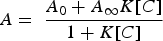
The association constant of organoruthenium complexes to ferriprotoporphyrin IX (Sigma-Aldrich) was measured as described previously (Egan et al. Reference Egan, Mavuso, Ross and Marques1997). Titration of a 2 mL solution (7·5 µ m of ferriprotoporphyrin IX in 40% of DMSO, pH 7·5) in presence of compound (500 µ m in 40% of DMSO, pH 7·5) was performed by UV absorbance at λ = 402 nm using Hewlett Packard spectrophotometer, diode array model 8452 (Santa Clara, CA). The volume of each titration was 5 µL and the relative molar ratio varied from 0 to 10 with regard to [FeIII-PPIX]. Spectra were recorded about 1 min after each addition. The absorption of all compounds was subtracted by adding the same amounts to the blank (40% of DMSO, pH 7·5). Fitting model with a 1:1 association using the equation described by Egan (Egan et al. Reference Egan, Mavuso, Ross and Marques1997):
 $$A = \; \displaystyle{{A_0 + A_\infty K[C]} \over {1 + K[C]}}$$
where, A 0 is the absorbance of hemin before addition of complex or free chloroquine, A ∞ is the absorbance for the drug-hemin adduct at saturation, A is the absorbance at each point of the titration, and K is the conditional association constant. Three independent experiments were performed.
Assessment of inhibition of β-hematin formation by infrared spectroscopy
Polymerization of hemin into β-hematin in acid acetate buffer was studied using the method previously described in the literature (Egan et al. Reference Egan, Ross and Adams1994). Briefly, 12 mg of hemin (Sigma-Aldrich) dissolved in 3 mL of NaOH 0·1 M were added in 0·3 mL of HCl 0·1 M and 1·7 mL of acetate buffer 10 M (pH 5), keeping the temperature at 60 °C during reaction. Primaquine and chloroquine were used as positive and negative controls, respectively. In a control test, after 0, 10, 30 and 60 min, 1 mL of each solution was collected, cooled on ice for 10 min, and then filtered over cellulose acetate (0·22 µm). The effect of the compounds was studied by performing the reaction as described above, adding three equivalents of each compound to the reaction mixture, where the reaction was stopped after 30 min. prior acidification. Pellets were thoroughly washed with water in order to remove acetate salts. Each solid was dried in silica gel and P2O5 for 48 h. Disks were mounted in KBr pellets and infrared spectra were analyzed in a Bomem–Michelson FT MB-102 instrument in the 4000–200 cm−1 region. Three independent experiments were performed.
Assessment of inhibition of β-hematin formation by UV-Vis spectroscopy
The assay was performed according to the method previously described in the literature (Parapini et al. Reference Parapini, Basilico, Pasini, Egan, Olliaro, Taramelli and Monti2000). A solution of hemin chloride (50 µL, 4 mm) dissolved in DMSO was distributed in 96-well plates. Different concentrations (1–100 mm) of each complex was dissolved in DMSO and added in triplicate (50 µL) to a final concentration of 1·25–25 mm well−1. Control contained water or DMSO. The formation of β-hematin was initiated by addition of acetate buffer (100 µL, 0·2 M, pH 4·4). The plates were incubated at 37 °C for 48 h and then centrifuged. After removing the supernatant, the solid was washed twice with DMSO and finally dissolved in NaOH (200 µL, 0·2 N). After diluting with NaOH (0·1 N), absorbance was measured at 405 nm in a spectrophotometer. The inhibition of β-hematin was calculated in comparison with negative control, plotted against drug concentration generated using GraphPad Prism 5·01. Three independent experiments were performed.
SYTO 61 staining of P. falciparum
100 µL of rings of P. falciparum 3D7 strain at 2% parasitemia and 2·0% hematocrit in RPMI were dispensed in a 96-well round bottom plate. Then, 100 µL of drugs previously suspended in RPMI were added to the respective wells. Each drug concentration was tested in triplicate. Untreated parasite received 100 µL of medium containing 0·5% DMSO. Plates were incubated for 48 h at 37 °C under 3% O2, 5% CO2, 91% N2 atmosphere. Plate was centrifuged with 200 µL of drug-free medium at 1500 rpm for 5 min. and 100 µL of SYTO 61 (Life) at 0·5 µ m suspended in medium was added to each well and incubated in the dark for 30 min. After washing and adding 400 µL of isoton diluent, samples were analyzed in a flow cytometer (LSRFortessa, BD, Franklin Lakes, NJ). Gate of infected cells was determined in comparison with uninfected control. At least 200·000 events were acquired in the allophycocyanin channel (633, 660 nm). The analysis was performed using FlowJo (LLC, Ashland, Covington, LA). Three independent experiments were recorded.
CM-H2-DCFDA and SYTO 61 staining of P. falciparum
A volume of 100 µL of rings of P. falciparum 3D7 strain at 3·0% parasitaemia and 1·0% hematocrit in RPMI were dispensed per well in a 96-well round bottom plate. A volume of 25 µL of CM-H2-DCFDA (Life) at 15 µ m suspended in media was added to each well and incubated in the dark for 20 min. Then, 100 µL of drugs previously suspended in RPMI were added to the respective wells. Each drug concentration was tested in triplicate. Untreated parasite received 100 µL of medium containing 0·5% DMSO. Plates were incubated for 3·5 h at 37 °C under 3% O2, 5% CO2, 91% N2 atmosphere. A volume of 25 µL of SYTO 61 (Life) at 0·5 µ m suspended in media was added to each well and incubated in the dark for 30 min. Plates were centrifuged at 1500 rpm for 5 min, supernatant was discarded and 200 µL of isoton diluent was added and samples were analyzed in a flow cytometer (LSRFortessa, BD). Gate of infected cells was determined in comparison with uninfected control. At least 200·000 events were acquired in the fluorescein isothiocyanate channel (488, 585 nm) for CM-H2-DCFDA and allophycocyanin channel (633, 660 nm) for SYTO 61. The analysis was performed using FlowJo (LLC), in three independent experiments.
In vivo blood schizontocidal activity
Male Swiss Webster mice (4–6 weeks) were infected by intraperitoneal injection of 106 NK65 strain P. berghei-infected erythrocytes and randomly divided into groups of n = 5. Each drug was solubilized in DMSO/saline (20:80 v/v) prior administration. Treatment was initiated within 3 h post-infection and given daily for 4 consecutive days orally by gavage or by intraperitoneal injection of 100 µL. Chloroquine treated mice were used as positive control group, while untreated infected mice were used as negative control group. The following parameters were evaluated: parasitaemia counted at 4, 5, 6, 7 and 8 days post-infection and 30 days post-infection animal survival. The per cent of parasitaemia reduction was calculated as follows ([mean vehicle group – mean treated group)/mean vehicle group] × 100%). Two independent experiments were performed.
Ex vivo drug-induced cardiotoxicity in rat hearts using Langendorff system
Male Wistar rats of 6–8 weeks-old were heparinized (800 IU kg−1, i.p.) and following 20 min anaesthetized with ketamine (90 mg kg−1, i.p.) and xylazine (10 mg kg−1, i.p.). Heart was quickly excised via thoracotomy and immediately cannulated through the aorta to retrograde perfusion on the Langendorff system apparatus with peristaltic pump Minipuls 3 (Peristaltic Pump, ADInstruments, Sydney, Australia) under constant flow 6·5 mL min−1. The heart was immersed in Krebs-Henseleit buffer (KHB) solution containing (in mM): 4·7 KCl, 1·2 KH2PO4, 118 NaCl, 25 Na2HCO3, 1·2 MgSO4, 1·75 CaCl2, 0·5 EDTA and 8·0 D-glucose, pH7·4; 37 °C; 5% CO2, warmed to 37 °C. Two electrodes were then positioned at atrium and ventricle to obtain optimal electrocardiographic recordings. These electrodes were connected to the differential inputs of a Bioamplifier (PowerLab 8/35, ADInstrument) and a third was connected to ground. Recordings were displayed on a computer. Experimental protocol consisted of taking control records for 20 min in Krebs’ solution, 10-min, perfusion period with MCQ or FCQ drugs (0·1; 1·0 and 10 µg mL−1) dissolved in DMSO and suspended in KHB solution and a return to Krebs’ solution for 20 min. Each drug was tested in triplicate at the three different concentrations. Electrocardiography recordings were registered as the presence of arrhythmias, heart rate, ventricular activation time, PR intervals, amplitude and time intervals of QT waves. Each drug was tested in at least three isolated hearts, LabChart Pro software including blood pressure and ECG analysis was used.
Statistical analyses
Non-linear regression analysis was used to calculate CC50 and IC50 values by using GraphPad Prism version 5.01 (Graph Pad Software, San Diego, USA), OriginPro version 8.5 (OriginLab, Northampton, USA) or Gen5 1·10 software provided with the Synergy4 plate reader (Biotek, Winooski, USA). One-way ANOVA analysis and Bonferroni multiple comparison tests were used. Results were considered statistically significant when P < 0·05 as analysed by GraphPad Prism version 5.01.
RESULTS
Chemical structure and stability of organoruthenium complexes
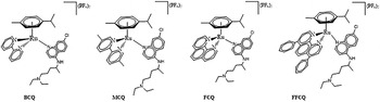
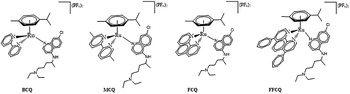
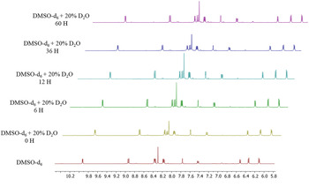
The structures of organoruthenium complexes with general formula [RuCQ(η6-C10H14)(N-N)]2+ studied in this work are shown in Fig. 1, where η6-C10H14 is α-phellandrene and N-N is 2′-bipyridine (BCQ), 5,5′-dimethyl-2,2′-bipyridine (MCQ), 1,10-phenanthroline (FCQ), 4,7-diphenyl-1,10-phenanthroline (FFCQ). An organoruthenium complex lacking CQ in its composition, named FCL, was used during pharmacological evaluation. All these complexes were previously characterized by usual chemical tools (Colina-Vegas et al. Reference Colina-Vegas, Villarreal, Navarro, de Oliveira, Graminha, Maia, Deflon, Ferreira, Cominetti and Batista2015). To assess whether these organoruthenium complexes are dissociating in cell culture media and releasing CQ, stability in 80% DMSO-d 6 and 20% D2O solution was monitored by 1H NMR at different times up to 60 h. Spectra of FCQ samples are shown in Fig. 2. Any modification in intensity and chemical shifts were observed in the spectra up 60 h of incubation time. The same profile was observed after the monitoring of MCQ samples (data not shown).
Chemical structures of organoruthenium complexes.
Organoruthenium complexes stability in aqueous solution in incubation up to 60 h. 1H-NMR spectra of FCQ complex in DMSO-d 6 containing 20% of D2O. The incubation times are displayed in the left side. At least two independent experiments were performed.
Organoruthenium complexes are active against asexual blood stages and display low toxicity against mammalian cells
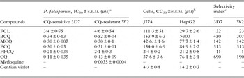
The mean IC50 values of the compounds were determined against CQ-sensitive and CQ-resistant strains of P. falciparum, while the CC50 values in mammalian cells were determined against J774 macrophages and HepG2 hepatocellular cells (Table 1). In the CQ-sensitive strain, all complexes were 2-fold less active than CQ. In the CQ-resistant strain, with the exception of compound FFCQ, the activity of complexes was similar to that of CQ. Apart from the FFCQ complex, susceptibility to drugs tested was in general similar for CQ–sensitive and CQ –resistant strains. Complexes did not either cause hemolysis in concentration up 100 µ m (Fig. S1, supporting information) or cardiotoxicity in ex vivo isolated rat hearts (Table S1, supporting information). Regarding mammalian cytotoxicity, CC50s for complexes were 5- to 10-fold higher than for the reference drug gentian violet. The only exception was observed for the FFCQ compound, which was cytotoxic in low micromolar concentrations. The cytotoxicity of complexes was similar or lower than that of CQ. Calculated selectivity index (SI) values of 513 and 450 were obtained for FCQ and BCQ, respectively, which are similar or higher than that of CQ. Of note, the FCL complex showed low antiparasitic activity and SI values several folds lower than CQ-ruthenium complexes.
Cytostatic activity against intraerythrocytic P. falciparum, mammalian cell cytotoxicity and selectivity indexes of organoruthenium complexes
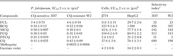
a Determined 48 h after incubation with compounds.
b Determined 72 h after incubation with compounds. Values were calculated as mean of three independent experiments.
c Determined as CC50 (J774 cells)/IC50. IC50, inhibitory concentration at 50%. CC50, cytotoxic concentration at 50%. s.e.m., standard error of the mean.
Organoruthenium complexes impair asexual parasite differentiation
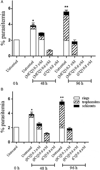
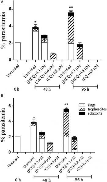
Once antiparasitic activity had been observed, the onset of action and the activity against erythrocytic parasite cycle was investigated for MCQ and FCQ complexes (Fig. 3). Treatment with an IC50 concentration of MCQ decreased parasitaemia at the onset of drug exposure, while an accumulation of parasites in the trophozoite stage and consequent impairment to schizont progression were observed 96 h post exposure. In the presence of approximately 2-fold the IC50 of MCQ, substantial accumulation of trophozoites was achieved 48 h post exposure, while progression in parasite growth and differentiation was abrogated 96 h post exposure. Similar treatments with FCQ led to a similar impairment on parasite cycle at the IC50 values of the drugs and complete blockage of the parasite cycle twice that concentration. A morphological examination of stained blood smears revealed that during treatment with the IC50 concentration of MCQ and FCQ, digestive vacuoles containing hemozoin crystals were absent in trophozoites (data not shown). This is typically observed for CQ, a well-known hemozoin inhibitor. At higher concentration of complexes, most parasites appeared as a picnotic mass.
Drug-susceptibility testing against blood stage W2 strain P. falciparum. Ring stage parasites (2% parasitaemia, 2·5% hematocrit) were incubated with vehicle (DMSO 0·5%) as a untreated control, CQ, MCQ (panel A) and FCQ (panel B) at 0 and 48 h. Quantification of intraerythrocytic stages at 48 and 96 after addition of the compounds are shown. Values are shown from one of two independent experiments. Error bars represent standard deviation. *p < 0·05 for quantification of rings vs untreated 0 h. **p < 0·05 for quantification of trophozoites vs untreated 48 h. CQ, chloroquine.
Organoruthenium complexes are fast-acting parasiticidal agents against asexual blood stages
To assess the parasiticidal activity of complexes, time- and concentration-dependent effects were studied in synchronized P. falciparum trophozoites. Growth inhibition by drug exposure for 6, 18, 24 or 48 h was monitored by microscopy examination. At 2 × the IC50 values of the FCQ and MCQ, parasite inhibition was detected after 18 h of compound exposure (data not shown). This incubation time was selected for the parasiticidal activity study. After 18 h of exposure to different drug concentrations, followed by extensive washing to remove drugs, the culture was maintained for 48 h and growth was quantified by hypoxanthine incorporation relative to untreated control. Artesunate was used as a positive control. Parasite clearance was estimated as 99 ± 1·0% parasite growth and expressed as MPC. Table 2 shows mean LC50 and MPC values. Complexes show parasiticidal activity, with LC50 values comparable with CQ. MPC values show that MCQ was more potent than FCQ. The parasiticidal potency of the complexes was lower than artesunate but higher than CQ, which did not lead to parasite clearance in concentrations up 10 µ m.
In vitro parasiticidal property of organoruthenium complexes against intraerythrocytic P. falciparum

a Activity determined in trophozoites incubated for 18 h with drugs then for 48 h in drug-free conditions.
b LC50, lethal concentration at 50%.
c MPC, minimal parasiticidal concentration.
Antiparasitic activity is not governed by inhibition of β-hematin formation
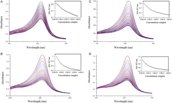
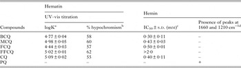
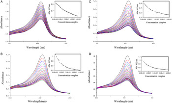
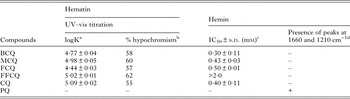
Given their potent in vitro growth inhibition against trophozoites, the effects of organoruthenium complexes within hemin polymerization as well as their interaction to ferriprotoporphyrin IX (α-hematin) were investigated. Firstly, we determined the binding constant to ferriprotoporphyrin IX by analyzing the absorption spectra at various concentrations of complexes. As shown in Fig. 4, increasing the concentration of complex caused a reduction in the absorption intensity, ranging from 60 to 70%. These titration curves were further fitted to a 1:1 association model to give the binding constant values expressed as logK (Table 3). LogK values for organoruthenium complexes were lower than the observed for CQ and in general, there was no great difference when the complexes were compared with each other.
Spectroscopic titration of Fe(III)PPIX at pH 7·4 with organoruthenium complexes. Panel A, FCQ complex; Panel B, FFCQ complex; Panel C, BCQ complex; Panel 4, MCQ complex.
Binding constant for hematin and inhibition of hemin polymerization for organoruthenium complexes
a Binding constant expressed as logK.
b Determined in comparison with untreated (no drug).
c Determined 48 h after drug incubation.
d Determined by infrared spectrum.
After measuring the interaction with hematin, the compounds’ ability to inhibit hemin aggregation into β-hematin was evaluated by UV-Vis spectroscopy analysis (Soret's porphyrin band). This experiment was performed in acetate buffer at pH 4·9 and determined 48 h after incubation (Table 3). Comparison of IC50 values revealed that FFCQ complex was unable to inhibit hemin aggregation process in concentration up 2·0 mm. In contrast, other organoruthenium complexes inhibited hemin aggregation with potency similar to CQ. Additionally, the hemin aggregation process was evaluated by infrared spectra. Figure S2 illustrates the infrared spectrum of hemin in the absence or presence of CQ, primaquine and organoruthenium complexes. Over time, hemin aggregates into β-hematin, leading to the formation of iron-carboxylate bonds observed as bands at 1·660 and 1·210 cm−1. Adding primaquine did not impair β-hematin formation, while adding CQ or its ruthenium complexes inhibited β-hematin formation.
Oxidative stress induced by organoruthenium treatment
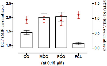
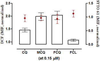
Firstly, we determined parasite viability in 3D7 strain trophozoites by staining with SYTO 61 accordingly to a previously described protocol (Fu et al. Reference Fu, Tilley, Kenny and Klonis2010) and analyzing by flow cytometry. Treatment at 1·25 µ m of CQ, MCQ and FCQ for 48 h incubation time led a population of non-viable parasites, while at 0·31 µ m we observed two populations: a non-viable population and another that is viable but with decreased SYTO 61 staining, possible due to delay in parasite growth (Fig. S3, supporting information). Trophozoites were treated with CQ or complexes and incubated for 4 h and co-labelled with ROS probe CM-H2DCFDA and SYTO 61 (Fig. S4, supporting information). Parasite viability following treatment at 0·15 µ m and monitored by SYTO 61 staining was not altered in comparison to untreated parasites (Fig. 5). In contrast, at the same incubation and treatment, MCQ and FCQ increased DCF fluorescence levels in approximately 2-fold in comparison with untreated parasites. CQ treatment at same concentration increased in approximately 50% DCF fluorescence, while the complex lacking CQ in its composition (FCL) did not increase DCF when compared with untreated parasites.
3D7 strain P. falciparum trophozoites (3·0% parasitaemia, 1% hematocrit) double stained with CM-H2-DCFDA (probe for reactive oxygen species) and SYTO 61 (probe for cell viability). Drugs were incubated for 4 h and analysed by flow cytometry. Bars represent DCF signal and red dots represent SYTO 61 signal. MIF were normalized from untreated control and obtained from pooling data gathered of two independent experiments. MIF, median intensity fluorescence. Error bars represent standard deviation.
Multistage action of organoruthenium complexes
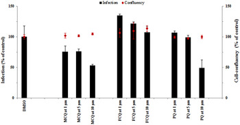
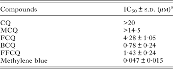
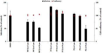
We next examined whether ruthenium complexes possess multistage activity. Firstly, the ability to inhibit Plasmodium hepatic infection was assessed in a well-established in vitro infection model, employing P. berghei-infected Huh7 cells, a human hepatoma cell line (Prudêncio et al. Reference Prudêncio, Mota and Mendes2011). Huh7 cells were cultured in the presence of each complex followed by addition of luciferase-expressing P. berghei sporozoites. Infection load and host cell cytotoxicity were analyzed 48 h post-infection. Primaquine, the only licensed drug against Plasmodium liver stages (Rodrigues et al. Reference Rodrigues, Prudêncio, Moreira, Mota and Lopes2012), was used as the reference drug. As shown in Fig. 6, the FCQ compound did not decrease infection in comparison to the untreated control. The MCQ compound inhibited approximately 50% infection at a 10 µ m concentration, without affecting host cell confluency. A similar concentration of primaquine inhibited infection by 50%. For comparison, CQ reduces hepatic infection by 50% at a 15 µ m concentration (Rodrigues et al. Reference Rodrigues, Prudêncio, Moreira, Mota and Lopes2012). We then assessed the transmission-blocking potential of each compound against stage V P. falciparum gametocytes. Inhibition of viability of stage V P. falciparum 3D7 gametocytes was evaluated and the compounds’ IC50 values were determined. Methylene blue was used as positive control (Table 4). In concentrations up 14 µ m, the MCQ complex did not inhibit gametocytes, while the FCQ and FFCQ complexes presented inhibitory activity. The BCQ complex was the most potent, exhibiting an IC50 value in the low micromolar range. Although less potent than methylene blue, this compound was active, while CQ is inactive in impairing gametocyte viability.
Drug-susceptibility testing against P. berghei sporozoites. Seeded Huh7 cells were treated with drugs and infected with sporozoites. Bioluminescence intensity was measured 48 h post-infection. Bars represent infection and red dots represent Huh7 cell confluency. Untreated control received only DMSO. PQ, Primaquine. Error bars represent standard deviation from each concentration in triplicate. Results of two independent experiments.
In vitro activity of organoruthenium complexes against stage V gametocytes of 3D7 P. falciparum
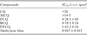
a Activity determined in gametocytes incubated for 72 h with drugs. Three independents experiments performed, each concentration in duplicate.
Organoruthenium complexes reduce parasitaemia in infected mice
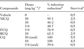
Before evaluating efficacy, compounds were examined regarding lethal doses in Swiss Webster mice (n = 3). A single intraperitoneal injection of MCQ or BCQ was not lethal at a dose of 50 mg kg−1 weight−1. FCQ was lethal at 50 mg kg−1, but safer at 5·0 mg kg−1. Due to its low SI, FFCQ was not tested in mice. By following 4-day treatment test in P. berghei-infected mice, parasitaemia suppression and survival rate were evaluated in comparison with untreated infected mice (vehicle). CQ was used as the reference drug (Table 5, Fig. S5). On day 8 post-infection, 50 mg kg−1 of MCQ caused a 95·1% reduction in parasitaemia in comparison with untreated group, with 40% of animal survival observed 30 days post-infection. When MCQ was used at 10 mg kg−1, a 46·2% reduction in parasitaemia was observed and no survival was achieved. In infected mice treated with FCQ, parasitaemia was reduced by 76·9% vs untreated group. Treatment with 10 mg kg−1 BCQ led to a 62·3% reduction in parasitaemia and 40% protection in animal survival vs untreated group. In comparison, treatment with CQ at same dose reduced in 100% the parasitaemia and conferred 80% protection in animal survival.
Summary of activities of organoruthenium complexes against blood stage in vivo infection
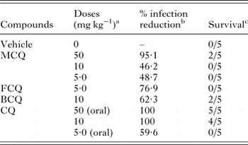
a Given for 4 days, by intraperitoneal route, with the exception for CQ at 50 and 5·0 mg kg−1, which were given orally. Vehicle = DMSO:saline (20:80 v/v).
b Determined on day 8 post infection.
c Number of mice alive 30 days post infection/(n = 5).
DISCUSSION
The insertion of CQ into organoruthenium complex resulted in compounds with antiparasitic activity, while a ruthenium complex lacking CQ did not present such property. Most complexes displayed potency and selectivity against the Plasmodium blood asexual stage with a magnitude similar to that of free CQ. Unspecific cytotoxicity was only observed for the FFCQ complex, possibly due to the presence of 4,7-diphenyl-1,10-phenanthroline ligand in its composition. Like CQ, complexes presented activity against all forms of blood asexual stages (rings, trophozoites and schizonts), which is an important characteristic to reduce a mixed parasite population during infection. A common feature of metal complexes is dissociation in solution, with concomitant ligand exchange (Peacock and Sadler, Reference Peacock and Sadler2008). Here we showed that these organoruthenium complexes do not release CQ upon solution in the same incubation time in the pharmacological evaluation, indicating that the entire organoruthenium complex is responsible for activity.
CQ-complexes present advantages in terms of pharmacological profile. Firstly, complexes inhibited the growth of all tested P. falciparum strains, irrespective of their drug resistance background. Secondly, they exhibited an onset of action detectable after 18 h drug exposure, which is faster than what is observed for CQ treatment. The LC50 values for complexes fall in the same micromolar range of IC50 values, indicating that compounds act as a cytotoxic rather than as a cytostatic drug. The MPC values revealed that complexes were able to clear parasites after 18 h drug exposure. Although these properties were not as potent as those of the fast-acting artesunate, they were superior to those observed for CQ. These findings show that ruthenium complexes induce parasite killing and this is achieved in short time. The fast parasite killing rate displayed by these complexes is attractive, especially because this can shorten treatment time as well as prevent parasite escape.
We further observed that ruthenium complexes present activity in the liver and in the blood sexual stages. In the liver-stage Plasmodium, MCQ complex displayed antiparasitic activity comparable with Primaquine and superior to CQ. Regarding the action in the sexual stage, we observed reduction of gametocyte viability in response to ruthenium complex treatment. Therefore, unlike CQ, which is classically an effective drug during blood schizogony, the ruthenium complexes show a multistage antiplasmodial profile. In fact, the effective concentrations of ruthenium complexes against sporozoites and gametocytes stages are higher than in the blood asexual stages. However, to the best of our knowledge, this is the first time that a multistage activity profile is demonstrated for metal complexes. This is desirable since drugs targeting different stages of the parasite vertebrate life cycle can work effectively in the prevention and against the relapse of malaria.
The strong activity of the complexes against trophozoites, where hemozoin formation takes place, led us to investigate whether organoruthenium complexes inhibit β-hematin formation. Except FFCQ, which was inactive, other complexes inhibited polymerization of hemin into β-hematin with potency similar to CQ. In contrast, all complexes presented lower binding constant to ferriprotoporphyrin IX (α-hematin) than CQ. These results argued that complexes are more potent in interaction with hemin rather than β-hematin, which is a finding also observed for other CQ-metal complexes (Martínez et al. Reference Martínez, Rajapakse, Jalloh, Dautriche and Sánchez-Delgado2009). CQ binds to free heme through its quinoline nitrogen, while in the CQ-ruthenium complexes this nitrogen is bound to ruthenium, indicating that these complexes interact with heme in a binding process different to CQ.
We demonstrated that the organoruthenium treatment increased ROS levels in trophozoites, where β-hematin formation is most active. Their effects on inducing oxidative stress were more pronounced than CQ-treatment and absence under treatment with CQ-lacking complex FCL. Importantly, these properties were observed in viable parasites, which reflect a primary mechanism of action of organoruthenium complexes rather than secondary consequences of cell death. Therefore, organoruthenium complexes achieved antiparasitic activity against asexual forms because they inhibit β-hematin formation, which cause an insult in parasites since it raises toxic free heme and consequently produces oxidative stress.
MCQ complex exhibited a dose-dependent effect and presented an optimal efficacy when treatment was given at 50 mg kg−1. This reduced blood parasitaemia and increased survival, showing that organometallic complexes are efficacious and kill parasites. A similar property was observed for BCQ treatment, while the FCQ complex presented narrow therapeutic window, restricting its evaluation. As a limitation, the efficacy of organoruthenium complexes is inferior to CQ treatment. Apart from FFCQ complex, unspecific cytotoxicity was not a concern since complexes were not toxic for two different cell linages (macrophages and hepatocellular cells). Absence of hemolysis of uninfected erythrocytes and of cardiotoxicity in isolated hearts suggests low toxicity for these organoruthenium complexes. Therefore, a major challenge that remains is to optimize the efficacy of this class of compounds to enable effective reduction of parasitaemia in vivo.
CONCLUSION
We have evaluated the antimalarial activity of organoruthenium complexes containing CQ. We showed that this class of compounds is stable in mixed DMSO-aqueous media without releasing CQ, and presented potency against blood asexual forms similar to CQ. Organoruthenium complexes exhibited moderate activity against liver stage and potent activity against sexual stage, while CQ is devoid of such properties. They operate by a mechanism of action that is distinct from that of CQ, by causing oxidative stress. Importantly, we demonstrated that organoruthenium complex treatment presented efficacy in inhibiting parasitaemia in mice, pointing out that further pharmacological investigation, as well as chemical modification are relevant to strengthen antimalarial drug development based on inorganic compounds.
SUPPLEMENTARY MATERIAL
The supplementary material for this article can be found at http://dx.doi.org/10.1017/S0031182016001153.
ACKNOWLEDGEMENTS
We are thankful to the flow cytometry facility of CPqGM (Brazil) and AVIS Comunale Milano (Italy) for providing blood samples for gametocyte culture.
FINANCIAL SUPPORT
This research was funded by FAPESB (grant PET0042/2013, Brazil) to M.B.P.S, FAPESP (grant 14/10516-7, Brazil) to A.A.B. and Fundação para a Ciência e Tecnologia (grant PTDC/SAU-MIC/117060/2010 Portugal) to M.P. A.A.B. and M.B.P.S. are recipients of senior fellowships by CNPq (Brazil).
TRANSPARENCY DECLARATIONS
The authors declare no competing financial interest.
CONTRIBUTING AUTHORS
A.A.B., D.R.M.M. and M.B.P.S. initiated the project and provided guidance for experimental design and interpretation of data. T.S.M. performed in vitro and in vivo drug susceptibility studies; M.D.P. assisted with cell culture; L.C.V. and M.N. synthesized and validated drugs, performed hematin assays; M.P. and M.M. performed liver stage drug activity assays; S.D. and NB performed drug susceptibility assays in gametocytes; P.C.M.O., B.C.B. and S.G.M. performed cardiotoxicity evaluation; D.R.M.M. initiated manuscript preparation. All authors have read and approved the final manuscript.