The importance of small dairy ruminants has increased significantly during recent years, especially in developing countries where they are an interesting and important alternative for the supply of dairy products for human consumption, whereas in developed countries they are considered as a healthier alternative to cows’ milk with significant organoleptic properties, either for direct consumption (e.g. Northern Europe) or cheese-making (e.g. Mediterranean basin). Additionally, it is often considered that dairy production in developing countries is an essential tool to overcome social and economic issues such as poverty and malnutrition, particularly in infants (McDermott et al. Reference McDermott, Staal, Freeman, Herrero and Van De Steeg2010). Therefore it is critical to understand the modifications associated with lactation in the mammary gland in order to develop strategies to improve milk yield or reduce the effect of diseases that decrease milk production and milk quality, with particular relevance to mastitis. Indeed, there are several factors related to this organ that may alter the lactation cycle, particularly the number of previous lactations or milking frequency.
The glandular parenchyma is responsible for milk production and is constituted by tubule-alveolar glands. Relative to its anatomical organisation, it has two main components, the parenchyma which includes the epithelial and myoepithelial cells, the stroma involving the non-cellular components, as collagen and elastin, smooth muscle cells and vessels and the ductal system (Nickel et al. Reference Nickel, Schummer and Seiferle1981). However, it is important to emphasise that mammary gland anatomy and histology are modified throughout the lactation cycle, mainly led by the neuroendocrine system. Indeed, there are three stages of mammary biology which characterise the gestation/lactation cycles: proliferation, secretion and involution. Although most proliferation occurs during pregnancy and most of the involution takes place after secretion has finished, these processes overlap: proliferation of secretory tissue continues during early lactation and involution begins during late lactation, concomitantly with secretion (Knight & Peaker, Reference Knight and Peaker1984).
Concerning the lactation period, it differs between small ruminant species. In sheep (Ovis aries) lactation last for 5 months with a peak between the weeks 3 and 4 (Zamiri et al. Reference Zamiri, Qotbi and Izadifard2001; Oravcová et al. Reference Oravcová, Peškovičová, Daňo, Milerski, Hetényi and Polák2006). In contrast, the lactation period in goats (Capra hircus) lasts for 10 months with a peak between weeks 5 and 10 (Salama et al. Reference Salama, Caja, Such, Casals and Albanell2005). These values are highly dependent on breed and nutritional status, among other factors (Snowder & Glimp, Reference Snowder and Glimp1991).
Considering the current importance of sheep and goat milk production, the aim of this review is to analyse the colostrogenesis and lactogenesis processes and the importance of the neuroendocrine system in mammary gland development, but most importantly to systematise and highlight the various morphological mammary gland patterns during lactation. Indeed, there is a strong relation between the lactation curve and different patterns in the mammary gland, raising the need to increase knowledge of, for instance, the association of specific periods of cell proliferation/decline with the lactation cycle itself. In this review we aim also to understand the effect of milking frequency, lactation number and different diets on mammary gland patterns, as well as the lactogenesis and colostrogenesis processes. Overall, these data will be important in supporting further studies aimed at improving lactation yield and mastitis prevention, among other factors, with benefits for this emerging dairy sector for both the industry and the consumer.
The importance of colostrogenesis
This complex process can be defined as the pre-partum transfer of components, mainly immunoglobulins, from maternal bloodstream into mammary secretions during a discrete and finite period (Barrington & Parish, Reference Barrington and Parish2001) since this process ceases abruptly immediately prior to parturition (Brandon et al. Reference Brandon, Watson and Lascelles1971). There are several factors that can affect the final colostrum composition, such as the species, breed, age, nutrition, litter size, length of dry period and health status (Csapó et al. Reference Csapó, Csapó-Kiss, Martin, Szentpeteri and Wolf1994; Awadeh et al. Reference Awadeh, Abdelrahman, Kincaid and Finley1998; Maunsell et al. Reference Maunsell, Morin, Constable, Hurley, McCoy, Kakoma and Isaacson1998), although colostrogenesis is still not fully understood (Barrington et al. Reference Barrington, McFadden, Huyler and Besser2001; Castro et al. Reference Castro, Capote, Batista, Bruckmaier and Argüello2011b). In general, colostrum is formed and stored in the mammary gland during late pregnancy (Linzell & Peaker, 1974) and there are several mechanisms that regulate input of colostrum secretion components into the mammary gland (Patton & Jensen, Reference Patton and Jensen1975) which are controlled by local and systemic factors (Castro et al. Reference Castro, Capote, Batista, Bruckmaier and Argüello2011b).
The importance of colostrum in newborn ruminants
The behaviour of the newborn ruminant and the mother after partum, as well as the management during that period, will deeply influence its chances of survival (Nowak et al. Reference Nowak, Porter, Lévy, Orgeur and Schaal2000). Mortality rates in newborn ruminants are high and variable depending on the farm, being 10–25% in lambs and 7–50% in goat kids (Mellor & Stafford, Reference Mellor and Stafford2004). As a consequence, not only is the animal welfare decreased in these conditions, but also the general benefits for farmers. It has been described that the immune system of ruminant neonates seems to be more or less unable to produce its own immunoglobulins during the first month of life (Logan & Pearson, Reference Logan and Pearson1978). For this reason, the importance of colostrum administration and newborn ruminant survival has been deeply studied (Argüello et al. Reference Argüello, Castro, Capote, Tyler and Holloway2004; Castro et al. Reference Castro, Capote, Álvares and Argüello2005, Reference Castro, Capote, Morales-delanuez, Rodríguez and Argüello2009, Reference Castro, Capote, Batista, Bruckmaier and Argüello2011b). Colostrum feeding is also important since it participates in the regulation of body temperature in newborn ruminants. Indeed, Dwyer & Morgan (Reference Dwyer and Morgan2006) observed that lambs that were active at birth, and therefore had better colostral nutrient supply to fuel thermoregulation, were better able to maintain their body temperature than less active lambs. Following this line, dos Santos et al. (Reference dos Santos, Bertolini, Prado, Macedo, Prado and Martins1994) observed that newborn goat kids increased their rectal temperature after colostrum feeding. In addition, colostrum is also important owing to its high magnesium content, playing an essential role in activation of peristalsis. Several authors have pointed out that colostrum promotes the ejection of meconium, avoiding the bacterial colonisation of the intestinal mucosa (García de Jalón et al. Reference García de Jalón, De las Heras, Ferrer and Sancho1990; Barza et al. Reference Barza, Marinescu and Blaga1993).
Colostrum composition
Colostrum contains a complex mixture of diverse components, such as fat, lactose, vitamins and minerals with high nutritional importance to the newborn ruminant (Ontsouka et al. Reference Ontsouka, Bruckmaier and Blum2003; Hernández-Castellano et al. Reference Hernández-Castellano, Almeida, Castro and Argüello2014a, Reference Hernández-Castellano, Almeida, Ventosa, Coelho, Castro and Argüellob). Despite its nutritional function, colostrum also contains a complex mixture of proteins that actively participate in protection of the neonate against pathogens and other post-partum environmental challenges (Bendixen et al. Reference Bendixen, Danielsen, Hollung, Gianazza and Miller2011). Moreover, colostrum also contains other components such as vitamins, hormones, growth factors, cytokines, enzymes (Blum & Hammon, Reference Blum and Hammon2000; Vetter et al. Reference Vetter, Argüello, Baumrucker and Bruckmaier2013) and metabolites derived from alveolar epithelial cells (Peaker & Linzell, Reference Linzell and Peaker1975) and immunocompetent cells (Lee et al. Reference Lee, Wooding and Kemp1980). Accordingly immunoglobulins are the most deeply studied proteins, as they play a crucial and key role in the protection of newborn ruminants against infections (Moreno-Indias et al. Reference Moreno-Indias, Sánchez-Macías, Castro, Moralez-delaNuez, Hernández-Castellano, Capote and Argüello2012). The most important immunoglogulins present in colostrum are IgG, IgM and IgA. As can be observed in Table 1, the concentration of different colostrum components varies among species.
Concentrations of immunoglobulins and chemical composition of sheep and goat milk and colostrum

† Adapted from 1Moreno-Indias et al. (Reference Moreno-Indias, Sánchez-Macías, Castro, Moralez-delaNuez, Hernández-Castellano, Capote and Argüello2012), 2Hernández-Castellano et al. (Reference Hernández-Castellano, Torres, Alavoine, Ruiz-Díaz and Argüello2011), 3Torres et al. (Reference Torres, Castro, Hernández-Castellano, Argüello and Capote2013a, Reference Torres, Castro, Argüello and Capoteb), 4Raynal-Ljutovac et al. (Reference Raynal-Ljutovac, Lagriffoul, Paccard, Guillet and Chilliard2008),5Ciuryk et al. (Reference Ciuryk, Molk, Kaczor and Bonczar2004) and 6Hurley & Theil (Reference Hurley and Theil2011)
Factors affecting colostrum composition
As described above, colostrum composition can be affected by several factors.
Gestation length has been highly correlated with the composition of colostrum in small ruminants. Accordingly Swanson et al. (Reference Swanson, Hammer, Luther, Carlson, Taylor, Redmer, Neville, Reed, Reynolds, Caton and Vonnahme2008) observed that ewes with short pregnancy periods (146·9 d), produced colostrum less concentrated in IgG (99·9 mg/ml) than others with 150-d gestations (127·7 mg/ml). In agreement with these results, Castro et al. (Reference Castro, Capote, Batista, Bruckmaier and Argüello2011a) showed that induction of parturition in dairy goats produced colostrum with a lower IgG concentration (∼90 mg/ml) than those produced by goats in which parturition was not induced (∼140 mg/ml).
Litter size is another important factor affecting colostrum composition. Nowak & Poindron (Reference Nowak and Poindron2006) reported that litter size is related with the amount of colostrum accumulated in the udder before partum. The authors described that twin-bearing ewes generally yielded more colostrum than single-bearing ewes, but the onset of lactation was slower in the former and they did not produced so much colostrum per lamb. In addition, Csapó et al. (Reference Csapó, Csapó-Kiss, Martin, Szentpeteri and Wolf1994) demonstrated that IgG concentration increased in ewes and goats bearing twins than in single bearing animals (118·4 vs. 98·7 and 132·4 vs. 112·2 mg/ml of IgG in colostrum from twin-bearing and single-bearing ewes and goats, respectively). Nevertheless, Argüello et al. (Reference Argüello, Castro, Álvarez and Capote2006) found no effect of the litter size on the colostrum composition of Majorera dairy goats. Similarly Romero et al. (Reference Romero, Beltrán, Rodríguez, De Olives and Molina2013) reported no differences in the colostrum IgG concentration of single- and multiple-partum goats, although these authors reported an increment of colostrum protein and lactose percentage in single-partum goats (9·10 and 3·92%, respectively) relative to the respective percentages in colostrum from multiple-partum goats (7·86 and 3·57%, respectively).
Nutrition during pregnancy is another well-studied factor that affects not only colostrum composition but also colostrum yield in small ruminants (Castro et al. Reference Castro, Capote, Batista, Bruckmaier and Argüello2011b). Meyer et al. (Reference Meyer, Reed, Neville, Thorson, Maddock-Carlin, Taylor, Reynolds, Redmet, Luther, Hammer, Vonnahme and Caton2011) studied the interactions of two Se supplementations (11·5 μg/kg of body weight (BW) and 77·0 μg/kg of BW) and three nutritional managements (60, 100 and 140% of the dietary requirements) on the sheep colostrum composition. Results showed that a higher Se supplementation (77·0 μg/kg of BW) and a diet providing 100% of recommended dietary requirements during pregnancy (NRC, 1985) increased the colostrum yield, as well as the fat, lactose and protein percentages. In agreement with these findings, Banchero et al. (Reference Banchero, Perez Clariget, Bencini, Lindsay, Milton and Martin2006) showed that ewes fed during pregnancy with 110% of metabolisable energy (ME) requirements had higher colostrum yield than those fed with 60% ME requirements. In contrast, Swanson et al. (Reference Swanson, Hammer, Luther, Carlson, Taylor, Redmer, Neville, Reed, Reynolds, Caton and Vonnahme2008) found that supplementing ewes with Se had no effect on colostrum yield or colostral IgG concentration.
When small ruminants are kept under poor grazing conditions, there is a general mobilisation of their body reserves during the last 6 weeks of gestation owing to rapid fetal growth and colostrum yield (Meyer et al. Reference Meyer, Reed, Neville, Thorson, Maddock-Carlin, Taylor, Reynolds, Redmet, Luther, Hammer, Vonnahme and Caton2011). In these conditions, Ramirez-Vera et al. (Reference Ramirez-Vera, Terrazas, Delgadillo, Serafín, Flores, Elizundia and Hernández2012) showed that corn-supplemented goats (0·6 kg/d during the last 12 d of pregnancy) had higher colostrum yield than non-supplemented goats.
The importance of lactogenesis
Lactogenesis may be defined as the initiation of milk secretion (Cowie et al. Reference Cowie, Forsyth and Hart2011). According to Linzell & Peaker (Reference Linzell and Peaker1975) this physiological process can be divided in two stages. The first stage occurs during pregnancy when the gland becomes sufficiently differentiated to secrete small quantities of specific milk components such as caseins and lactose. The second stage can be defined as the onset of copious milk secretion associated with parturition.
Milk composition
After colostrum, milk is the primary source of nutrition for young mammals before they are able to digest other types of food. As can be observed in Table 1 milk from goats and sheep contains lower immunoglobulin concentrations than the corresponding colostrum. Furthermore, their milk also contains lower fat and protein percentages, although the lactose percentage is higher compared with that of colostrum. When milk from both species is compared, sheep milk content is higher in fat and protein percentages (6·82 and 5·59%, respectively) than goat milk (3·70 and 3·57%, respectively). Nevertheless, milk from both species contains a similar percentage of lactose (4·88 and 4·92% in sheep and goats milk, respectively).
Factors affecting milk composition
As described above, colostrum and milk yields can be affected by several factors such as nutrition, litter size, pregnancy period etc. However, these factors can also produce alterations of the milk composition and therefore milk quality. This is an important factor to be considered as milk quality is one of the criteria used for pricing, thereby ensuring good quality of dairy products (Raynal-Ljutovac et al. Reference Raynal-Ljutovac, Gaborit and Lauret2005; Gross et al. Reference Gross, van Dorland, Bruckmaier and Schwarz2011; Sanchez-Macias et al. Reference Sanchez-Macias, Morales-delaNuez, Torres, Hernández-Castellano, Jiménez-Flores, Castro and Argüello2013).
Morand-Fehr et al. (Reference Morand-Fehr, Fedele, Decandia and Le Frileux2007) reviewed the effect of feeding system (pasture, indoor and their combinations) on milk composition of sheep and goat. The authors described that an increased supply of concentrate increased milk yield in goats and sheep but in contrast fat content was decreased probably owing to its dilution. On the other hand, small ruminants in pasture systems increased their milk fat content owing to the increased fibre content of their diet. Such milks had also other micro-components (fatty acids, vitamins) and volatile compounds (flavours, terpenes) favourable to human nutrition and health (Morand-Fehr et al. Reference Morand-Fehr, Fedele, Decandia and Le Frileux2007).
Photoperiod has been studied as another factor that may alter the milk composition in small ruminants but this is a controversial factor and its effect on the milk composition is still not clear. As described by Molik et al. (Reference Molik, Bonczar, Zebrowska, Misztal, Pustkowiak and Zieba2011) seasonal variations in milk composition cannot be overridden by farming through diet or breeding system. They reported that melatonin secretion (higher in short days) not only decreased lactose concentration in sheep milk, but also caused a significant deterioration in the fatty acid profile of milk. Conversely, Salama et al. (Reference Salama, Such, Caja, Rovai, Casals, Albanell, Marín and Martí2003) and Flores et al. (Reference Flores, Flores, Elizundia, Mejía, Delgadillo and Hernández2011) observed that the exposure to long-day photoperiod did not affect milk concentrations of fat, protein or lactose.
According to Carnicella et al. (Reference Carnicella, Dario, Consuelo Caribe, Laudadio and Dario2008), parity affects milk fat and protein concentrations and milk yield of dairy goats, and milk production from primiparous dairy goats had a lower weight (257·8 kg/lactation) than for multiparous dairy goats (302·1 kg/lactation). Zeng et al. (Reference Zeng, Zhang, Wiggans, Clay, LaCroix, Wang and Gipson2008) reported that the goat milk fat and protein concentrations were similar among the first five parities. In contrast to these findings, Sevi et al. (Reference Sevi, Taibi, Albenzio, Muscio and Annichiarico2000) found that parity affected the milk yield of dairy sheep (1·41, 1·44 and 1·48 kg/d during the first, second and third parturition, respectively) but did not affect the milk protein percentage (5·59, 5·68 and 5·99% during the first, second and third parturition, respectively) as well as the milk fat percentage (5·13, 5·60 and 6·05% during the first, second and third parturition, respectively). Finally, Carnicella et al. (Reference Carnicella, Dario, Consuelo Caribe, Laudadio and Dario2008) observed that litter size affected the milk yield in dairy goats; single-kidding goats had a lower milk yield (280·5 kg) than twin-kidding goats (288·6 kg). Similarly, Delgado-Pertíñez et al. (Reference Delgado-Pertíñez, Guzmán-Guerrero, Caravaca, Castel, Ruiz, González-Redondo and Alcalde2009) found that during the entire lactation period (210 d) single-kidding goats showed lower milk yield (1·89 kg/d) than twin-kidding goats (2·31 kg/d). Nevertheless, these authors also found that during the first 5 weeks after birth, milk from single-partum goats had a higher percentage of fat and protein (5·68 and 3·77%, respectively) than twin-partum goats (5·32 and 3·59%, respectively). Hence, it appears that physiological differences such as the mammary gland development during gestation have a greater impact on milk production than differences in stimulation of lactation by suckling in goats (Browning et al. Reference Browning, Leite-Browning and Sahlu1995; Goetsch et al. Reference Goetsch, Zeng and Gipson2011). However, litter size seems not to affect the protein and fat percentages in sheep milk. Oravcová et al. (Reference Oravcová, Peškovičová, Daňo, Milerski, Hetényi and Polák2007) studied this effect on three sheep breeds (Tsigai, Improved Valachian and Lacaune), showing that neither fat nor protein percentage in milk was affected.
The neuroendocrine system during lactation and mammary gland development
It has been long established that the structure and function of the mammary gland from the development of the gland through the milk ejection is orchestrated by the neuroendocrine system. The major role of the endocrine system is to synchronise mammary development and function with the reproductive stage of the animal, whereas the main role of the nervous system is to trigger the mechanism of milk removal. These two systems are connected in the hypothalamic-pituitary axis. Indeed, these two organs in close anatomic relation manage the entire process of milk production through the release of several products (prolactin, oxytocin, growth hormone, etc.) and by coordinating the function of other hormone-releasing organs (i.e. ovaries, placenta, mammary gland, etc.) (Klein, Reference Klein2013).
Reproductive hormones
These hormones include oestrogen, progesterone, placental lactogen, prolactin and oxytocin and they change with reproductive state, having direct actions on the mammary gland. Ellis et al. (Reference Ellis, McFadden and Akers1998) showed that the ovaries were not required for allometric growth of the mammary parenchyma through the first 13 weeks of age. The authors suggested that the endocrine control of ovine mammogenesis is distinctly different from that in other species. Short-term administration of high doses of oestrogen to the pre-pubertal ewe (Ellis et al. Reference Ellis, McFadden and Akers1998) increased DNA synthesis by the mammary epithelium. In contrast, mammary development in goats during a similar pre-pubertal interval apparently does require ovarian activity (Dessauge et al. Reference Dessauge, Finot, Wiart, Aubry and Ellis2009); the authors showed that pre-pubertal mammary development, epithelial cell proliferation and gene expression in goats were negatively impacted by ovariectomy. In goats, oestrogen initiates lactation (Tucker, Reference Tucker2000). Indeed, Peaker & Linzell (1974) investigated the effects of oestrogen on caprine lactation and they described how administration of oestrogen in late pregnancy caused a significant decline in milk yield.
Placental lactogen is structurally related to prolactin and, depending upon the species, to growth hormone (Tucker, Reference Tucker2000). Extensive mammary development has been observed after removal of the pituitary in ewes and goats (Forsyth, Reference Forsyth1986). Additional evidence for placental regulation of mammogenesis is provided by the fact that a positive relationship exists between the number of offspring and milk yield in goats and sheep, where plasma placental lactogen levels in sheep increase by day 50 of gestation and reach maximum levels just prior to parturition (Forsyth, Reference Forsyth1986). A relationship between litter size and milk yield in goats has been reported by Hayden et al. (Reference Hayden, Thomas and Forsyth1979). Serveley et al. (Reference Serveley, Emane, Houdebine, Djiane, Delouis and Kelly1983) suggested that the prolactin-like activity of sheep may have little physiological significance in comparison with its growth hormone-like properties. It is however clear that when prolactin is absent or suppressed in sheep or goats this hormone can fulfil a role as a stimulator of mammary epithelial cell differentiation (Forsyth, Reference Forsyth1986). In normal pregnancies there is a temporal relationship between the onset of lactogenesis stage I and rising prolactin concentrations. In goats, prolactin concentrations rise and lactogenesis stage I begins on day 70–80 of pregnancy (Hayden et al. Reference Hayden, Thomas and Forsyth1979). In sheep both events are delayed until day 100. The relationship among concentrations of this hormone in late pregnancy, udder size pre-partum, and milk yield post partum implicate the role of goat and sheep prolactin in the increase of cell number in the mammary gland (Forsyth, Reference Forsyth1986).
Proliferation of mammary tissue during lactation may be stimulated by prolactin, a known mammogenic hormone secreted in response to the milking stimulus. However, other components of the normal mammogenic complex of hormones are either totally absent during lactation (e.g. placental lactogen) or only present in small amounts or at specific times (e.g. oestrogen) (Knight & Wilde, Reference Knight and Wilde1987). Suppression of prolactin secretion in goats (Hart, Reference Hart1973) and in sheep (Hooley et al. Reference Hooley, Campbell and Findlay1978) had no effect in goats and only partially in sheep lactation. According to Flint & Knight (Reference Flint and Knight1997) prolactin is at least as important as growth hormone in maintaining goat milk yield. Administration of bromocriptine (prolactin inhibitor) to pre-pubertal ewe lambs had no effect on their mammary development (Johnsson et al. Reference Johnsson, Hart and Turvey1986). Treatment of post-pubertal ewes with progesterone suppressed epithelial proliferation (Schams et al. Reference Schams, Rüsse, Schallenberger, Prokopp and Chan1984).
The completion of tubuloalveolar development in ewes ultimately requires oestrogen and progesterone in the presence of endogenous prolactin (Schams et al. Reference Schams, Rüsse, Schallenberger, Prokopp and Chan1984). One of the classical roles assigned to oxytocin is milk ejection from the mammary gland. Although it is mainly associated with milk ejection, treatment with exogenous oxytocin was associated with increased milk production in sheep (Zamiri et al. Reference Zamiri, Qotbi and Izadifard2001).
In the goat, most of the milk in the udder is stored in voluminous cisterns whence it can be withdrawn by suction applied to the nipple. Therefore, a milk ejection reflex is not essential for the feeding of the young, though it could facilitate the process (Lincoln & Paisley, Reference Lincoln and Paisley1982; Olsson & Högberg, Reference Olsson and Högberg2009). In fact it is possible to identify goats with very high milk yield and either strong milk flow rate that have no appreciable increases in plasma oxytocin concentrations during milking (Marnet & McKusick, Reference Marnet and McKusick2001). Perhaps this finding is indicative of a lower dependency on oxytocin for milk removal in goats.
The metabolic hormones include growth hormone which plays a role in ductal development, glucocorticoids and thyroid hormone which are engaged in the milk secretion, and insulin. They may alter mammary responsiveness to reproductive hormones and indirectly regulate milk synthesis and secretion by altering nutrient flux to the lactating mammary gland (Neville et al. Reference Neville, McFadden and Forsyth2002). Administration of growth hormone to pre-pubertal and pubertal ewe lambs stimulated parenchymal growth (Johnsson et al. Reference Johnsson, Hart and Turvey1986). There is a positive effect of this hormone on lactation, since it increases milk yield in ruminants. Nowadays, the use of growth hormone in dairy herds is commercialised under the name bovine somatotropin (Neville et al. Reference Neville, McFadden and Forsyth2002). Bovine somatotropin (bST) maintained cell number in lactating goat mammary glands (Knight et al. Reference Knight, Fowler and Wilde1990) and data suggest that bST increases mammary cell proliferation during late gestation in sheep (Stelwagen et al. Reference Stelwagen, Grieve, Walton, Ball and McBride1993). Administration of growth hormone causes an increase in the volume and/or mass of the udder in the goat (Boutinaud et al. Reference Boutinaud, Rousseau, Keisler and Jammes2003) but it appears that growth hormone limits progressive cell loss (Knight et al. Reference Knight, Fowler and Wilde1990). Bovine somatotropin administration to dairy goats in the late stage of lactation can also increase milk yield and delay involution. Even in very late lactation, bST administration can modulate its activity and function (Baldi et al. Reference Baldi, Modina, Cheli, Gandolfi, Pinotti, Scesi, Fantuz and Dell'Orto2002). This is consistent with the findings of previous studies in dairy goats (Gallo et al. Reference Gallo, Bailoni, Schiavon, Carnier, Ramazin, Andrighetto and Bittante1997) and dairy sheep (Dell'Orto et al. Reference Dell'Orto, Chiofalo, Savoini, Zumbo and Sgoifo Rossi1996; Chiofalo et al. Reference Chiofalo, Baldi, Savoini, Polidori, Dell'Orto and Politis1999). During early lactation, treatment with exogenous growth hormone had no effect on mammary cell proliferation in goats (Sejrsen et al. Reference Sejrsen, Purup, Vestergaard, Weber and Knight1999). However, when administered during mid-lactation, this hormone was associated with an increase in total volume of secretory tissue in goats (Knight et al. Reference Knight, Fowler and Wilde1990)
The mammary hormones include growth hormone, parathyroid hormone-related peptide (PTHrP), leptin and prolactin. This is due to the fact that the mammary gland is itself considered to be an endocrine organ, synthesising and secreting hormones which may have local or systemic roles in mammary biology (Neville et al. Reference Neville, McFadden and Forsyth2002).
Nervous system
Secretion of the mammary glands is triggered when the offspring begins to suck on the nipple. Stimulation of tactile receptors at that site generates sensory impulses that are transmitted from the nipples to the spinal cord and then to the secretory oxytocinergic neurons in the hypothalamus. The milk ejection reflex involves an ascending neuronal pathway from the nipples to the hypothalamus and a descending vascular link that conveys pituitary hormones, especially oxytocin, to the mammary gland. Oxytocin-secreting neurons are located in the paraventricular nucleus and supraoptic hypothalamic nuclei projecting to the posterior lobe of hypophysis. Upon nipple stimulation, oxytocin is released into the bloodstream leading to contraction of the myoepithelial cells surrounding the mammary alveoli, and subsequently expelling the stored milk from the alveoli to become available for the young or the milking machine (Tsingotjidou & Papadopoulos, Reference Tsingotjidou and Papadopoulos2008).
When complete denervation of the mammary gland was performed in ewes, retention of 20% of the total volume of milk was observed. This retention was not due to an opposing effect on the galactopoietic hormones but rather due to local mechanisms that inhibited milk synthesis (Marnet & McKusick, Reference Marnet and McKusick2001). When the mammary gland was subjected to unilateral denervation so that both udders were receiving the same systemic endocrine stimuli, but only the non-denervated half was receiving local stimuli, both udder halves had an increased milk flow rate and decreased residual milk (Laboussière et al. Reference Laboussière, Combaud and Petrequin1978). These observations suggest an underlying nervous tone that can interfere with the milk ejection process. Similar results are been obtained in goats when the udder is auto-transplanted to the neck region (Linzell, Reference Linzell1963).
Mammary gland morphology during lactation: macroscopic and microscopic patterns
From a histological point of view, the mammary gland is composed of two tissues: the parenchyma and stroma. The first corresponds to the secretory part of the gland and originates from the ectoderm of the embryo. The stroma has a mesodermic origin and is constituted by blood and lymph vessels and adipose, connective and nervous tissues (Delouis & Richard, Reference Delouis, Richard, Thibault and Levasseur1991). During gestation and throughout lactation, the mammary gland undergoes several alterations, not only of its volume, but also of its internal structure and composition, which have a strong impact in milk production in terms of both quantity and quality (Fowler et al. Reference Fowler, Knight, Cameron and Foster1990). This section reviews such major changes in both small ruminant species, from the macroscopic and microscopic points of view.
Macroscopic patterns during lactation
For many years, it was considered that a positive correlation existed between milk yield and size of the mammary gland (Linzell, Reference Linzell1966; Boutinaud et al. Reference Boutinaud, Guinard-Flament and Jammes2004). Despite its relation, there are several other factors that influence udder morphology, particularly genotype, lactation stage and number, breeding system, etc. (Milerski et al. Reference Milerski, Margetín, Čapistrák, Apolen, Špánik and Oravcová2006).
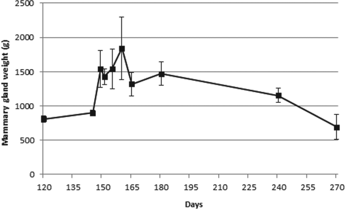
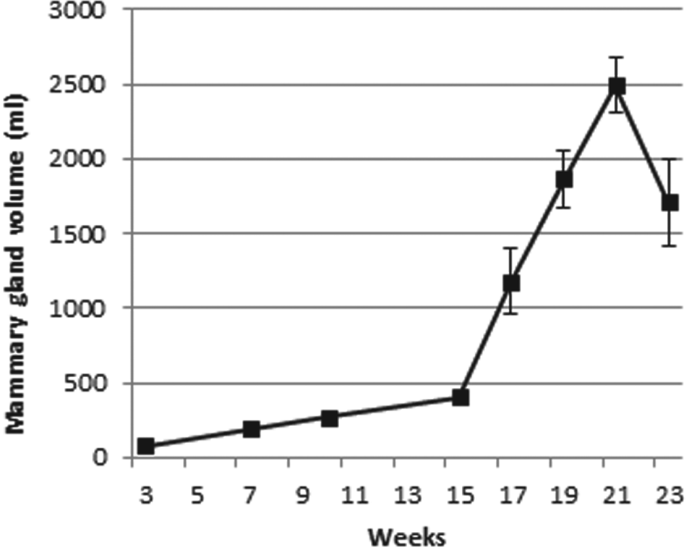
Anderson & Wahab (Reference Anderson and Wahab1990) assessed udder modifications during various stages of pregnancy, lactation and involution considering groups with goats of four breeds: Toggenburg, Nubian, Saanen and French Alpine. The authors reported, during pregnancy, no differences in udder weights until day 120, when values started to increase significantly. Indeed, the majority of udder growth occurred between the last 30 d of pregnancy and the first 10 d of lactation (Fig. 1). Considering mammary gland volume changes during late gestation and the first 2 weeks of lactation, Fowler et al. (Reference Fowler, Knight, Cameron and Foster1990), using Saanen goats, had similar results to those by Anderson & Wahab (Reference Anderson and Wahab1990) (Fig. 2). From Figs. 1 & 2, a slight reduction of the mammary gland is visible with the onset of lactation, related to milking.
Mammary gland weight changes during late gestation (days 120–150) and lactation (days 150–270) in goats. Adapted from Anderson & Wahab (Reference Anderson and Wahab1990).

Mammary gland volume changes during late gestation (weeks 3–21) and early lactation (week 23) in goats. Adapted from Fowler et al. (Reference Fowler, Knight, Cameron and Foster1990).
In addition, Fowler et al. (Reference Fowler, Knight, Cameron and Foster1990) reported an alteration of mammary gland tissue composition during gestation and lactation, as well as for the first 15 d of gestation, where parenchyma fatty tissue proportion decreased and fluid-rich tissue increased. Thereafter, mammary gland composition remained constant throughout late gestation and the entire lactation period. Alterations in parenchyma composition can be directly related to the increment of milk secretion and fluid accumulation in the gland.
More recently, Peris et al. (Reference Peris, Caja and Such1999) using Murciano-Granadina goats reported a marked reduction of mammary gland volume during mid-lactation, which is comparable with the previously mentioned studies, as the majority of udder growth occurs during early lactation. Additionally, these authors showed that an elevated number of parities was correlated to a higher reduction of the udder volume during the course of lactation, but also to a higher volume at the onset of lactation. Interestingly, in Toggenburg goats a significant increment in udder volume at the onset of lactation occurs with increasing parity number (Wang, Reference Wang1989). Moreover, the authors also reported that goats with twins had a significantly more voluminous udder (40% higher). Interestingly, since the udder volume has been positively correlated to higher milk yields, the association between twin occurrence and larger udder volume appears to be an adaptation related to kid survival.
It is also important to emphasise that both Anderson & Wahab (Reference Anderson and Wahab1990) and Fowler et al. (Reference Fowler, Knight, Cameron and Foster1990) showed the existence of a specific period of exponential mammary gland tissue growth, which begins before lactation. As expected, the phenomenon of exponential mammary gland growth occurred before the lactation peak, since the increment of mammary gland cells must arise to augment the milk production. Also, the whole mammary gland tissue undergoes pronounced alterations in its constitution, with the replacement of adipose tissue by secretory tissue (Fowler et al. Reference Fowler, Knight, Cameron and Foster1990) which further indicates the growth of mammary gland cells during this period. Additionally, it has to be stressed that the increase in udder volume due to the presence of twins (Peris et al. Reference Peris, Caja and Such1999) was associated by Hayden et al. (Reference Hayden, Thomas and Forsyth1979) and Byatt et al. (Reference Byatt, Eppard, Munyakazi, Jorbet, Veenhuizen, Curran and Collier1992) with placental development and a higher milk production. Following this argument, a heavier placenta may produce a higher level of lactogen hormone, which is responsible for mammary tissue development during gestation (Knight & Peaker, Reference Knight and Peaker1982; Peris et al. Reference Peris, Caja and Such1999).
Fernandez et al. (Reference Fernandez, Alvarez, San Primitivo and de la Fuente1995) used Churra sheep to study the influence of parity and milk yield on the udder volume throughout lactation. Their results show that not only was the udder volume significantly increased between the first and following parities, but also that milk yield was positively correlated with udder size. Similarly, Rovai et al. (Reference Rovai, Such, Piedrafita, Caja and Pujol1999) observed in Manchega and Lacaune sheep that udder volume reached its maximum during the week 1 of lactation and thereafter progressively reduced. Additionally, Fernandez et al. (Reference Fernandez, Alvarez, San Primitivo and de la Fuente1995) also demonstrated a positive correlation between udder size and milk yield during lactation. A similar correlation was also reported by other authors for Sicilo-Sarde (Ayadi et al. Reference Ayadi, Such, Ezzehizi, Zouarie, Najard, Ben M'Rad and Casal2011) and Bergamasca (Emediato et al. Reference Emediato, Siqueira, Stradiotto, Maestá and Fernandes2008) sheep.
According to the previously described studies for both sheep and goat, there is a general pattern for udder volume change with the different phases of the reproductive cycle. In both species there is an increment of udder volume during late gestation, which continues during early lactation, reducing thereafter. Moreover several studies show a positive correlation between both milk yield and prolificacy with udder size.
Nevertheless it should be emphasised that the small number of studies regarding this topic and also the variability between breeds concerning udder dimensions and milk yields may influence the changes of udder size. On the other hand, the genetic component i.e. the heritability of possible genes associated with traits like higher milk yields and/or larger udders was not considered in these studies. Finally, it has to be stressed that there is a need to improve our knowledge to understand the correlation not only between udder volume and milk yield, but also to understand the genetic mechanisms behind the milk yield increase with a higher prolificacy.
Microscopic patterns: a general overview
Mammary remodelling is determined by a combination of cell differentiation, proliferation and programmed death (Wilde et al. Reference Wilde, Quarrie, Tonner, Flint and Peaker1997) controlled not only by systemic hormones, but also by proteins produced either in the stromal or in the epithelial compartments (Hennighausen & Robinson, Reference Hennighausen and Robinson2001).
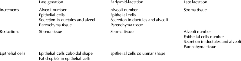
For goats, several authors investigated histological modifications of the mammary gland during the course of lactation. Li et al. (Reference Li, Wilde, Finch, Fernig and Rudland1999b) observed alveoli development as a result of cellular proliferation, as well as the presence of secretions inside alveoli and ducts in Saanen goats during both gestation and lactation periods. Additionally, these authors reported the presence of large fat droplets in epithelial cells’ apical membranes throughout gestation, and the reduction of stromal tissue percentage due to its substitution by the secretory tissue (parenchyma). Further, with the onset of lactation epithelial cells assume a columnar morphology, alveoli are surrounded by a thin layer of fibroblasts and capillaries and there is the presence of highly distended alveoli. All these alterations can be related to the preparation of the mammary gland for milk production. Indeed, development of the lobular-alveolar system is obviously essential to produce and store milk, which replaces stroma, reducing its amount in the mammary gland, whereas the alteration of the epithelial cells to a columnar shape is probably due to their proliferation. More recently, Elsayed et al. (Reference Elsayed, EL-Shafie, Saifelnasr and Abu El-Ella2009) studied mammary gland patterns throughout lactation in Damascus goats, also observing an increase in alveolar secretory cells number during early and mid-lactation and a diminishment in late lactation. Likewise, the alveoli area was smaller during late lactation compared with early and mid-stages. Additionally, alveoli numbers were reduced and stroma tissue increased.
As expected, an expansion of secretory tissue occurs through the increase in alveoli number and size during gestation and early lactation, corresponding to a macroscopic increase of udder volume. In addition, and with a macroscopic correlation, during mid and late lactation, the alveoli number and size are reduced and stroma amount increased as a consequence of the reduction of secretory tissue itself as detailed in Table 2.
Mammary gland histological patterns during late gestation and lactation for goats and sheep†
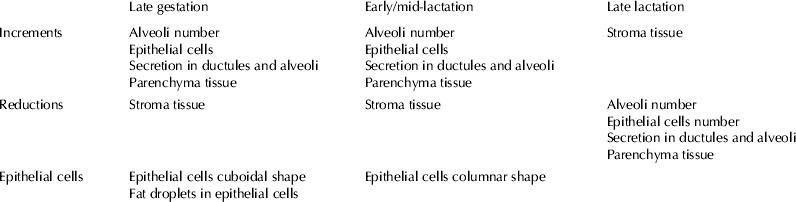
† Adapted from Li et al. (Reference Li, Wilde, Finch, Fernig and Rudland1999b) and Colitti & Farinacci (Reference Colitti and Farinacci2009)
In relation to sheep, there are studies concerning histological and macroscopical changes through lactation. Carretero et al. (Reference Carretero, Ruberte, Caja, Pérez-Aparicio, Such, Peris, Manesse and Navarro1999) studied mammary gland histological differences in two breeds, Manchega and Lacaune, with different milk yields between weeks 1 (suckling) and 5 (start of milking). In both breeds the authors observed an alveolar growth with the same development pattern, despite the different milk yield. The authors also reported, at week 1, an increase in tubule number, originating from the pre-existing ones. At week 5, the presence of fully developed alveoli together with others in the first phase of development was observed. More recently, Nørgaard et al. (Reference Nørgaard, Nielsen, Theil, Sørensen, Safayi and Sejrsen2008) studying mammary gland development in Shropshire sheep during late gestation and lactation, reported an increment of mammary parenchymal tissue weight from day 38 to day 6 pre-partum (epithelium area was 5 and 19%, respectively). It was also observed that the amount of parenchyma and its histological composition had their major changes between day 38 and day 6 pre-partum, which suggests that mammary gland growth and differentiation occurs mainly during late gestation, slightly differently from goats.
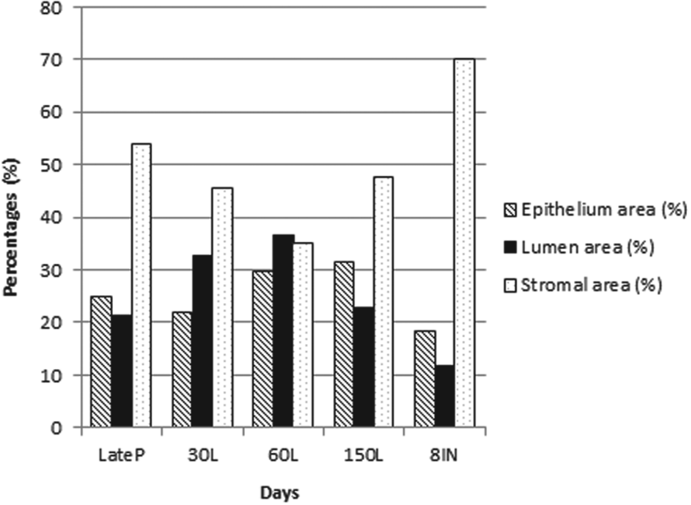
Colitti & Farinacci (Reference Colitti and Farinacci2009) performed a study in Sardinian sheep, reporting the presence of immature alveoli with a thin lumen and cuboidal epithelial cells with a large nucleus during late gestation. They also observed the presence of some alveoli filled with secretion and lipid droplets in the epithelial cells. As a consequence, the mammary gland underwent structural alterations in its alveoli at the end of the gestation period, which passed from immature to mature at early lactation. These results agree with previous reports in lactating cows (Akers, Reference Akers2002). Indeed, Colitti & Farinacci (Reference Colitti and Farinacci2009) observed that at day 30 of lactation (early lactation) the mammary gland had developed fully differentiated alveoli constituted by epithelial cells with vacuoles in their apical membrane, filled with secretion and there was also the presence of interalveolar connective tissue. Regarding the vacuoles present in the epithelial cells, probably they correspond to the previously described fat droplets that were incorporated in milk. Relatively to mid and late lactation (days 60 and 150, respectively) the alveolar lumen became larger and epithelial cells had their apical ends with a high prominence corresponding to secretory vesicles. Regarding the amount of stroma, its lowest value was obtained at day 60 of lactation and the highest at day 8 of involution (Fig. 3).
Mammary gland stroma, lumen and epithelium areas during late pregnancy and lactation in sheep. Late P, late pregnancy; L, lactation days; IN, involution. Adapted from Colitti & Farinacci (Reference Colitti and Farinacci2009).
Finally, from Fig. 3, it is interesting to observe the remarkable reduction of stromal area from late pregnancy to mid-lactation, which is progressively replaced by lumen area. Indeed, at the beginning of lactation, the epithelium percentage is slightly reduced owing to the lumen being filled with secretion.
In Turcana sheep, Cadar et al. (Reference Cadar, Mireşan, Lujerdean and Răducu2012) described a non-homogeneous aspect of the mammary gland during early lactation, with some large alveoli filled with secretion and smaller ones without secretion. Moreover, these authors also observed epithelial cells with a large nucleus, vacuoles in their cytoplasm and apical prominences corresponding to lipid droplets.
Hence, considering the studies performed in sheep, it is once more evident that secretory tissue growth occurs essentially during late gestation, as well as in early lactation (Carretero et al. Reference Carretero, Ruberte, Caja, Pérez-Aparicio, Such, Peris, Manesse and Navarro1999; Colitti & Farinacci, Reference Colitti and Farinacci2009; Cadar et al. Reference Cadar, Mireşan, Lujerdean and Răducu2012). Comparing the studies performed in goat and sheep species, a common pattern was seen through the several phases considered, since there was a mutual increment of lobule-alveolar system throughout gestation, as well as a reduced amount of stroma tissue. Additionally, during early and mid-lactation there was an increment of alveolus number, size and differentiation as well as the presence of epithelial cells with a columnar shape and secretory vesicles. During late gestation, a reduction of epithelial cells number was observed (Table 2) as seen in Figs. 4 & 5 for goat and sheep, respectively. As expected, all these histological processes can be associated with the different stages of the lactation cycle. Indeed, during late gestation and lactation, it is necessary to progressively increase the alveoli size/number and lumen area, which replace the stroma tissue, in order to have enough secretory cells to start producing milk after partum. The modification of the epithelial cells shape and the presence of secretory vesicles is also an adaptation essential for milk production.
Transverse section of mammary gland parenchyma in goat stained with HE showing distended alveoli with the characteristic secretory epithelium. Alveoli are surrounded by a layer of connective tissue with capillaries that forms the stroma of the gland and there is also the presence of secretory vesicles.
Transverse section of mammary gland parenchyma in sheep stained with HE showing distended alveoli with the characteristic secretory epithelium. Alveoli are surrounded by a layer of connective tissue with capillaries that forms the stroma of the gland and there is also the presence of secretory vesicles.
Bearing in mind the small number of studies of histological alterations of the mammary gland through gestation and lactation, it is clear that further studies in this area are needed.
Effect of different milking frequencies
In both dairy and non-dairy breeds, an increased milking frequency is often associated with a higher milk yield (Linzell & Peaker, Reference Linzell and Peaker1971; Henderson et al. Reference Henderson, Blatchford and Peaker1985). Indeed, goats milked thrice daily instead of twice daily had an increment in milk yield of 10–20% (Amos et al. Reference Amos, Kiser and Loewenstein1985; Knight, Reference Knight1992; Campos et al. Reference Campos, Wilcox, Head, Webb and Hayen1994; Boutinaud et al. Reference Boutinaud, Rousseau, Keisler and Jammes2003) whereas goats milked once a day had a milk yield reduction of 10–30% (Wilde et al. Reference Wilde, Henderson, Knight, Blatchford, Faulkner and Vernon1987; Carruthers et al. Reference Carruthers, Davis and Norton1991; Lynch et al. Reference Lynch, Hunt and Mackenzie1991; Boutinaud et al. Reference Boutinaud, Rousseau, Keisler and Jammes2003). However, the increase of milking frequency in dairy goats negatively affects the concentration of immune components in milk and therefore the susceptibility of mastitis is increased (Hernández-Castellano et al. Reference Hernández-Castellano, Torres, Alavoine, Ruiz-Díaz and Argüello2011). Despite these studies, the correlation between milking frequency and mammary gland growth has been addressed in relatively few experiments.
In goats, Li et al. (Reference Li, Rudland, Fernig, Finch and Wilde1999a) investigated the effect of differential milking frequencies using the Saanen breed. Animals were milked twice-daily until weeks 20–22 of lactation and, afterwards, one gland was milked thrice-daily for 10 weeks and the other once daily (half-udder design). The results showed that after 4 weeks of differential milking, once-daily milked glands had smaller alveoli and fewer alveolar cells per alveolus. After 10 weeks, once-daily milked glands registered a heterogeneous composition, as there were some alveoli that appeared to be involuting (small, regressing alveoli, constituted by alveolar cells without a columnar shape) and others still with the lactating morphology. In thrice-daily milked mammary gland, most of the tissue had the typical secretory alveoli at the same period of time, mainly composed of closely packed alveoli separated by small amounts of interstitial tissue, being the alveolar cells columnar and with a large apical secretory vesicle. In addition, the authors recorded the presence of apoptotic bodies in alveolar cells, but only in the once-daily milked glands. These results suggest the possibility that mammary gland growth can occur during established lactation and not only in pregnancy and early lactation; also mammary gland growth is locally controlled.
Boutinaud et al. (Reference Boutinaud, Rousseau, Keisler and Jammes2003) used Saanen goats in week 32 of lactation in a similar experiment to the previous study. Their results were similar to those of Li et al. (Reference Li, Rudland, Fernig, Finch and Wilde1999a) in finding that milking frequency was positively correlated with mammary gland weight. Additionally, Wilde et al. (Reference Wilde, Henderson, Knight, Blatchford, Faulkner and Vernon1987) reported that total RNA content (gram per udder-half), an indicator of overall transcriptional activity, had a tendency to become higher with increasing frequency of milking. This result showed that along with the higher number of epithelial cells, there was also an increase in secretory activity. Concerning morphological pattern, the number of epithelial cells per alveolus and per alveolar diameter was higher in thrice-milked udder halves, similarly to the previous described study. In contrast, Suárez-Trujillo et al. (Reference Suárez-Trujillo, Capote, Argüello, Castro, Morales-DelaNuez, Torres, Morales and Rivero2013) also applying the half-udder design for 6 weeks in Palmera, Majorera and Tinerfeña goats, observed no influence of the milking frequency in the mammary histological parameters. These differences between studies were probably due to the use of different breeds and different evaluation period.
In another study, Capote et al. (Reference Capote, Argüello, Castro, López and Caja2006) used two groups of Tinerfeña goats, one milked once daily and the other milked twice daily during 5 d of week 21 of lactation to study udder morphology. In contrast to the previous studies, these authors observed a smaller volume, in twice-daily milked mammary glands than in those milked once daily. However, it must be recognised that the milking period was significantly shorter and this fact may be the main reason for such a difference.
More recently, Torres et al. (Reference Torres, Castro, Hernández-Castellano, Argüello and Capote2013a) in three different dairy goat breeds (Palmera, Majorera and Tinerfeña) examined the effect of different milking frequencies during 5 weeks using the half-udder design as previously described by Li et al. (Reference Li, Rudland, Fernig, Finch and Wilde1999a). The results showed no differences in udder volume due to either the breed or milking frequency. Again, in another study performed by the same authors, the milking frequency had no effect on the percentage of cisternal or alveolar milk (Torres et al. Reference Torres, Castro, Argüello and Capote2013b). In general, the previously mentioned studies demonstrated that less frequent milking reduces the mammary gland weight and RNA concentration, which is directly correlated with cell activity, the number of epithelial cells, alveolar size and epithelial cells per alveolus. Finally, Li et al. (Reference Li, Rudland, Fernig, Finch and Wilde1999a) also reported the presence of apoptotic bodies, demonstrating that less frequent milking also induces mammary gland cells apoptosis.
Nevertheless and as previously highlighted, the number of studies on this topic is small and it has to be remembered that the wide variation between studies with respect to breed and lactation period renders comparisons difficult.
Effect of lactation number
There are several factors that affect milk yield during lactation, such as the number of epithelial cells (Capuco et al. Reference Capuco, Ellis, Hale, Long, Erdman, Zhao and Paape2003), their secretory activity (Akers et al. Reference Akers, Capuco and Keys2006) or the mammary gland vascular system, essential for the provision of nutrients and removal of metabolic waste products, among others. Importantly, these factors can be influenced by lactation number, as primiparous animals have to use their nutrients not only for lactation, but also for their own growth (Wathes et al. Reference Wathes, Cheng, Bourne, Taylor, Coffey and Brotherstone2007; Safayi et al. Reference Safayi, Theil, Elbrønd, Hou, Engbaek, Nørgaard, Sejrsen and Nielsen2010).
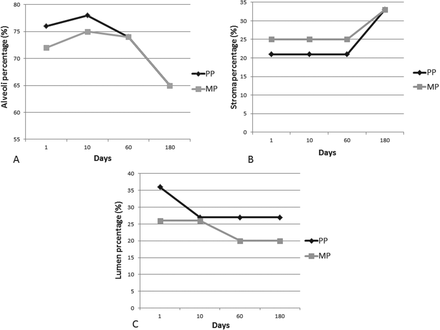
Peris et al. (Reference Peris, Caja and Such1999) investigated udder volume in Murciano-Granadina dairy goats with respect to different lactation numbers (first, second, third or more) and lactation stages (early lactation in weeks 1–2 and mid-lactation in weeks 16 and 17). Udder volume decreased as lactation progressed in all lactations, but the reduction was higher in goats that were in their 3rd or more lactation. In multiparous goats, part of the alveoli developed in the previous lactation had not regressed entirely, but were added to those which grew in the following lactation, increasing the udder volume, especially the secretory parenchyma tissue. More recently, Safayi et al. (Reference Safayi, Theil, Elbrønd, Hou, Engbaek, Nørgaard, Sejrsen and Nielsen2010) used Danish crossbred Landrace-Saanen dairy goats to study the different morphological pattern during lactation between primiparous and multiparous animals. They found a higher alveoli number and stroma tissue proportion but a lower alveoli proportion throughout the whole lactation period. In the first 10 d of lactation, the authors observed an increase in alveoli proportion and mammary epithelial cells for both multiparous and primiparous glands (Fig. 6a), whereas interstitial tissue remained almost at the same level as in parturition (25 and 21% for multiparous and primiparous, respectively) (Fig. 6b). Relative lumen percentage was reduced in the primiparous glands until reaching the same value as the multiparous glands (27%) (Fig. 6c).
Mammary gland alveoli percentage (A), stroma percentage (B) and lumen percentage (C) during lactation in primiparous and multiparous goats. PP, primiparous; MP, multiparous. Adapted from Safayi et al. (Reference Safayi, Theil, Elbrønd, Hou, Engbaek, Nørgaard, Sejrsen and Nielsen2010).
Considering the period between early (day 10) and mid to late lactation (days 60–180), Safayi et al. (Reference Safayi, Theil, Elbrønd, Hou, Engbaek, Nørgaard, Sejrsen and Nielsen2010) identified a reduction of alveoli proportion for both glands. In the case of lumen percentage, it stabilised (26–27%) and started to decrease in multiparous glands to its lowest level (20%) in late lactation. Nevertheless, interstitial proportion increased until reaching a peak in late lactation (33%).
So, in general, there was an increment in alveoli proportion during early lactation, which is probably the cause of the reduction of lumen percentage during the same period, and then a reduction of those values, coinciding with changes in milk yield in both groups. Interestingly, during early lactation the lumen area and alveoli proportions were higher for primiparous glands compared with multiparous. Indeed, this was probably due to the higher percentage of stromal tissue in the multiparous glands, since the authors showed that the multiparous glands had a higher number of alveoli per slide.
Considering these studies, it has been shown that the secretory structures probably do not regress totally during involution, as multiparous glands had a higher alveoli number, stroma amount and udder volume. Indeed, the positive correlation between parity and udder volume was previously reported in goats by Wang (Reference Wang1989) in the Toggenburg breed and in Murciano-Granadina by Peris et al. (Reference Peris, Caja and Such1999), as described before.
Effect of nutrition
In many dairy production systems, it is common to use high-concentrate diets to increase milk production, as described for goats (Rapetti et al. Reference Rapetti, Bava, Tamburini and Crovetto2005; Serment et al. Reference Serment, Schmidely, Giger-Reverdin, Chapoutot and Sauvant2011) or cows (Yang & Beauchemin, Reference Yang and Beauchemin2007; Agle et al. Reference Agle, Hristov, Zaman, Schneider, Ndegwa and Vaddella2010). However, animals fed with those kinds of diets may suffer from sub-acute ruminal acidosis and off-feed periods can be observed (Serment & Giger-Reverdin, Reference Serment and Giger-Reverdin2012) which demonstrates the impact of nutrition in animal production. Additionally, nutrition can have effects not only in milk composition and yield (Ollier et al. Reference Ollier, Leroux, de la Foye, Bernard, Rouel and Chilliard2009; Lérias et al. Reference Lérias, Hernández-Castellano, Morales-delaNuez, Araújo, Castro, Argüello, Capote and Almeida2013) but also on mammary gland apoptosis, which affects mammary gland involution (Colitti et al. Reference Colitti, Stradaioli and Stefanon2000).
Regarding sheep, Colitti et al. (Reference Colitti, Stradaioli and Stefanon2000) studied the effect of α-tocopherol (vitamin E) deprivation on mammary gland morphology and apoptosis rate during involution. In this experiment, the authors used two groups, a control group receiving α-tocopherol supplementation and an experimental group with no supplementation. Comparing both apoptotic rates, the authors reported no significant differences between treatments, as in both diets the apoptosis index increased along the course of involution (0·12 to 4·06%, respectively from days 1 to 8). In addition, the presence of apoptotic cell clusters were seen in both groups, demonstrating that α-tocopherol deprivation had no effect in apoptosis regulation.
More recently, Colitti et al. (Reference Colitti, Stradaioli and Stefanon2005) evaluated the effect of diet composition on mammary gland cell turnover in ewes during mid-lactation, using three groups fed with different diets: a control diet, which contained 22% of starch and 3% of fat and the others with a high starch (HS) or a high fat (HFa) diets. The HS diet contained 15% of starch and 3% of fat and the HFa 14·4% of starch and 5·4% of fat. With reference to apoptotic cells number, the HFa group had a higher level within the same alveolus compared with the other groups. Considering proliferating cells, they were homogeneously distributed in control and HS groups, whereas in HFa group the proliferating cells appeared as single cells. Additionally, the authors observed some myoepithelial cells in proliferation, but only in control and HS groups. In relation to the proliferation per apoptosis ratio, it was lower in HFa (0·11) compared with control and HS groups (0·66 and 0·64 respectively). From these results, a higher level of apoptosis and lower level of proliferation was evident in the HFa group. Indeed, it is considered that diets with high lipid levels stimulate the production of conjugated linoleic acid (CLA) in rumen and mammary gland (AbuGhazaleh et al. Reference AbuGhazaleh, Schingoethe, Hippen and Kalscheur2003), inhibiting the transition from G1 to S-phase in mitosis (Kemp et al. Reference Kemp, Jeffy and Romagnolo2003). Possibly the CLA action overcame the paracrine regulation of the fat pad, resulting in the larger levels of apoptosis observed in this group.
Finally, the effect of feed restriction was examined by Nørgaard et al. (Reference Nørgaard, Nielsen, Theil, Sørensen, Safayi and Sejrsen2008) using Shropshire sheep divided in two experimental groups, the control group fed ad libitum and the other fed with a diet that would only supply 50% of total energy requirements. Results showed a high mammary parenchymal tissue growth during gestation from day 38 and day 16 pre-partum, respectively for control and fed restricted groups, to day 6 pre-partum (14 and 19% for fed restricted and ad-libitum groups, respectively). However, the authors observed no significant differences between 6 pre-partum samples from both groups either in parenchyma amount or in its distribution between tissue types. Concerning epithelial cell proliferation, there was, once more, an increment of those values from day 38 to day 6 pre-partum, but with no significant differences between both groups.
In general, the above-mentioned studies demonstrate that nutrition, whether considering animal total energy intake or a specific nutrient, had no significant effect on sheep mammary gland morphology. However, again it has to be emphasised that the small number of studies and variability in the nutrient considered indicates a need for more experiments in this field.
Concluding remarks
Analysis of mammary gland morphological patterns during the various stages of the reproductive cycle is essential to understand its functionality as a milk production organ. Indeed, with the progressive increase in the importance of small ruminants in milk production, it is essential to understand mammary gland modification pattern during lactation in the two small ruminant species, goat and sheep. Nonetheless, it is apparent that there is indeed a similar mammary gland pattern between both species along the whole lactation cycle. As a general rule for small ruminants, early/mid-lactation is defined by the occurrence of cell proliferation, a higher level of cell activity and alveoli number and an increase in udder volume. In addition, altogether they are correlated with higher milk yields that characterise this lactation period. During mid/late lactation, a reduction of epithelial cell number, an increased detection of apoptotic bodies, a reduction in alveolar size and number and a diminishment of udder volume occurs, in all cases correlated with the reduction in milk yield.
As herein discussed, it is essential to study further the mammary gland in dairy animals, since future progress in dairy animal production depend on the improvement of our knowledge regarding mammary gland modifications throughout lactation. Additionally, the so-called novel ‘omics’ tools, such as proteomics, transcriptomics or lipidomics (Ferreira et al. Reference Ferreira, Bislev, Bendixen and Almeida2014) can be used to enhance detailed study of mammary gland patterns throughout lactation. Indeed, applying these tools to dairy animal industries may be the key to their improvement.
The authors acknowledge the financial support of Fundação para a Ciência e a Tecnologia (Lisbon, Portugal) through research project PTDC/CVT/116499/2010 – Lactation and milk production in Goat (Capra hircus): identifying molecular markers underlying adaptation to seasonal weight loss, and SFRH/BPD/90916/2012 grant that finances author A.M. Almeida. Author A. Suárez-Trujillo acknowledges FPU 12/06079 scholarship from the Ministry of Education, Culture and Sports of the Spanish Government (Madrid, Spain).