





Introduction
Cryptosporidium species are single-celled parasites that must live inside host cells (obligate intracellular protozoa) and cause enteric (intestinal) disease in a wide range of vertebrate hosts, including humans and livestock (Fayer and Xiao, Reference Fayer and Xiao2008). In cattle, infection is closely associated with neonatal diarrhoea, reduced growth rates and production losses. (Shaw et al., Reference Shaw, Innes, Morrison, Katzer and Wells2020) Additionally, the shedding of oocysts (the infectious stage) into the environment contributes substantially to waterborne and foodborne transmission pathways relevant to human health (Xiao and Feng, Reference Xiao and Feng2008). Cryptosporidium parvum is the most epidemiologically important Cryptosporidium species infecting cattle due to its zoonotic potential and capacity to cause severe disease in both calves and humans (Amer et al., Reference Amer, Honma, Ikarashi, Tada, Fukuda, Suyama and Nakai2010; Abeywardena et al., Reference Abeywardena, Jex, Nolan, Haydon, Stevens, McAnulty and Gasser2012; Chen et al., Reference Chen, Huang, Qin, Wang, Li and Zhang2023). Other species, including Cryptosporidium bovis and Cryptosporidium ryanae, are also frequently detected in cattle (Taylan-Ozkan et al., Reference Taylan-Ozkan, Yasa-duru, Usluca, Lysen, Ye, Roellig, Feng and Xiao2016; Lichtmannsperger et al., Reference Lichtmannsperger, Harl, Freudenthaler, Hinney, Wittek and Joachim2020). These species are generally regarded as host-adapted and of lower zoonotic concern, but their epidemiology, transmission dynamics and contribution to environmental oocyst contamination remain incompletely characterized (Bulumulla et al., Reference Bulumulla, Xiao, Feng, Ash, Aleri, Ryan and Barbosa2025).
Molecular identification of Cryptosporidium infections is routinely performed using nested PCR and Sanger sequencing of the 18S rRNA small subunit gene (Xiao et al., Reference Xiao, Escalante, Yang, Sulaiman, Escalante, Montali, Fayer and Lal1999). While this approach is widely used, it typically yields a single consensus sequence and may fail to detect mixed infections where multiple species or subtypes co-occur. Increasing evidence indicates that co-infection is common in livestock systems, particularly under conditions of intensive production and high environmental oocyst burden (Maddox-Hyttel et al., Reference Maddox-Hyttel, Langkjær, Enemark and Vigre2006; Langkjær et al., Reference Langkjær, Vigre, Enemark and Maddox-Hyttel2007; Rialch et al., Reference Rialch, Shivani, Kaushal, Mal, Banerjee, Sankar, Singh, Sharma, Gopinath, Jairath, Katoch, S and Thakur2026). In contrast, high-throughput amplicon sequencing, also known as metabarcoding, offers an alternative approach that can detect minority taxa, resolve mixed infections and provide quantitative read-based profiles by generating thousands of reads per sample. High-throughput metabarcoding of amplicon loci provides an alternative framework for Cryptosporidium surveillance, allowing detection of multiple amplicon sequence variants (ASVs) within the same sample (Paparini et al., Reference Paparini, Gofton, Yang, White, Bunce and Ryan2015; Turner et al., Reference Turner, Naguib, Pierce, Li, Valente, Glenn Travis, Rosenthal Benjamin, Kissinger Jessica and Khan2025). Metabarcoding approaches have been increasingly adopted for molecular epidemiology and One Health surveillance of protozoan parasites (Zahedi et al., Reference Zahedi, Monis, Gofton, Oskam, Ball, Bath, Bartkow, Robertson and Ryan2018; Garcia-R et al., Reference Garcia-R, Ogbuigwe, Pita, Velathanthiri, Knox, Biggs, French and Hayman2021; Ogbuigwe et al., Reference Ogbuigwe, Biggs, Garcia-Ramirez, Knox, Pita, Velathanthiri, French and Hayman2022, Reference Ogbuigwe, Biggs, Garcia-Ramirez, Knox, Pita, Velathanthiri, French and Hayman2025), enabling improved resolution of infection complexity and population structure.
Subtype characterization using the gp60 (60 kDa glycoprotein) locus is widely used for C. parvum and C. hominis to support outbreak investigations, source attribution and transmission studies (Chalmers and Katzer, Reference Chalmers and Katzer2013; Garcia-R and Hayman, Reference Garcia-R and Hayman2017; Garcia-R et al., Reference Garcia-R, Cox and Hayman2020a, Reference Garcia-R, Pita, Velathanthiri, French and Hayman2020b; Ogbuigwe et al., Reference Ogbuigwe, Biggs, Garcia-Ramirez, Knox, Pita, Velathanthiri, French and Hayman2025). Although gp60 subtyping has become a cornerstone of Cryptosporidium molecular epidemiology, its application to C. bovis remains limited, partly due to the absence of suitable primers and significant differences in the gp60 gene sequence compared to the well-studied C. parvum and C. hominis. Recent studies have overcome some of these challenges by developing C. bovis-specific gp60 assays and identifying multiple subtype families (XXVIa–XXVIf), suggesting that substantial diversity may exist within this species (Wang et al., Reference Wang, Wan, Yang, Li, Xiao, Feng and Guo2021; Feng et al., Reference Feng, Li, Huang, Chen, Wen, Wang, Ryan, Xiao, Feng and Guo2023). Nevertheless, mixed infections involving multiple gp60 subtypes of C. bovis remain poorly characterised. Application of gp60 metabarcoding to C. bovis could therefore provide a significant methodological advance, providing improved resolution for molecular epidemiology in cattle.
Despite New Zealand’s large dairy cattle population, and its highdairy cattle densities per unit land area (Foote et al., Reference Foote, Joy and Death2015; Grout et al., Reference Grout, Marshall, Hales, Baker and French2022), high-resolution molecular data on bovine Cryptosporidium diversity remain limited. Previous surveys have reported farm-level prevalence of C. parvum at 50.5% and C. bovis at 6.0% across dairy farms, predominantly in calves (Winkworth et al., Reference Winkworth, Matthaei and Townsend2008; Al Mawly et al., Reference Al Mawly, Grinberg, Prattley, Moffat and French2014, Reference Al Mawly, Grinberg, Velathanthiri and French2015), with gp60 subtype families IIa and IId most frequently identified in C. parvum-positive calves (Garcia-R et al., Reference Garcia-R, French, Pita, Velathanthiri, Shrestha and Hayman2017). However, these studies relied exclusively on conventional Sanger sequencing and therefore could not resolve mixed infections. Here, we applied a combined approach of conventional 18S rRNA PCR/Sanger sequencing, 18S amplicon metabarcoding and gp60 metabarcoding to characterize Cryptosporidium species composition, broader apicomplexan co-infection profiles and C. bovis gp60 subtype family diversity. The novelty of this work lies in the integration of high-throughput metabarcoding at both the 18S and gp60 loci to improve detection of mixed Cryptosporidium infections and provide high-resolution subtyping in New Zealand dairy cattle.
Materials and methods
Sample collection and DNA extraction
Faecal samples were collected from dairy cattle across Canterbury (South Island) and Manawatu (North Island) farms in New Zealand as part of an opportunistic molecular surveillance programme. Specimens were obtained from participating collaborators when available. Fresh faecal pat samples were collected by convenience sampling from young (ages 2–20 weeks) cattle during the Spring calving season in 2022 and the non-calving period in December 2023. No Animal Ethics approval was required as sampling was non-invasive. DNA was extracted from approximately 200 mg of faecal material using the Zymo Quick-DNA Fecal/Soil Microbe kit (Irvine, California, USA) supplemented with an additional bead-beating disruption step to enhance oocyst lysis, following the manufacturer’s protocol. Extracted DNA was stored at −20 °C until further analysis.
PCR and Sanger sequencing
Faecal DNA extracts were initially screened for Cryptosporidium using a nested PCR targeting an approximately 600 bp fragment of the 18S rRNA small subunit gene. PCR amplifications were carried out using FIREPol® Master Mix and outer (SSU_F1 5′-GATTAAGCCATGCATGTCTAA-3′ and SSU_R1 5′-TTCCATGCTGGAGTATTCAAG-3′) and inner primers (SSU_F2 5′-CAGTTATAGTTTACTTGATAATC-3′ and SSU_R2 5′-CCTGCTTTAAGCACTCTAATTTTC-3′) as described by Lindergard et al. (Reference Lindergard, Nydam, Wade, Schaaf and Mohammed2003). Cycling conditions consisted of an initial denaturation at 94 °C for 2 min, followed by 30 cycles of 94 °C for 50 sec, 58 °C for 50 sec and 72 °C for 1 min, with a final extension at 72 °C for 10 min. The secondary (nested) PCR used 2 µL of the primary PCR product as the template under the same conditions, except that the annealing temperature was decreased to 56 °C. Samples confirmed as C. parvum by 18S rRNA gene analysis were subsequently subtyped at the gp60 locus as previously described (Garcia-R et al., Reference Garcia-R, French, Pita, Velathanthiri, Shrestha and Hayman2017, Reference Garcia-R, Cox and Hayman2020a, Reference Garcia-R, Pita, Velathanthiri, French and Hayman2020b). Purified PCR products were Sanger-sequenced in both directions using BigDye Terminator v3.1 chemistry on an ABI 3730XL automated sequencer (Applied Biosystems, Foster City, CA, USA).
Next-generation sequencing
All samples confirmed positive by nested 18S rRNA PCR were subjected to Illumina-based 18S metabarcoding. A nested PCR was performed using the same outer primers as above, followed by a second PCR using the internal primers 18SiF (5′- AGTGACAAGAAATAACAATACAGG-3′) and 18SiR (5′-CCTGCTTTAAGCACTCTAATTTTC-3′), described by Morgan et al. (Reference Morgan, Constantine, Forbes and Thompson1997). These primers were modified to incorporate Illumina MiSeq adapter sequences at the 5′ end, following a 2-step amplicon library preparation protocol. PCR contamination controls were used, including negative controls. Successful amplification of the expected ∼300 bp fragment was confirmed by agarose gel electrophoresis before library preparation. Samples identified as C. bovis by 18S Sanger sequencing were additionally subjected to gp60 metabarcoding using the primers and amplification conditions described by Wang et al. (Reference Wang, Wan, Yang, Li, Xiao, Feng and Guo2021). Due to the large size of the gp60 amplicon (>1 kb), which exceeded the range for reliable paired-end read merging on the Illumina MiSeq platform, downstream analyses were restricted to forward reads, which consistently exhibited higher sequence quality and read retention than reverse reads. Negative PCR controls were included in every run. PCR products were purified according to Illumina recommendations, quantified on a Qubit fluorometer (Thermo Fisher Scientific) and normalized prior to pooling. Sequencing was performed on an Illumina MiSeq using 500-cycle v2 chemistry (2 × 250 bp paired-end reads). An Illumina PhiX library was spiked at 20% as an internal sequencing control. Raw sequence reads and Sanger sequences are deposited in the NCBI Sequence Read Archive under BioProject accession PRJNA1476777.
Analyses
Consensus Sanger sequences were assembled from forward and reverse reads and edited manually in Geneious Pro 10.2.6 (Kearse et al., Reference Kearse, Moir, Wilson, Stones-havas, Cheung and Sturrock2012). BLAST software (http://www.ncbi.nlm.nih.gov/blast/) was used for species identification against the NCBI GenBank nucleotide database. Raw Illumina reads for both 18S and gp60 datasets were processed within the Quantitative Insights Into Microbial Ecology 2 (QIIME 2) environment (Bolyen et al., Reference Bolyen, Rideout, Dillon, Bokulich, Abnet, Al-ghalith, Alexander, Alm, Arumugam, Asnicar, Bai, Bisanz, Bittinger, Brejnrod, Brislawn, Brown, Callahan, Caraballo-rodríguez, Chase, Cope, Da Silva, Diener, Dorrestein, Douglas, Durall, Duvallet, Edwardson, Ernst, Estaki, Fouquier, Gauglitz, Gibbons, Gibson, Gonzalez, Gorlick, Guo, Hillmann, Holmes, Holste, Huttenhower, Huttley, Janssen, Jarmusch, Jiang, Kaehler, Kang, Keefe, Keim, Kelley, Knights, Koester, Kosciolek, Kreps, Langille, Lee, Ley, Liu, Loftfield, Lozupone, Maher, Marotz, Martin, McDonald, McIver, Melnik, Metcalf, Morgan, Morton, Naimey, Navas-molina, Nothias, Orchanian, Pearson, Peoples, Petras, Preuss, Pruesse, Rasmussen, Rivers, Robeson, Rosenthal, Segata, Shaffer, Shiffer, Sinha, Song, Spear, Swafford, Thompson, Torres, Trinh, Tripathi, Turnbaugh, Ul-hasan, van der Hooft, Vargas, Vázquez-baeza, Vogtmann, von Hippel, Walters, Wan, Wang, Warren, Weber, Williamson, Willis, Xu, Zaneveld, Zhang, Zhu, Knight and Caporaso2019). DADA2 (Callahan et al., Reference Callahan, McMurdie, Rosen, Han, Johnson and Holmes2016) was used for filtering and trimming, dereplication, merging paired reads, removing chimaeras and denoising into ASVs. Paired-end reads were merged for the 18S data, whereas only forward reads were processed for gp60 due to the longer amplicon size. All high-quality ASVs were taxonomically assigned by BLAST comparison against the NCBI nt database (accessed 10 April 2026), applying thresholds of ≥95% nucleotide identity and ≥100% query coverage. ASV count tables and taxonomy assignments were imported into the R package phyloseq (McMurdie and Holmes, Reference McMurdie and Holmes2013) for downstream analysis and visualization. Relative abundance was calculated as the read count for each taxon divided by the total read count per sample. Stacked barplots were generated to visualize species and subtype family distributions across samples. Cryptosporidium bovis gp60 ASVs were assigned to subtype families by similarity matching against published XXVI subtype reference sequences (Wang et al., Reference Wang, Wan, Yang, Li, Xiao, Feng and Guo2021).
Results
Of 98 faecal samples screened by nested 18S rRNA PCR, 45 (46%, 95% confidence interval: 37–57%) were confirmed positive for Cryptosporidium (Supplementary Table S1). Species identification by Sanger sequencing indicated that C. parvum was predominant (33/45, 73.3%), followed by C. bovis (8/45, 17.8%) and C. ryanae (4/45, 8.9%). At 2 weeks of age, most infected calves were positive for C. parvum (n = 22), with fewer infections caused by C. bovis (n = 2). Among older calves C. parvum remained the most frequently detected species (n = 7), followed by C. bovis (n = 6) and C. ryanae (n = 4). The age of 4 calves infected with C. parvum was unknown. Gp60 subtyping revealed that 22 C. parvum isolates belonged to subtype IIaA18G3R1.
Metabarcoding showed that Cryptosporidium-positive cattle samples frequently contained diverse apicomplexan communities. Metabarcoding provided evidence of mixed Cryptosporidium infections in a subset of samples, including co-detection of C. parvum with C. bovis and/or C. ryanae. In most cases, the dominant Cryptosporidium species detected by metabarcoding was consistent with the Sanger-based assignment, while minority taxa were detectable only through metabarcoding. In addition to mixed Cryptosporidium infections, substantial read counts were assigned to other apicomplexan taxa, including Eimeria cylindrica, E. subspherica, E. ferrisi and Isospora sp. and unidentified apicomplexan, as well as fungal species Mortierella polygonia and Saitozyma podzolica (Figure 1).
Relative abundance of taxa detected by 18S metabarcoding (taxonomy cutoff ≥95% nucleotide identity, 100% query coverage). Stacked barplots show proportional read abundance of Cryptosporidium spp., Eimeria spp., Isospora sp., fungi, and uncharacterised apicomplexan lineages across all Cryptosporidium-positive samples.

Figure 1 Long description
Relative abundance of taxa detected by 18S metabarcoding (taxonomy cutoff greater than or equal to 95% nucleotide identity, 100% query coverage). Stacked barplots show proportional read abundance of Cryptosporidium spp., Eimeria spp., Isospora sp., fungi and uncharacterised apicomplexan lineages across all Cryptosporidium-positive samples.
In some samples, Eimeria-associated reads constituted the dominant fraction of total apicomplexan reads, indicating that bovine coccidia are frequently present alongside Cryptosporidium infections. The Isospora sp. was detected in a single sample co-infected with C. bovis, where it constituted approximately 50% of total reads.
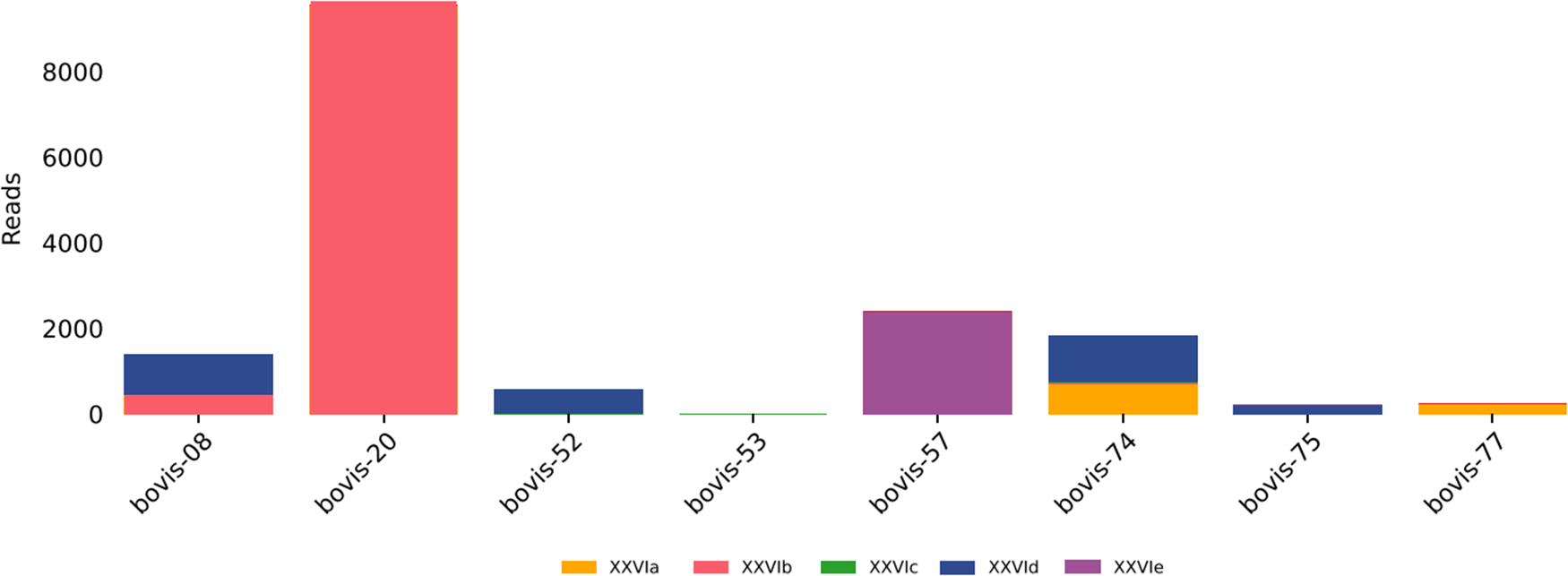
The 8 samples identified as C. bovis by 18S Sanger sequencing were subjected to gp60 metabarcoding. Multiple XXVI subtype families were detected across these samples, including XXVIa, XXVIb, XXVIc, XXVId and XXVIe (Figure 2). Importantly, some individual samples contained reads assigned to more than 1 subtype family, providing direct evidence of mixed gp60 subtype-family infections within single animals.
gp60 metabarcoding subtype family profiles for Cryptosporidium bovis-positive samples. Multiple XXVI subtype families (XXVIa–XXVIe) and evidence of mixed subtype-family infections within individual samples are shown.

Figure 2 Long description
gp60 metabarcoding subtype family profiles for Cryptosporidium bovis-positive samples. Multiple XXVI subtype families (XXVIa to XXVIe) and evidence of mixed subtype-family infections within individual samples are shown.
Discussion
This study demonstrates that integrating conventional 18S rRNA PCR/Sanger sequencing with high-throughput metabarcoding provides improved resolution for molecular surveillance of bovine Cryptosporidium. Conventional 18S rRNA PCR identified C. parvum as the dominant species (73.3%), and gp60 analysis demonstrated the predominance of subtype IIaA18G3R1. Members of the IIa subtype family are widely recognized as the major cattle-associated C. parvum lineages and are among the most frequently reported zoonotic subtypes worldwide. These findings therefore align with the established epidemiology of C. parvum as a leading cause of diarrhoeal disease in young calves and an important source of environmental contamination and zoonotic transmission (Chalmers and Katzer, Reference Chalmers and Katzer2013; Edwards et al., Reference Edwards, Katzer, Bartley, Roden and Shaw2025). Detection of C. bovis (17.8%) and C. ryanae (8.9%) is consistent with their established association with cattle and highlights their contribution to bovine Cryptosporidium diversity. These species are more frequently encountered in post-weaning calves and adult animals in Europe, Australia and North America (Santı́n et al., Reference Santı́n, Trout, Xiao, Zhou, Greiner and Fayer2004; Pinto et al., Reference Pinto, Ribeiro, Hoque, Hammouma, Leruste, Détriché, Canniere, Daandels, Dellevoet, Roemen, Barbier Bourgeois, Kváč, Follet and Tsaousis2021).
A central finding of this study is that 18S metabarcoding revealed complex apicomplexan communities within Cryptosporidium-positive cattle samples. Frequent detection of E. cylindrica, E. subspherica, E. ferrisi and uncultured apicomplexan lineages indicates that cattle commonly harbour multiple protozoan parasites simultaneously. This finding is epidemiologically relevant because clinical diarrhoea in calves can result from multi-parasite interactions rather than single-agent infections, and Eimeria co-infections may influence parasite shedding dynamics and host immune responses. There is evidence that concurrent apicomplexan infections can exacerbate intestinal pathology and impair immune clearance of individual pathogens (Wilhelm and Yarovinsky, Reference Wilhelm and Yarovinsky2014; Grondin et al., Reference Grondin, Jamal, Mowna, Seto and Khan2024; Chen et al., Reference Chen, Rehman, He, Li, Jian, Zhang and Huang2025). Eimeria cylindrica and E. subspherica are bovine-specific coccidian species with well-characterized host ranges restricted to cattle (Daugschies and Najdrowski, Reference Daugschies and Najdrowski2005). Both species are regarded as non-pathogenic or minimally pathogenic under typical herd conditions, in contrast to the highly pathogenic species E. bovis and E. zuernii that are the primary causes of clinical bovine eimeriosis and clinical outbreaks (Daugschies and Najdrowski, Reference Daugschies and Najdrowski2005; Lassen et al., Reference Lassen, Viltrop, Raaperi and Järvis2009; Kabir et al., Reference Kabir, Murakoshi, Fukuda, Itoh, Kano, Sato, Otomaru, Fujikura, Endo, Yamamura, Goto, Shibano, Mochizuki, Fumiaki, Yosuke, Miura, Bandai and Kato2026). Eimeria ferrisi (GenBank reference sequence accession number MH751962; 99% nucleotide identity, 100% query coverage), however, was originally described in rodents (Levine and Ivens, Reference Levine and Ivens1965; Jarquín-Díaz et al., Reference Jarquín-Díaz, Balard, Ferreira, Mittné, Murata and Heitlinger2022), and its detection in cattle could be due to cross-species infection or environmental oocyst contamination. This finding should be interpreted cautiously and warrants confirmatory sequencing.
The detection of an Isospora sp. (GenBank reference sequence accession number PP660167; 99% nucleotide identity, 100% query coverage) in a single C. bovis-positive sample and presenting approximately 50% of reads is unusual. Isospora (sensu stricto, excluding the now-reclassified mammalian Cystoisospora spp.) is primarily associated with avian hosts (Samarasinghe et al., Reference Samarasinghe, Johnson and Ryan2008; Rahman et al., Reference Rahman, Ullah, Begum, Ahmad and Rehman2026), and its occurrence as a dominant read fraction in a bovine faecal sample is unexpected. Several explanations can be considered for this result: (i) environmental contamination of faecal material with avian oocysts in a pasture setting; (ii) a genuine but incidental bovine infection; or (iii) an artefact of database representation, where the closest available BLAST reference may not reflect the true taxonomic identity of a divergent bovine coccidian. Given that the accession PP660167 is of recent deposition and the taxonomic context remains unclear, this finding should be regarded as provisional. Nevertheless, it illustrates that metabarcoding can generate unexpected biological detections that may not be identified by species-targeted PCR assays. Furthermore, 2 fungal species were detected, likely representing off-target amplification of environmental fungi present in the faecal matrix, given the broad taxonomic reach of 18S rRNA amplification. Saitozyma podzolica and M. polygonia are soil-associated fungi of no known clinical relevance to cattle (Wagner et al., Reference Wagner, Stielow, Hoffmann, Petkovits, Papp, Vágvölgyi, de Hoog, Verkley and Voigt2013), and their detection most probably reflects pasture-derived environmental contamination. The rDNA Internal Transcribed Spacer (ITS) region is the most commonly used target in fungal systematics as a DNA barcode marker, and its use would likely identify a much greater fungal diversity (Kauserud, Reference Kauserud2023). The detection of Eimeria and other taxa represents further methodological limitations (Turner et al., Reference Turner, Naguib, Pierce, Li, Valente, Glenn Travis, Rosenthal Benjamin, Kissinger Jessica and Khan2025). The 18S primers used in this study amplified other pathogens, which can lead to both under-representation of Cryptosporidium reads and over-representation of dominant taxa due to competitive amplification (Burki et al., Reference Burki, Sandin and Jamy2021; Vaulot et al., Reference Vaulot, Geisen, Mahé and Bass2022).
Because gp60 subtype diversity in C. bovis remains poorly characterized compared with C. parvum, and recent studies have suggested the existence of multiple divergent XXVI subtype families within cattle populations, we specifically targeted C. bovis-positive samples to explore intra-species diversity using a high-resolution metabarcoding approach. The gp60 metabarcoding component provides novel evidence of substantial subtype family diversity within C. bovis infections. Multiple XXVI subtype families were detected (XXVIa–XXVIe) across 8 samples, and some samples contained more than 1 subtype family, providing the first metabarcoding-based evidence of intra-host gp60 subtype heterogeneity in C. bovis. These results suggest that C. bovis infections may frequently represent genetically heterogeneous populations. Although gp60 is a well-established marker for C. parvum epidemiology, its application to C. bovis has been limited, and the present findings support metabarcoding as a scalable approach for C. bovis subtype surveillance. The biological significance of C. bovis gp60 subtype diversity remains unclear in the absence of evidence for zoonotic transmission; however, the rapid expansion of the described XXVI subtype families (Wang et al., Reference Wang, Wang, Sun, Zhang, Jian, Qi, Ning and Xiao2011, Reference Wang, Wan, Yang, Li, Xiao, Feng and Guo2021; Feng et al., Reference Feng, Li, Huang, Chen, Wen, Wang, Ryan, Xiao, Feng and Guo2023) suggests that the epidemiology of C. bovis is more complex than previously appreciated. A technical limitation of gp60 metabarcoding for C. bovis is the long amplicon length using an Illumina platform, which prevents standard paired-end read merging. The feasibility of independent forward analysis as a substitute for merged read analysis represents a practical workflow solution for gp60-based surveillance when long-read sequencing platforms are not available.
This study has several limitations that should inform the interpretation of the results. First, the convenience sampling design introduces ascertainment bias that precludes prevalence estimation in the wider New Zealand dairy cattle population. Second, sampling metadata (e.g. farm location and management system) were not available for this analysis, preventing any evaluation of geographic clustering or farm-level risk factors. Third, sampling was not distributed across the year, limiting our ability to assess potential seasonal variation in infection patterns. Fourth, the small number of C. bovis-positive samples (n = 8) subjected to gp60 metabarcoding limits statistical generalization of the subtype family findings. Nevertheless, this study demonstrates that metabarcoding substantially enhances molecular surveillance of bovine Cryptosporidium and provides novel insight into gp60 subtype family diversity in C. bovis. Future studies incorporating complete farm-level metadata, longitudinal sampling, C. parvum gp60 subtyping, and integration with environmental and human isolates will be essential for strengthening One Health inference in New Zealand’s intensive dairy farming landscape.
Supplementary material
The supplementary material for this article can be found at https://doi.org/10.1017/S0031182026102856.
Data availability statement
Sanger sequences and raw sequencing reads are deposited in the NCBI Sequence Read Archive (SRA) under BioProject PRJNA1476777.
Acknowledgements
We would like to thank Ahmed Fayaz, Natalia Gonzalez, Ben Davidson and Christie Balcomb for their assistance with sample collection.
Author contributions
Conceptualization: J.C.G.R. and M.K. Data gathering/Lab work: J.C.G.R. and M.K. Formal analysis: J.C.G.R. and D.H. Writing – original draft: J.C.G.R. Writing – review & editing: all authors. All authors have read and approved the final manuscript.
Financial support
J.C.G.R. was supported by Massey University Research Fund (MURF) and Lewis Fitch funds; M.K. by the SREF fund; and D.T.S.H. by the Massey Foundation and the Percival Carmine Chair in Epidemiology and Public Health.
Competing interests
The authors declare no conflict of interest.
Ethical standards
Not applicable.



 Open access

