Pulmonary valve stenosis is among the most common forms of CHD occurring in ~0.7/1000 live births.Reference Hoffman and Kaplan 1 Pulmonary balloon valvuloplasty is the treatment of choice for children with moderate, severe, and critical pulmonary valve stenosis and is widely considered a safe and effective treatment.Reference O’Connor, Beekman, Lindauer and Rocchini 2 – Reference Rome 8 A few studies have evaluated the long-term outcomes following pulmonary balloon valvuloplasty for pulmonary valve stenosis, particularly the incidence of pulmonary regurgitation. The purpose of this study was to evaluate outcomes in children after more than 1 year following pulmonary balloon valvuloplasty for isolated pulmonary valve stenosis at a single centre, specifically focussing on the degree of pulmonary valve stenosis, pulmonary regurgitation, and the need for subsequent valve repair or replacement.
Materials and methods
A retrospective analysis was performed at our institution to evaluate the outcomes of pulmonary balloon valvuloplasty in children with isolated pulmonary valve stenosis. The cardiac catheterisation database was searched for all patients who underwent pulmonary balloon valvuloplasty between January, 2000 and July, 2007. Inclusion criteria were as follows: patients with isolated pulmonary valve stenosis, aged younger than 19 years, and at least one echocardiogram performed at least 1 year after pulmonary balloon valvuloplasty. Patients were excluded if they had other CHDs affecting the right ventricular outflow. Clinical reports, echocardiograms, and cardiac catheterisation data were reviewed. Comparison of pre- and post-catheterisation data and echocardiographic data included the following: peak-to-peak pressure gradient across the pulmonary valve (catheterisation) and peak gradient and pulmonary regurgitation severity (echocardiography).
Pulmonary balloon valvuloplasty was performed in the standard manner. Initial right heart haemodynamic and oximetric data were obtained. Angiographic images were acquired by means of a power injection cine angiogram of the right ventricle and right ventricular outflow tract. Appropriate balloon sizes were selected for intervention with diameters between 120 and 140% of the diameter of the pulmonary valve annulus measured in both the anteroposteior with right anterior oblique angulation and the lateral views. Post-intervention angiography to assess resolution of obstruction and to detect extravasation of contrast from the right ventricular outflow tract rupture is a standard practice, as well as repeat measurement of the right ventricular outflow tract gradient.
The primary outcome measure was comparison of pulmonary valve peak instantaneous gradient by continuous wave echo Doppler at three time points: before pulmonary balloon valvuloplasty, first echocardiogram after pulmonary balloon valvuloplasty, and at the most recent follow-up. Pressure gradients were recorded from generated echocardiogram reports, and these measurements were corroborated by the recorded images of the Doppler tracings. The secondary outcome was the degree of pulmonary regurgitation, evaluated at the same time points. Pulmonary regurgitation severity was assessed by evaluating a combination of factors including the width of the pulmonary regurgitation jet using color Doppler, the degree of right ventricular dilation, and the degree of flow reversal in the main and branched pulmonary arteries. Pulmonary regurgitation was severe if colour flow reversal in the pulmonary arteries extended to the branch vessels in the setting of moderate right ventricular dilation. Pulmonary regurgitation was moderate if flow reversal was isolated to the main pulmonary artery and right if ventricular dilation was mild. Mild pulmonary regurgitation is defined as colour flow evidence of regurgitation in the absence of flow reversal in the pulmonary arteries and no right ventricular dilation.
Additional secondary outcomes included the need for re-intervention and adverse events related to the pulmonary balloon valvuloplasty procedure. Peak-to-peak pressure gradients by cardiac catheterisation across the pulmonary valve both before pulmonary balloon valvuloplasty and after pulmonary balloon valvuloplasty were obtained from the cardiac catheterisation database.
Statistical methods
Descriptive summaries are presented as means with standard deviations or as medians with ranges when appropriate for continuous variables. The categorical outcome, degree of pulmonary regurgitation, is summarised as frequencies and percentages at each follow-up time point.
To investigate significant differences in pulmonary valve gradient outcomes over time, linear mixed-effect models with repeated-measures analysis were performed. Pulmonary valve peak gradients by echocardiogram were compared at two pairs of time points – that is, before to after pulmonary balloon valvuloplasty and after pulmonary balloon valvuloplasty to the recent follow-up – whereas peak-to-peak pulmonary valve gradients by catheterisation were compared at one pair of time points – that is, before and after pulmonary balloon valvuloplasty. The Cochran–Mantel–Haenszel tests were used to identify differences in the degree of pulmonary regurgitation before and after pulmonary balloon valvuloplasty as well as after pulmonary balloon valvuloplasty and the recent follow-up by comparing each patient’s pulmonary regurgitaiton levels over time. No adjustments were made for multiple comparisons. All statistical tests were conducted as two-sided tests using a 5% significance level.
Results
A total of 105 patients underwent pulmonary balloon valvuloplasty during the study period. Among them, 53 (29 male, 24 female) patients met inclusion criteria. The median age at the time of pulmonary balloon valvuloplasty was 0.4 years (0.01–10.6 years). The median weight was 6.8 kg (2.3–29.2 kg). Only five patients had known genetic syndromes, including two patients with Noonan’s syndrome, one patient with Pierre–Robin sequence, and one patient with Beckwith–Wiedemann syndrome. The last follow-up was 4.8±2.3 years after pulmonary balloon valvuloplasty.
Pulmonary stenosis
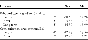
Descriptive summaries of pulmonary valve pressure gradients by echocardiography and cardiac catheterisation for all time points are presented in Table 1. Of the 53 study patients, six (11.3%) pre-pulmonary balloon valvuloplasty pulmonary valve gradients/measurements by catheterisation were not available.
Pulmonary valve gradient (mean, standard deviation) measured by echocardiography and cardiac catheterisation at different time points.

The peak-to-peak gradient by catheterisation of 42.5±19.6 mmHg before pulmonary balloon valvuloplasty was reduced to 13±7.7 mmHg after pulmonary balloon valvuloplasty (p<0.0001). The pre-pulmonary balloon valvuloplasty peak gradient by echocardiography was 60.6±14.7 mmHg. The first echocardiogram was performed at a median of 2 days (0–142 days) following pulmonary balloon valvuloplasty. A total of five patients presented with critical pulmonary stenosis determined by a requisite need for immediate intervention or prostaglandin dependence. The peak gradient by echocardiography after pulmonary balloon valvuloplasty was reduced to 25.5±12.1 mmHg (p<0.0001). At the most recent follow-up (4.8±2.3 years after pulmonary balloon valvuloplasty), the average peak gradient further decreased to 14.8±16 mmHg from the first post-pulmonary balloon valvuloplasty echocardiogram (p<0.0001).
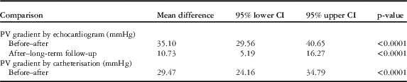
The estimated differences in pulmonary valve gradient by echocardiogram and by catheterisation with 95% confidence interval are presented in Table 2. As reported, results from the mixed model analysis indicated differences in mean pulmonary valve peak gradient by echocardiography before pulmonary balloon valvuloplasty to after pulmonary balloon valvuloplasty as well as after pulmonary balloon valvuloplasty to the most recent follow-up (both, p<0.0001). There was also a difference in peak-to-peak pulmonary valve gradient by catheterisation from before pulmonary balloon valvuloplasty to after pulmonary balloon valvuloplasty (p<0.0001).
Comparison of mean difference in pulmonary valve (PV) gradient between time points by echocardiography and cardiac catheterisation.

CI=confidence interval
Pulmonary regurgitation
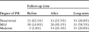
A summary of the degree of pulmonary regurgitation by time points is presented in Table 3. None of the patients had severe pulmonary regurgitation. Before pulmonary balloon valvuloplasty, 62% of patients had no/trivial pulmonary regurgitation, and 21% had no/trivial pulmonary regurgitation at long-term follow-up; however, 79% of patients had mild pulmonary regurgitation or less at long-term follow-up. Following-up each patient over time, the Cochran–Mantel–Haenszel test indicated a difference in the degree of pulmonary regurgitation before and after pulmonary balloon valvuloplasty (p<0.0001), but no difference between the post-pulmonary balloon valvuloplasty level and the most recent follow-up level (p=0.52).
Pulmonary regurgitation (PR) severity at three time points (before pulmonary balloon valvuloplasty, after pulmonary balloon valvuloplasty, and at long-term follow-up).

Re-intervention
There were no significant procedural complications. Only three patients (6%) required re-intervention for worsening re-stenosis (two surgical and one pulmonary balloon valvuloplasty). The patient with repeat pulmonary balloon valvuloplasty was asymptomatic with mild residual stenosis (10–15 mmHg) 5 years after repeat pulmonary balloon valvuloplasty with mild-to-moderate pulmonary regurgitation; one surgical patient was asymptomatic with mild pulmonary stenosis and mild pulmonary regurgitation 4 years after surgery. The other surgical patient was lost to follow-up.
Discussion
There was an immediate improvement in pulmonary valve gradient following pulmonary balloon valvuloplasty in children with isolated pulmonary valve stenosis. This study demonstrates that these patients have an ongoing reduction in their pulmonary valve peak instantaneous gradient by echocardiography, decreasing by 10 mmHg over 5 years. These findings support those reported 20 years earlier by O’Connor et al. and Masura et al.Reference O’Connor, Beekman, Lindauer and Rocchini 2 , Reference Masura, Burch, Deanfield and Sullivan 3 Moreover, these data demonstrate that there are both immediate and ongoing benefits of pulmonary balloon valvuloplasty. The continued improvement in the transvalvar gradient is likely attributed to the decrease in infundibular obstruction as right ventricular hypertrophy improves following resolution of valvar stenosis.
Pulmonary regurgitation
A known consequence of balloon valvuloplasty procedures is the possibility of producing or worsening valve insufficiency. Previous studies have established that the pulmonary valve can be effectively and safely dilated if the balloon diameter is selected between 120 and 140% of the pulmonary annulus diameter. This has been shown to maximise the reduction of the valvar gradient without introducing significant pulmonary regurgitation and other procedural complications.Reference Radtke, Keane, Fellows, Lang and Lock 9 , Reference McCrindle 10
We found an increase in patients with higher degrees of pulmonary regurgitation following pulmonary balloon valvuloplasty; however, this has not progressed over time. More importantly, none of the patients in our study required intervention for pulmonary regurgitation. This is in contrast with the results reported by Poon et al.Reference Poon and Menahem 11 who found a significant increase in the number of patients with moderate pulmonary regurgitation at long-term follow-up (median 5.64 years) compared with immediately after pulmonary balloon valvuloplasty. Rao et al.Reference Rao, Galal, Patnana, Buck and Wilson 12 found that the overall incidence gradually increased from immediately after pulmonary balloon valvuloplasty to intermediate-term follow-up – that is, <2 years following pulmonary balloon valvuloplasty – and at long-term follow-up (median 7 years). Similar late follow-up results were reported by Ganty et al.Reference Garty, Veldtman, Lee and Benson 13
Study limitations
This study was limited by its retrospective nature. As a result, uniformity of timing of echocardiographic evaluation was not possible. Standardised follow-up intervals have not been established, and therefore discreet comparisons of follow-up cannot be performed.
Conclusions
Pulmonary balloon valvuloplasty remains a safe and effective treatment for children with isolated pulmonary valve stenosis with excellent outcomes and no mortality; 94% of patients require no further intervention, and residual stenosis improves with later follow-up. Although there is a mild increase in pulmonary regurgitation following pulmonary balloon valvuloplasty, the degree of pulmonary regurgitation does not progress at long-term follow-up. Furthermore, significant pulmonary regurgitation requiring pulmonary valve replacement was not observed in our patients, with a few requiring re-intervention for valve re-stenosis.
Acknowledgements
The authors have no acknowledgements. Authors’ Contributions: J.P., M.R., E.B., and J.B. all contributed to the study design; acquisition, analysis, and interpretation of data; drafting the paper and revising it critically; and approved the final versions.
Financial Support
This research received no specific grant from any funding agency, commercial, or not-for-profit sectors.
Conflicts of Interest
None.
Ethical Standards
The authors assert that all procedures contributing to this study complied with the ethical standards of the relevant national guidelines on human experimentation and with the Helsinki Declaration of 1975, as revised in 2008, and has been approved by our Institutional Review Board.