It was in 1888 that Fallot emphasised the frequent co-existence of four morphological features in hearts of patients presenting with “la maladie bleu”, suggesting that these features constituted a tetralogy.Reference Fallot 1 It was Abbott,Reference Abbott 2 however, who suggested that the tetralogy should be described in his name. Several decades later, Van Praagh et alReference Van Praagh, Van Praagh, Nebesar, Muster, Sinha and Paul 3 suggested that the tetralogy, in reality, was a monology, whose essence was the underdevelopment of the subpulmonary infundibulum. Anderson and Tynan subsequently endorsed the notion of a monology, but rather than concentrating on underdevelopment of the subpulmonary infundibulum, as proposed by Van Praagh et al,Reference Van Praagh, Van Praagh, Nebesar, Muster, Sinha and Paul 3 they chose to emphasise the feature of the antero-cephalad deviation of the outlet septum relative to the septomarginal trabeculation.Reference Anderson and Tynan 4 It subsequently emerged, however, that such antero-cephalad deviation of the muscular outlet septum was insufficient, in itself, to serve as the phenotypic feature of tetralogy, as a comparable arrangement was to be found in the so-called “Eisenmenger malformation”. The additional feature required to differentiate tetralogy from the Eisenmenger lesion was a “squeeze” at the mouth of the infundibulum between the outlet septum and abnormal septoparietal trabeculations.Reference Anderson and Weinberg 5 , Reference Anderson, Spicer, Giroud and Mohun 6 In this regard, clinicians have suggested that, in some patients initially having the Eisenmenger malformation, subsequent development of infundibular stenosis can produce an eventual picture of tetralogy of Fallot.Reference Fukuda, Suzuki and Ito 7 With this in mind, and as we had previously described our findings in a series of hearts obtained from patients known to have the Eisenmenger malformation,Reference Restivo, di Gioia, Anderson, Carletti and Gallo 8 we have now compared those findings with the comparable features as seen in a series of hearts obtained from patients diagnosed with tetralogy of Fallot. We analysed in detail the features of the subpulmonary infundibular anatomy, concentrating on the mode of insertion of the muscular outlet septum relative to the limbs of the septomarginal trabeculation, also known as septal band. In a previous study, Goor et alReference Goor, Lillehey and Edwards 9 have already noted variations in this feature, but we are unaware of others who have assessed these aspects of the lesion. Furthermore, we analysed the morphological sequels of these two modes of insertion. In addition to assessing the mode of insertion of the outlet septum, we also analysed the extent of rightward position of the aortic root, using the same methodolgy as described in our earlier study.Reference Restivo, di Gioia, Anderson, Carletti and Gallo 8 We also assessed the longitudinal extension of the subpulmonary infundibulum and we more specifically assessed the extension of the subpulmonary infundibular sleeve by a morphometric analysis, comparing the features in tetralogy with those found in the Eisenmenger malformation.Reference Restivo, di Gioia, Anderson, Carletti and Gallo 8 On this basis, we hope to adjudicate on the phenotypic relationship between tetralogy of Fallot and the Eisenmenger malformation.
Materials and methods
We examined 43 heart specimens previously catalogued in the cardiac archive of the Museum of Pathologic Anatomy, Policlinico Umberto I, University of Rome “La Sapienza”, as having tetralogy of Fallot. The criteria used to make the diagnosis were consistent with those initially described by Fallot himselfReference Fallot 1 – namely, antero-cephalad deviation of the outlet septum, biventricular connection of the overriding aortic root, an interventricular communication opening to the outlet of the right ventricle between the limbs of the septomarginal trabeculation, and narrowing of the subpulmonary infundibulum. These features were present in all our specimens except one, in which the aorta arose exclusively from the right ventricle. We also found the “squeeze” at the mouth of the subpulmonary infundibulum between the deviated outlet septum and abnormal septoparietal trabeculations in all specimens. In one specimen, originally catalogued as tetralogy of Fallot, however, we found no evidence of the squeeze at the mouth of the subpulmonary infundibulum, nor diffuse narrowing of the infundibulum itself, but instead we found isolated pulmonary valvar stenosis. This heart, therefore, because of the unequivocal overriding of the aortic root, was re-catalogued as an uncommon variant of the Eisenmenger malformation with pulmonary valvar stenosis. For the subsequent purposes of comparison, we added this case to the 11 previously identified as exhibiting the Eisenmenger malformation.Reference Restivo, di Gioia, Anderson, Carletti and Gallo 8 The specimen in which the aortic root was exclusively supported above the morphologically right ventricle, but in association with the phenotypic feature of the infundibular squeeze, was retained as exhibiting tetralogy. The concomitant right ventricular hypertrophy, again as outlined by Fallot,Reference Fallot 1 was constant but of variable severity. Among the 43 examples deemed to exhibit tetralogy of Fallot, six had pulmonary atresia rather than stenosis. All specimens, some consisting of heart–lung blocks, had been opened in the conventional manner, thus permitting examination of all four cardiac chambers at the time of autopsy. In several of them, it proved necessary to make additional dissections so as to expose the posterior surface of the component of the aortic root supported by the right ventricle and the anterior component that remained supported by the left ventricle. All specimens had been fixed in 10% formalin. The age of death of the patients from whom the hearts had been obtained ranged from 3 months to 12 years. Whenever necessary, for purposes of illustration, the margins of cut areas of the specimens were reconstituted without suturing. We also observed several additional intracardiac anomalies, as reported below. Although our purpose was to make morphological, rather than morphometric, observations, we did measure, in each specimen, the distance between the most proximal attachment of the pulmonary valvar leaflets and the inferior margin of the outlet septum anteriorly, thus providing the length of the subpulmonary infundibulum, and the distance between the attachments of the aortic valvar leaflets and the margin of the outlet septum posteriorly, with this measurement giving the length of the subaortic outlet septum. We then considered that the difference between these two measurements correlated with the length of the free-standing subpulmonary infundibular sleeve. This then permitted us, as had been the case in our previous study of the Eisenmenger malformation,Reference Restivo, di Gioia, Anderson, Carletti and Gallo 8 to make inferences regarding the length of the muscular outlet septum relative to the extent of the free-standing subpulmonary infundibular sleeve. Further inferences regarding the extent of the subpulmonary infundibulum were derived by assessing the attachment of the inferior margin of the outlet septum relative to the cranial base of the septomarginal trabeculation.
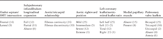
As in our earlier study,Reference Restivo, di Gioia, Anderson, Carletti and Gallo 8 we also quantified the degree of anatomical rightward rotation of the aortic root by assessing the relationship of the left coronary leaflet of the aortic valve relative to the aortic leaflet of the mitral valve (Table 1). In this regard, it should be noted that the rightward rotation of the aortic root, as assessed anatomically or clinically, does not coincide with the rotation of the aortic root observed during the embryonic development. During normal development, the aortic root rotates in the opposite direction – that is, leftwards – in order to be transferred from the right to the left ventricle.Reference Pernkopf and Wirtinger 10 – Reference Bajolle, Zaffran and Kelly 13 For this reason, we consider it more appropriate to describe the “rightward position” of the aortic root, both in the settings of tetralogy of Fallot and the Eisenmenger malformation.
Summary of the relevant features of the 43 examined hearts with tetralogy of Fallot.

SMT=septomarginal trabeculation
Statistical analysis
Data are expressed as mean±standard deviation, with median values. Differences between the parameters were determined using Student’s t-test and were considered significant at p<0.05.
Results
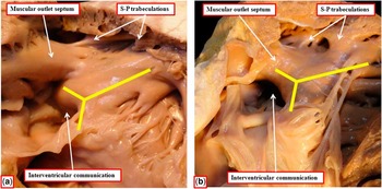
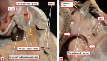
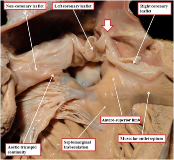
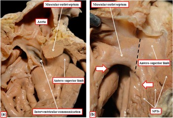
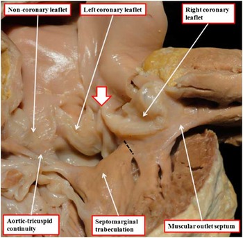
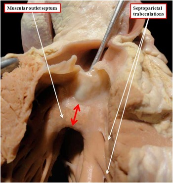
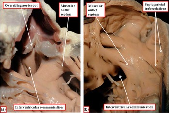
Our overall findings are summarised in Tables 1 and 2. The distinctive phenotypic feature of tetralogy of Fallot, namely, antero-cephalad deviation of the outlet septum relative to the limbs of the septomarginal trabeculation, along with a squeeze at the mouth of the subpulmonary infundibulum due to abnormal septoparietal trabeculations, and with poor circular expansion of the subpulmonary infundibulum, was present in all hearts examined, including our retained specimen in which the aortic root was supported exclusively above the cavity of the right ventricle (see below). The outlet septum itself was muscular in all our specimens. As had been observed by Goor et al,Reference Goor, Lillehey and Edwards 9 we found two discrete arrangements between the septal attachment of the muscular outlet septum and the septomarginal trabeculation. We distinguished between these as a “frontal” insertion (Fig 1a) as opposed to a “lateral” insertion (Fig 1b). In the majority of cases, 34 in all, the septal attachment of the outlet septum was displaced “in front” of the anterosuperior limb of the septomarginal trabeculation (Fig 2a and b). In Figure 3, we show the subaortic view of the heart illustrated in Figure 2b, emphasising this frontal insertion. In a minority of nine cases, the outlet septum attached to the right posterior margin of the anterosuperior limb of the septomarginal trabeculation in “lateral” fashion (Fig 4a and b). In hearts with the outlet septum attached in the frontal fashion, the septoparietal trabeculations combined to produce a relatively discrete narrowing at the mouth of the infundibulum (Fig 2b). In those with a lateral attachment, the septoparietal trabeculations were attached cranially to the left anterior margin of the anterosuperior limb of the septomarginal trabeculation itself, but more caudally to the inferior margin of the outlet septum (Fig 4b). In those with frontal attachment, when viewed from the aspect of the subaortic component of the overriding aortic root, the right posterior margin of the anterosuperior limb of the septomarginal trabeculation was visible as a discrete entity (Fig 3). In contrast, in specimens showing lateral insertions, this margin of the anterosuperior limb was not visible, as it was fused with the outlet septum (Fig 4a). In the majority of cases, the bicuspid pulmonary valve was found (Table 1).
This figure shows the two types of insertion of the outlet septum relative to the anterosuperior limb of the septomarginal trabeculation. Panel ( a ) shows frontal insertion. The cranial limbs of the yellow Y indicate the anterosuperior and postero-inferior limbs of the septomarginal trabeculation. Note the location of the right posterior margin of the anterosuperior limb of the septomarginal trabeculation behind the outlet septum. Panel ( b ) shows the lateral insertion, in which the inferior margin of the outlet septum extends caudally to meet the cranial base of the septomarginal trabeculation, thus creating an arch. In this latter arrangement, it is no longer possible to identify the right margin of the anterosuperior limb, as it is fused with the outlet septum itself. S-P=septo-parietal.

The images show the features of the frontal attachment of the outlet septum to the anterosuperior limb of the septomarginal trabeculation, or septal band. Panel ( a ) shows the view towards the interventricular communication, which opens between the limbs of the septomarginal trabeculation. Note the squeeze between the outlet septum and the septoparietal trabeculations (SPTs), which is the phenotypic feature of tetralogy of Fallot, together with circular underdevelopment of the infundibulum, as in the majority of cases. Note also the unattached right posterior margin of the anterosuperior limb of the septomarginal trabeculation behind the outlet septum (compare with Figs 1b and 4a) and also the almost equal extension of the subpulmonary and subaortic surfaces of the outlet septum and, consequently, the absence of the subpulmonary infundibular sleeve. Panel ( b ) shows a different heart with the view as seen from the subpulmonary infundibulum. The arrows show the narrowed mouth of the infundibulum, which is elongated.

The image shows the subaortic aspect of the heart shown in Figure 2b. The muscular outlet septum is displaced in front of the anterosuperior limb of the septomarginal trabeculation. The commissure between the two coronary aortic valvar leaflets (white arrow with red borders) is in line with the right posterior margin of the anterosuperior limb of the septomarginal trabeculation. The interventricular communication is perimembranous due to the aortic-tricuspid valvar fibrous continuity.

The images show the features of the lateral attachment of the outlet septum to the anterosuperior limb of the septomarginal trabeculation. Panel ( a ) shows the view towards the interventricular communication. Note that the outlet septum is attached to the right posterior margin of the anterosuperior limb of the septomarginal trabeculation (compare with Fig 2a). Panel ( b ) shows the lateral attachment in a different heart, as seen from its infundibular aspect (compare with Fig 2b). The deviation of the outlet septum, together with the abnormal septoparietal trabeculations (SPTs) and the poor circular expansion of the infundibulum itself, as in the majority of cases, again produce the phenotypic feature of tetralogy of Fallot. SPTs are fused in part with the inferior edge of the outlet septum and in part with the anterosuperior limb of the septomarginal trabeculation. The arrows indicate the narrow infundibular mouth, although the walls of the specimen are purposely and artificially dilated for photographic reasons. The dotted line indicates the approximate junctional borderline between the outlet septum and the anterosuperior limb of the septomarginal trabeculation.

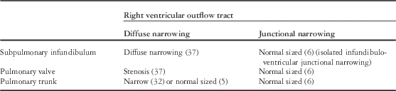
Forty-three cases of tetralogy of Fallot subdivided in two groups in terms of the right ventricular outflow tract.

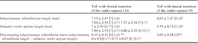
In hearts with the lateral attachment having bicuspid pulmonary valves, the support of the posterior valvar leaflet, or the left posterior leaflet in the tricuspid cases, was shared by both the muscular outlet septum and the anterosuperior limb of the septomarginal trabeculation (Fig 4b). Comparison with the height of the attachment of the right coronary leaflet of the aortic valve along the dorsal surface of the outlet septum revealed the presence of a free-standing subpulmonary infundibular sleeve, as was also the case in our earlier study of the Eisenmenger malformation;Reference Restivo, di Gioia, Anderson, Carletti and Gallo 8 the sleeve is seen to a greater extent in the normal heart. We show, indeed, in Figure 5 that the subaortic side of the outlet septum was shorter in these hearts when compared with the overall length of the subpulmonary infundibulum (Fig 4b). This is not the case in hearts with bicuspid pulmonary valves when the outlet septum inserted frontally, as seen in the majority of hearts (Fig 2b). In these specimens, the posterior valvar leaflet was supported exclusively by the outlet septum. In these hearts, therefore, in part because of the lack of support by the anterosuperior limb of the septomarginal trabeculation, both sides of the outlet septum were of equal, or approximately equal, length (Fig 2a). The subpulmonary infundibular sleeve, therefore, was relatively extensive in the hearts with a lateral attachment of the outlet septum (Fig 4a and b). The sleeve itself, in contrast, was absent (Fig 2a), or extremely short, in the hearts with the outlet septum attached in frontal fashion. We have summarised in Table 3 the morphometric data concerning the effective length of the outlet septum versus the overall length of the subpulmonary infundibulum. We have also introduced, in the same table, the morphometric data relative to the length of the free-standing subpulmonary infundibular sleeve, obtained as the difference between the subpulmonary infundibulum and the subaortic outlet septum. Assessment of the data included in Table 3 shows that the feature underscoring the differences is the length of the subaortic outlet septum. In Figure 6, we illustrate a specimen with lateral insertion of the outlet septum, but with a significantly shorter subpulmonary infundibulum.
The image shows the subaortic right ventricular view of the heart shown in Figure 4b. There is a continuous lateral intersection between the outlet septum and the anterosuperior limb of the septomarginal trabeculation. The commissure between the two coronary aortic valvar leaflet (white arrow with red borders) is more posterior and leftward and displaced away from the septomarginal trabeculation when compared with the arrangement seen in Figure 3. The dotted line indicates, as in Figure 4b, the approximate junction between the outlet septum and the anterosuperior limb of the septomarginal trabeculation. Note that the entire block, including outlet septum and the septoparietal trabeculations fusing with the inferior margin of the outlet septum itself, is totally overturned in order to show the subaortic view. Note also the short length of the subaortic side of the outlet septum as compared with the whole subpulmonary infundibular length (Fig 4b).

In this heart, there is a relatively short subpulmonary infundibulum with lateral insertion of the outlet septum. The red arrow refers to the developmental extension of the outlet septum.

Morphometric analysis of the outlet septal morphology in 43 cases of tetralogy of Fallot (ToF).

The values are expressed as mean±SD (median); the number of examined hearts is reported in cursive style. In the group with frontal insertion of the outlet septum (34), there was no significant statistical difference between the lengths of the subpulmonary infundibulum and the subaortic outlet septum. More specifically, in the same group, 27 cases showed identical length on the subpulmonary and subaortic sides of the outlet septum, with no existence of a subpulmonary infundibular sleeve; in seven cases, there was a minimal, but not significant, difference of length with an extremely short subpulmonary infundibular sleeve. In the nine cases with a lateral insertion of the outlet septum, there was a significant difference between the lengths of the subpulmonary infundibulum and the subaortic outlet septum (p<0.00025). As a consequence, there was a significant difference in the length of the free-standing subpulmonary infundibular sleeve between the frontal and the lateral insertion groups (p<0.00025).
*, **=p<0.00025
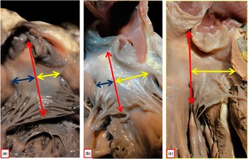
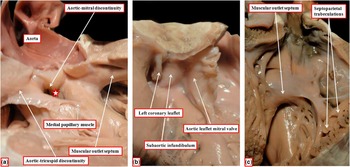
In terms of rightward position of the overriding aortic root, which was supported in biventricular fashion in all hearts except one, we used the same criteria to determine mild, intermediate, or severe rightward position as for our previous study of Eisenmenger defects.Reference Restivo, di Gioia, Anderson, Carletti and Gallo 8 We found mild rightward position in 25 hearts (Fig 7a), an intermediate position in five hearts (Fig 7b), and severe rightward position in 12 hearts (Fig 7c). In the remaining heart, the rightward position was such that the aortic root was supported exclusively above the right ventricle (Fig 8a). This specimen also had discontinuity between the leaflets of the aortic and mitral valves (Fig 8b); in other words, there were bilateral infundibulums or conuses. The heart, nonetheless, retained the phenotypic infundibular morphology of tetralogy of Fallot (Fig 8c). As in the Eisenmenger malformation,Reference Restivo, di Gioia, Anderson, Carletti and Gallo 8 in the specimens with mild and intermediate degrees of aortic rightward position, we considered the ventriculoarterial connections to be concordant, whereas in the hearts with severe aortic rightward position, where right ventricular predominance of the aortic overriding occurred, the ventriculoarterial connection was that of double-outlet right ventricle.
The images show mild ( a ), intermediate ( b ), and severe ( c ) rightward rotation of the aortic root as assessed on the basis of the perpendicular line drawn from the commissure between the left coronary and non-coronary leaflets of the aortic valve (red double-headed arrow) and the inferior margin of the aortic leaflet of the mitral valve. The yellow and blue double-headed arrows show the proportion of the mitral leaflet to the left and right sides of the perpendicular line, respectively. Note that, in the severe arrangement ( c ), the left coronary leaflet is entirely related to the mitral leaflet.

The images show the extreme aortic rightward position in which the aortic root is supported exclusively by the right ventricle, with panel ( a ) showing the subaortic view of the right ventricle, panel ( b ) showing the left ventricular view, and panel ( c ) showing the subpulmonary view of the right ventricle. The characteristic squeeze between the deviated outlet septum and the septoparietal trabeculations provides the phenotypic feature of tetralogy of Fallot ( c ). In panel ( a ) the interventricular communication (star) can be seen, also showing a muscular postero-inferior rim producing aortic-tricuspid discontinuity. The medial papillary muscle is also present, along with aortic-mitral discontinuity, meaning that there is a completely muscular subaortic infundibulum.

By assessing the relationships between the leaflets of the aortic and tricuspid valves, we noted fibrous continuity postero-inferiorly in 24 hearts, making the interventricular communication perimembranous (Fig 3). In the remaining 19 hearts, the postero-inferior limb of the septomarginal trabeculation had fused with the inner heart curvature, producing a muscular postero-inferior rim to the septal defect (Fig 8a). In all the cases with fibrous continuity, we were able to identify an abortive remnant of the interventricular membranous septum, also known as the membranous flap. In cases with the postero-inferior muscular rim, an intact and fully developed membranous septum was present caudal to the rim.
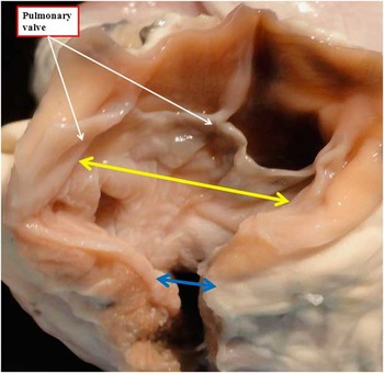
The medial papillary muscle, also known as the muscle of Lancisi, was identified in only eight of the 43 hearts (Table 1; Fig 8a). The outlet septum itself was poorly developed in four of these eight cases. In the majority of cases, 25 in all, the pulmonary valve was bifoliate and dysplastic. In 10 cases, the valve had three well-developed leaflets. In two hearts, the valve showed a dysplastic unicommissural arrangement. The remaining six specimens had pulmonary valvar atresia. In 37 of the hearts, the subpulmonary infundibulum was diffusely narrowed, with concomitant narrowing of the pulmonary valve, including six cases with pulmonary valvar atresia (Table 2). In five of the 37 hearts, nonetheless, despite the diffuse narrowing of the infundibulum, the pulmonary trunk was relatively normally sized (Table 2). In the remaining six hearts (Table 2), the subpulmonary infundibulum was normally and widely expanded, showing only a stricture at the mouth of the infundibulum itself (Fig 9). In all six of these cases, nonetheless, the infundibulum had significant length, with the outlet septum being frontally displaced. In four, there was mild aortic rightward position, with one each showing intermediate and severe rightward position. In the specimen excluded from the group deemed to have tetralogy of Fallot, rather than a diffuse subpulmonary narrowing and a muscular squeeze at the mouth of the subpulmonary infundibulum, there was only valvar pulmonary stenosis, meaning that the appropriate diagnosis was the Eisenmenger malformation (Fig 10), rather than tetralogy of Fallot.
The figure shows the features of a normally well-expanded infundibulum, with a pulmonary valve of good size (double-headed yellow arrow), but a severe narrowing at the mouth of the infundibulum (double-headed blue arrow).

The images show the features of the heart initially diagnosed as tetralogy of Fallot, but re-categorised as being an example of the Eisenmenger malformation with pulmonary valve stenosis. Panel ( a ) showing the view from the inlet of the right ventricle, the overriding aortic root, and the deviated outlet septum. Panel ( b ) shows the unobstructed and wide subpulmonary infundibulum, despite the presence of mild hypertrophy of the septoparietal trabeculations. Pulmonary valve stenosis can be seen.

Discussion
Despite extensive studies conducted by morphologists on the lesion initially described by Fallot, some of its more subtle features remain controversial. It has long been recognised that the essence of the anomaly is antero-cephalad deviation of the muscular outlet septum. It was often thought that this finding, in itself, was sufficient to serve as its phenotypic feature.Reference Anderson and Weinberg 5 , Reference Anderson, Spicer, Giroud and Mohun 6 A similar antero-cephalad deviation of the muscular outlet septum, however, is also to be found in the so-called Eisenmenger malformation, but in the absence of subpulmonary infundibular narrowing.Reference Eisenmenger 14 In this regard, some authorsReference Fukuda, Suzuki and Ito 7 , Reference Altshuler 15 , Reference Gasul, Dillon, Vrla and Hait 16 studying patients with the Eisenmenger malformation, who subsequently developed hypertrophic obstruction of the right ventricular outflow tract and cyanosis, were inclined to hypothesise that the Eisenmenger malformation might, in certain circumstances, undergo transformation into tetralogy of Fallot. The findings of our present study do not support this hypothesis. Indeed, as we had previously conducted an extensive morphological study identifying the essential features of the Eisenmenger malformation,Reference Restivo, di Gioia, Anderson, Carletti and Gallo 8 we were able to compare the findings from that study with the present ones, obtained using hearts from patients already diagnosed as exhibiting tetralogy of Fallot. We were able to confirm that the phenotypic feature of tetralogy, over and above the antero-cephald deviation of the outlet septum, was the squeeze produced at the mouth of the subpulmonary infundibulum by the combination of the outlet septum with the septoparietal trabeculations. In most of the specimens currently examined, this feature also co-existed with poor circular expansion of the entirety of the subpulmonary infundibulum, contrasting with the arrangement observed in the Eisenmenger malformation, where despite the anterior deviation of the outlet septum itself, and a certain degree of hypertrophy of the septoparietal trabeculations, the infundibulum was well expanded (Fig 10b). In a minority of cases (six cases) with the phenotypic feature of tetralogy of Fallot, nonetheless, the infundibulum was well expanded, with a pulmonary valvar orifice of almost normal dimensions, despite the obvious squeeze at the mouth of the infundibulum (Fig 9). All six of these cases, furthermore, showed a frontal insertion of the outlet septum above the anterosuperior limb of the septomarginal trabeculation, indicating a greater degree of anterior displacement of the outlet septum, as compared with those deemed to have the lateral arrangement of insertion. Despite that, the infundibulum was well expanded (Fig 9). It follows, therefore, that a more anterior deviation of the outlet septum does not correlate with the degree of infundibular narrowing. We also found no evidence to support the notion that a superior and anterior insertion of the parietal end of the embryonic outlet septum might be the cause for the poor expansion of the parietal wall of the subpulmonary infundibulum.
We were able to confirm, nonetheless, that as previously described by Goor et al,Reference Goor, Lillehey and Edwards 9 some of the hearts with the obvious phenotypic feature of the infundibular squeeze, and therefore qualifying as examples of tetralogy, exhibited a lateral rather than a frontal insertion of the outlet septum relative to the anterosuperior limb of the septomarginal trabeculation. Goor et alReference Goor, Lillehey and Edwards 9 had distinguished these groups as having “in-row” and “T-shaped” junctions, which we have described as our “lateral” and “frontal” arrangements. The majority of our specimens showed the frontal arrangement (Table 1), in keeping with their finding of most hearts having a “T-shaped” junction. It was then noteworthy that, despite all of the hearts diagnosed as tetralogy having the outlet septum attached in lateral fashion, and also having the unequivocal feature of the infundibular squeeze, the attachments of the arterial valvar leaflets on either side of the deviated muscular outlet septum were comparable with the arrangement found in the hearts with the Eisenmenger malformation,Reference Restivo, di Gioia, Anderson, Carletti and Gallo 8 including the additional case examined in our current series with valvar rather than muscular infundibular stenosis (Fig 10a and b). In our previous series of specimens with the Eisenmenger malformation,Reference Restivo, di Gioia, Anderson, Carletti and Gallo 8 and in the one heart now included in the present study, the insertion of the outlet septum was constantly found in lateral arrangement. This lateral arrangement of insertion of the supraventricular crest into the anterosuperior limb of the septomarginal trabeculation also occurs in the normal heart.
The cases also showed the comparable variation in terms of the rightward deviation of the aortic root.Reference Goor, Lillehey and Edwards 9 , Reference Isaaz, Cloez, Marcon, Worms and Pernot 17 Thus, in those with frontal attachment of the outlet septum, the anterior commissure between the coronary leaflets of the overriding aortic valve, which was established to represent the axis of rightward rotation of the aortic root,Reference Goor, Lillehey and Edwards 9 , Reference Isaaz, Cloez, Marcon, Worms and Pernot 17 was in line cranially with the right posterior surface of the anterosuperior limb of the septomarginal trabeculation (Fig 3). This frontal insertion of the outlet septum and the position of the anterior aortic valvar commissure were maintained independently of the extent of rightward position of the aortic root (compare Figs 3 and 8a). In contrast, in those with the lateral attachment of the outlet septum, the commissure occupied a more cranial, leftward, and posterior position, being discrete from the anterosuperior limb of the septal band (Figs 5 and 10a), in concomitance with the more posterior position of the aortic root when compared with the hearts with frontal arrangement of the outlet septum. Thus, the fixed and unchanged axis of aortic rotationReference Goor, Lillehey and Edwards 9 , Reference Isaaz, Cloez, Marcon, Worms and Pernot 17 applies only, according to our findings, in cases having a frontal insertion of the outlet septum. In hearts with the lateral arrangement, in contrast, this axis moves following the anatomical rightward rotation of the aortic root.Reference Restivo, di Gioia, Anderson, Carletti and Gallo 8 When assessing the nine cases of tetralogy of Fallot with lateral insertion of the outlet septum, in only one case was the rightward position of the aortic root found to be severe. It was intermediate in three, with the remaining five cases having only mild rotation. Thus, among the 25 cases overall with a mild rightward position of the aortic root, 20 had a frontal insertion of the outlet septum, with only five placed in the lateral group. We suggest that these findings show that the specific intersection of the outlet septum relative to the limbs of the septomarginal trabeculation does not correlate with the extent of rightward positioning of the aortic root.
We then found further correlations in terms of the inferred components of the subpulmonary infundibulum. It is now well established that, in the normal heart, the larger part of the subpulmonary infundibulum is composed of the free-standing infundibular sleeve, with little, if any, of the infundibulum made up of a true muscular outlet septum. It was not possible in our specimens to section the septal aspect of the subpulmonary infundibulum so as to determine precisely, in the malformed hearts, the infundibular components made up by the muscular septum as opposed to the infundibular sleeve. We were able to estimate these values, however, by assessing the attachments of the proximal hinges of the valvar leaflets on the subaortic and subpulmonary aspects of the muscular outlet septum, and by comparing the length of the free-standing outlet septum as assessed on the subaortic aspect with the overall measurement of the length of the subpulmonary infundibulum (Table 3), as had been achieved in our previous study devoted to the Eisenmenger malformation.Reference Restivo, di Gioia, Anderson, Carletti and Gallo 8 When we compared the findings in the different tetralogy groups, the free-standing component of the muscular outlet septum was always found to be consistently reduced in those with a lateral junctional arrangement, including all the Eisenmenger malformation cases that always showed this sort of lateral arrangement,Reference Restivo, di Gioia, Anderson, Carletti and Gallo 8 when compared with those with the frontal arrangement (Table 3); concomitantly, and in contrast, the free-standing subpulmonary infundibular sleeve was always longer in the setting of these malformations with the lateral junction of the outlet septum (Table 3), although not to the extent as seen in the normal heart.Reference Anderson and Weinberg 5 , Reference Anderson, Spicer, Giroud and Mohun 6 , Reference Anderson and Becker 18 When we assess the morphometric data as shown in Table 3, it is our opinion that the feature underscoring the differences found is the length of the subaortic side of the outlet septum. This feature might be of interest in directing future investigations by embryologists and developmental biologists. Taken together, and in keeping with the opinion of Van Praagh et al,Reference Van Praagh, Van Praagh, Nebesar, Muster, Sinha and Paul 3 we believe that our findings show that there is, indeed, a phenotypic difference between hearts diagnosed as tetralogy of Fallot when compared with those with the Eisenmenger malformation. As discussed above, therefore, we doubt whether patients having the features of the Eisenmenger defect can progress to become examples of tetralogy of Fallot. We accept, nonetheless, that it is not possible to be certain of this interpretation, as morphologists are constrained by their ability to study hearts only at a solitary point in the natural history of the patient from whom they were obtained. Only clinical investigations, using our suggested phenotypic features to distinguish between tetralogy of Fallot and the Eisenmenger malformation, will produce the definitive answer to this ongoing conundrum.
If we return to our specific findings in the group of hearts with tetralogy of Fallot, we found that, when considered to the findings in the overall group, the subpulmonary infundibulum was relatively short in one-quarter of the series. This is not unexpected, as the outlet septum can also be fibrous, and therefore exceedingly short when deviated in the antero-cephalad fashion in association with abnormal septoparietal trabeculations.Reference Anderson and Weinberg 5 , Reference Anderson, Spicer, Giroud and Mohun 6 These hearts have doubly committed and juxta-arterial interventricular communications.Reference Griffin, Sullivan, Anderson and Macartney 19 On this basis, doubt was expressed as to whether such hearts should be considered as representing tetralogy.Reference Griffin, Sullivan, Anderson and Macartney 19 Had the endocardial cushions that divide the developing outflow tract muscularised, however, there is little doubt that the hearts would have shown unequivocal evidence of tetralogy of Fallot. There is, therefore, a spectrum in terms of the length of the subpulmonary infundibulum, but when assessed relative to the overall length of the right ventricle the infundibulum is longer in the majority of tetralogy cases than in the normal heart.Reference Becker, Connor and Anderson 20 , Reference Howell, Ho, Anderson and Elliott 21 Our study provides no support for the notion that the subpulmonary infundibulum is always short.Reference Van Praagh, Van Praagh, Nebesar, Muster, Sinha and Paul 3 The subpulmonary infundibulum, therefore, is arguably better developed in the setting of tetralogy of Fallot, although always of lower volume due to its narrowness. In this regard, it was also noteworthy that the infundibulums were uniformly long and narrow in most of the hearts with frontal attachment of the outlet septum, but relatively less well longitudinally developed in those with a lateral arrangement. These findings suggest that development of the cardiac outflow tract might be under different molecular controls, regulating its circular as opposed to longitudinal expansion. The fact that the infundibulum is well expanded in some examples of tetralogy of Fallot adds additional value to this notion.
Among all, two hearts identified in the course of our current investigation are worthy of further comment. In the first, we found a completely muscular subaortic infundibulum (see Fig 8a and b). This finding lends further support to the fact that discontinuity between the leaflets of the aortic and mitral valves does not preclude the diagnosis of tetralogy of Fallot.Reference Dickinson, Wilkinson, Smith, Hamilton and Anderson 22 Therefore, it is hardly surprising that in our initial study we also encountered an example of Eisenmenger’s malformation with bilateral infundibulums and mild aortic rightward position.Reference Restivo, di Gioia, Anderson, Carletti and Gallo 8 As Van Praagh et alReference Van Praagh, Davidoff, Chin, Shiel, Reynolds and Van Praagh 23 have emphasised, lesions should be diagnosed on the basis of their intrinsic morphology and not according to other features than themselves, which may be variable. The obvious logic of this principle, dubbed the morphological method, rules out the use of the bilateral conus as a feature of double-outlet right ventricle, since the bilateral infundibulums are themselves another variable feature. In the second case, we identified a heart with the obvious infundibular squeeze, making it an example of tetralogy of Fallot, yet with the aortic root was supported exclusively by the right ventricle. Adopting the principle of the morphological method, the presence of the phenotypic infundibular squeeze validates the diagnosis of this heart as showing tetralogy of Fallot despite the lack of aortic valvar overriding. It cannot be coincidental, therefore, that Fallot also reported this feature in one of the hearts described in his initial series.
As far as the medial papillary muscle is concerned, this muscle was identified only in eight cases out of 43. Among these eight cases, the outlet septum was underdeveloped in half of the cases. These findings, therefore, do not support the viewReference Van Mierop and Wiglesworth 24 that the medial papillary muscle develops normally when the outlet septum undergoes full development, whereas it fails to develop when the outlet septum is proximally deficient. Indeed, the outlet septum is no more than a virtual entity in the normal heart.
Our findings are also of potential surgical significance, as we found a perimembranous interventricular communication in three-fifths of our specimens. In all of these hearts, we also found evidence of the membranous flap.Reference Suzuki, Yo, Anderson and Deanfield 25 As was emphasised by Kurosawa et al,Reference Kurosawa, Morita, Yamagishi, Shimizu, Becker and Anderson 26 when present, this flap can be used to secure safe anchorage of the patch that restores septal integrity without fear of damaging the atrioventricular conduction axis. It is more usual in this setting, however, that surgeons will deviate the stitches caudally in the postero-inferior quadrant of the area requiring closure, thus avoiding the site of penetration of the atrioventricular bundle. In two-fifths of our hearts; nonetheless, the right ventricular aspect of the channel between the ventricles had exclusively muscular borders. In these hearts, because the muscular postero-inferior rim separates an intact membranous septum from the margin of the defect, sutures can judiciously be placed in the rim without fear of producing iatrogenic heart block.
In conclusion, we propose that our investigation lends further support to the notion that, although hearts exhibiting tetralogy of Fallot and the Eisenmenger malformation share the feature of variable rightward position of the aortic root, they are phenotypically discrete entities. We suggest that emphasis on the distinguishing phenotypic features will provide important specific morphological information to embryologists and molecular biologists involved in establishing the morphogenesis of this group of outflow tract malformations.
Acknowledgements
None.
Financial Support
This research received no specific grant from any funding agency, commercial, or not-for-profit sectors.
Conflicts of Interest
None.