Isolation of the right subclavian artery is a rare congenital aortic arch anomaly, which may be associated with other anomalies such as tetralogy of Fallot, interruption of the aortic arch, transposition of the great arteries, and with syndromes such as Williams–Beuren, Peutz–Jeghers, and 22q11 deletion.Reference McElhinney, Silverman, Brook, Reddy and Hanley 1 – Reference Hokari, Kuroda, Furukawa, Houkin and Iwasaki 4 The subclavian artery is either connected to a patent arterial duct or to the arterial ligament. Asymptomatic patients are diagnosed clinically with low-volume arm pulses, radio-radial pulse delay, and blood pressure differences between the two arms. If there is significant “steal” phenomenon, the patients present with cerebrovascular insufficiency and/or ipsilateral arm claudication.Reference Hokari, Kuroda, Furukawa, Houkin and Iwasaki 4 – Reference Schreiber, Cleuziov, Eicken and Lange 6 Surgical reimplantation of the subclavian artery to the aorta is performed in such patients.Reference McElhinney, Silverman, Brook, Reddy and Hanley 1 , Reference Schreiber, Cleuziov, Eicken and Lange 6 We report three patients with isolation of the right subclavian artery who had good improvement in the blood supply to the arm after successful interventional closure of the patent arterial duct.
Patients, methods, and results
Patient 1
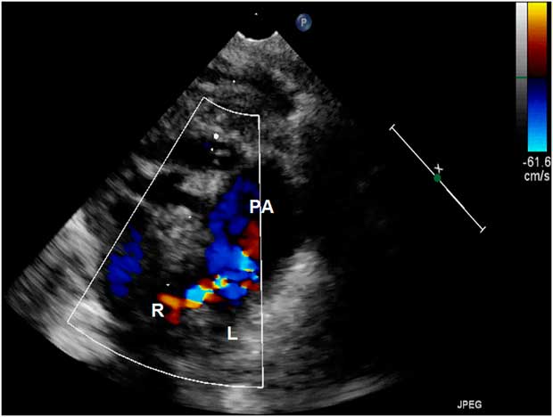
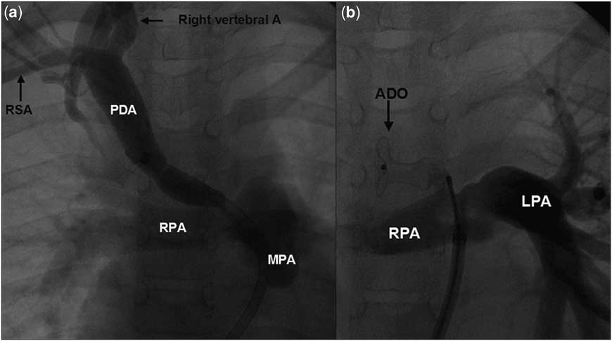
A 10-year-old girl presented with right arm claudication, recurrent chest infections, poor somatic growth, and congestive cardiac failure. Clinical examination showed low-volume right arm pulses, right arm blood pressure of 50 mm of mercury lower than the left arm, and a loud continuous murmur along the right upper sternal border. The continuous colour Doppler flow from the duct into the right pulmonary artery on the parasternal short-axis view of the echocardiogram was observed (Fig 1; Movie clip S1). The aortic arch was left sided, but the aortic origin of the duct was not visualised on the echocardiogram. An aortic arch angiogram identified isolation of the right subclavian artery, which filled late from the right vertebral artery. There was a large patent arterial duct of 8 mm, which filled the right pulmonary artery, and there was a faint opacification of the subclavian artery beyond the first rib. From the femoral venous approach, the arterial duct was entered into from the right pulmonary artery using a catheter, exchanged with a long 8-Fr sheath, and a right subclavian arteriogram was performed (Fig 2; Movie clip S2). This confirmed the “steal” from the right subclavian artery to the pulmonary artery, through the large patent arterial duct. On performing balloon occlusion of the arterial duct, there was improvement of the pulse volume of the right radial artery. The right arm systolic blood pressure improved from 80 to 110 mm of mercury, but was still 20 mm of mercury lower than the left arm blood pressure. The arterial duct was closed using a 12/10 Amplatzer duct occluder device (AGA Medical, Plymouth, Minnesota, United States of America; Fig 2).
Colour Doppler interrogation of the duct on the high parasternal short-axis view (Movie clip 1) shows a dilated main PA and abnormal entry of the duct on the mediastinal part of the R. This echocardiographic finding along with a lack of visualisation of the ductal ampulla in the distal arch may give clues for the diagnosis of the isolation of the right subclavian artery. L = left pulmonary artery; PA = pulmonary artery; R = right pulmonary artery.

Angiogram ( a ) (Movie clip 2) was performed after a venous sheath was advanced from the MPA into the isolated RSA through the PDA. Duct closure with a large duct occluder device is shown in ( b ). LPA = left pulmonary artery; MPA = main pulmonary artery; PDA = patent arterial duct; RPA = right pulmonary artery; RSA = right subclavian artery.

Patient 2
A 9-year-old girl with sickle cell anaemia was evaluated for cardiac murmur. Clinical examination revealed a continuous murmur along the right parasternal border. The echocardiogram showed a continuous flow into the right pulmonary artery from a patent arterial duct. The aortic arch was left sided. During cardiac catheterisation, an aortogram showed delayed opacification of the right subclavian artery from which the pulmonary arteries were opacified. Collaterals from the left internal mammary artery filled the right internal mammary artery, which later filled the right subclavian artery. It was decided to close the right-sided patent arterial duct to prevent run-off from the right subclavian artery into the pulmonary artery. A 7-Fr long Mullins sheath was placed in the pulmonary artery using the femoral venous approach, and a 6-Fr Judkins right coronary catheter was used to cross the duct using a 0.035″ hydrophilic guidewire. A right subclavian artery injection showed a 2.5-mm tortuous patent arterial duct with significant run-off into the pulmonary artery. A controlled-release 6.5-mm-diameter 4-loop Flipper stainless steel Dacron-fibred embolisation coil (Cook Medical, Bloomington, Indiana, United States of America) was deployed and released. The delayed phase of the right carotid artery angiogram showed good opacification of the right subclavian artery and complete closure of the arterial duct.
Patient 3
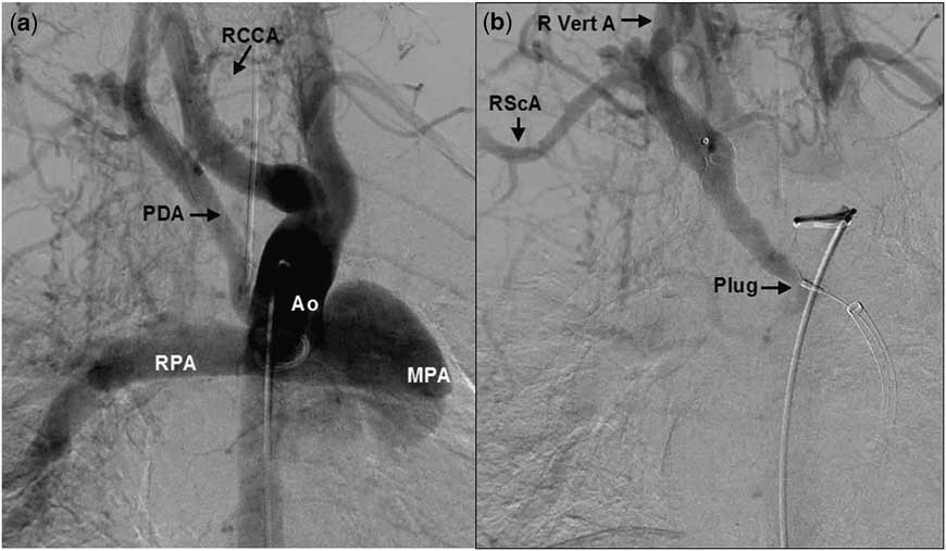
A 10-year-old girl presented at the age of 6 days with duct-dependent systemic circulation, and the echocardiogram showed aortic arch interruption (type B) and ventricular septal defect. She underwent urgent surgical repair of the interrupted arch combined with patch closure of the ventricular septal defect. She was also found to have renal cysts. During the subsequent follow-up, at the age of 8 years, an echocardiogram and colour Doppler showed a bicuspid aortic valve with a Doppler gradient of 50 mmHg and abnormal continuous flow into the right pulmonary artery. A magnetic resonance scan showed excellent repair of the left-sided aortic arch, a left-to-right shunt of 1.3:1, an additional right pulmonary artery filling from a right-sided patent arterial duct, and a small right subclavian artery. At the age of 10 years, catheter closure was attempted using femoral venous and arterial access. An ascending aortogram showed late filling of a right-sided arterial duct opacifying the right pulmonary artery, as well as a small right subclavian artery (Fig 3). The aortic pressure was 89/56, mean 71 mmHg, and the main pulmonary artery pressure was 34/17, mean 25 mmHg. From the right femoral vein and the pulmonary artery, the arterial duct was entered into using an 8-Fr-long sheath. A 14-mm-diameter Amplatzer Vascular Plug (AVP II, AGA Medical) was delivered and released within the arterial duct. After the procedure, the aortogram showed satisfactory occlusion of the arterial duct and improved opacification of the right subclavian artery (Fig 3).
Digital subtraction angiogram ( a ) showing the ascending Ao with its first branch RCCA. There is a delayed filling of the PDA, MPA, and RPA through the right vertebral artery. aortogram ( b ) after closure of the duct with a plug shows delayed but improved filling of the RScA through the R Vert A. Ao = aorta; MPA = main pulmonary artery; PDA = patent arterial duct; RCCA = right common carotid artery; RPA = right pulmonary artery; RScA = right subclavian artery; R Vert A = right vertebral artery.

Discussion
Although isolation of the left subclavian artery with a right aortic arch has been reported, a left aortic arch with isolation of the right subclavian artery is rare.Reference McElhinney, Silverman, Brook, Reddy and Hanley 1 – Reference Edwards 8 Mathieson et al were the first to describe the isolation of the right subclavian artery as a distinct entity.Reference Mathieson, Silver and Gordon Culham 7 Embryological explanations are based on the regressions at two locations within the ipsilateral fourth aortic arch.Reference Edwards 8 These regressions are located proximal and distal to the subclavian artery, resulting in its isolation. In this entity, the distal right dorsal aorta involutes after cephalad migration of the right seventh intersegmental (subclavian) artery to the level at which the right sixth (ductal) arch normally joins the proximal dorsal aorta. This together with involution of the right fourth arch leaves the subclavian artery isolated from the aortic arch but connected to the pulmonary artery via the right-sided arterial duct. This regression of the ipsilateral fourth aortic arch and persistence of the ipsilateral sixth distal aortic arch results in isolation of the right subclavian artery. During foetal life, the pulmonary artery supplies the isolation of the right subclavian artery because of the higher pulmonary vascular resistance. After postnatal decrease of the pulmonary vascular resistance, persistent flow through the patent arterial duct may result in a significant run-off from the subclavian artery to the ipsilateral pulmonary artery.Reference Nath 9
In patients presenting with clinical features of a patent arterial duct, the presence of a murmur along the right sternal border and weak right arm pulses may provide clinical clues for the diagnosis of isolation of the right subclavian artery. Once these clinical signs are observed, colour Doppler interrogation of the pulmonary arteries on the parasternal short-axis view will indicate an unusually rightward location of the ductal insertion site in the right pulmonary artery. An additional echocardiographic finding is the failure to identify the aortic origin of the duct from the suprasternal long-axis view of the left aortic arch.Reference McElhinney, Silverman, Brook, Reddy and Hanley 1
Surgical reimplantation of the isolated right subclavian artery to the aorta either directly or using an interposition graft is a possible solution in symptomatic patients, if it is a part of the surgical correction for associated congenital cardiac defects and indeed if it is recognised pre-operatively.Reference Schreiber, Cleuziov, Eicken and Lange 6 There was one patient with an aortic arch interruption and a ventricular septal defect, which were repaired during the neonatal period. However, isolation of the right subclavian artery was diagnosed several years later. The entire ductal tissue may have to be removed, as it may not be ideal to leave any ductal tissue in the region of the surgical anastomosis. Removal of the ductal tissue may necessitate the use of an interposition graft in many instances. If an interposition graft is used to reimplant the isolated right subclavian artery to the aorta, long-term antiplatelet therapy may be needed to maintain graft patency.Reference McElhinney, Silverman, Brook, Reddy and Hanley 1 When the ductal tissue is very narrow and long, a carotid-to-subclavian artery bypass is carried out for the relief of the subclavian steal syndrome.Reference Hokari, Kuroda, Furukawa, Houkin and Iwasaki 4 Owing to the absence of other associated structural cardiac defects requiring surgical correction and to minimise the chances of sickle cell crises in one of our patients, we did not opt for surgical reimplantation of the subclavian artery. A search of the literature revealed one case report of percutaneous closure of an arterial duct in a patient with a right aortic arch with isolation of the left subclavian artery.Reference Jones, Garabedian and Grifka 10
We used transcatheter closure of the patent arterial ducts in all of our patients to prevent “steal” from the right subclavian artery into the pulmonary artery. The improvement of the blood pressure in the right arm after device closure of the duct in the first patient confirmed the improved arm vascularity as a result of the procedure. Even though ductal “steal” into the pulmonary artery ceases after the ductal closure, subclavian “steal” from the circle of Willis may persist. The long-term consequences of marginally lower blood pressures in the arm remain unknown and warrant long-term follow-up. Selected patients with isolation of the subclavian artery with a patent arterial duct may benefit from transcatheter duct closure.
Conclusion
Isolation of the right subclavian artery is a rare aortic arch anomaly with a peculiar embryological basis. There are some distinct clinical signs and specific echocardiographic clues that may aid in the diagnosis of this lesion even before angiography. Haemodynamical isolation of the right subclavian artery may result in the dual subclavian and ductal “steal” phenomenon. Transcatheter closure of patent arterial duct can be performed in selected patients with isolation of the right subclavian artery to minimise this “steal”.
Supplementary materials
For supplementary material referred to in this article, please visit http://dx.doi.org/10.1017/S1047951112002314